Effects of Seaweeds on Matrix Metalloproteinases Derived from Normal Human Dermal Fibroblasts and Human Fibrosarcoma Cells
In-Hwan Park
1, Sang-Hoon Lee
2, Se-Kwon Kim
3, Dai-Nghiep Ngo
4, You-Jin Jeon
5and Moon-Moo Kim
1*
1
Department of Chemistry, Dong-Eui University, Busan 614-714, Korea
2
Korea Food Research Institute, Sungnam-Si, Gyeonggi-Do, 463-746, Korea
3
Department of Chemistry, Pukyong National University, Busan 608-737, Korea
4
Department of Biochemistry, Faculty of Biology, University of Science, Vietnam National University, HoChiMinh city, Vietman
5
Faculty of Biomedical Science, Cheju National University, Jeju 690-756, Korea
Received August 18, 2011 /Revised November 14, 2011 /Accepted November 17, 2011
In recent years novel potential pharmocological candidates have been looked for in animal, seaweed, sponge, fungi and marine bacteria resources. In this study, matrix metalloproteinases (MMPs) that play an important role in metastasis, arthritis, chronic inflammation and wrinkle formation were used as target enzymes to screen therapeutic agents. The inhibitory effects of several marine algae including green algae (5 species), red algae (18 species) and brown algae (4 species) methanolic extracts on MMPs were investigated in human dermal fibroblasts and human fibrosarcoma cell line (HT1080 cells) using gelatin zymography. In human dermal fibroblasts, the inhibition of MMP-2 was observed in Laurencia okamurae, Polysiphonia japonica, Grateloupia lanceolate and Sinkoraena lancifolia of red algae. In contrast, MMP-2 activation was enhanced in Enteromorpha compressa and E. linza of green algae, and Peltaronia bighamiae and Sargassum thunbergii of brown algae. In human fibrosarcoma cells, MMP-9 activation was decreased in the presence of S. thunbergii of brown algae, Polysiphonia japonica in red algae and E. com- pressa and E. linza of green algae. The interesting finding is that E. compressa and E. linza of green algae, and S. thunbergii of brown algae exhibited a positive effect on MMP-2 in normal cells, but a negative effect on MMP-9 in cancer cell lines. These results suggest that E. compressa and E. linza of green algae, and S. thunbergii of brown algae contain potential therapeutic ingredients for cancer treatment.
Key words : Matrix metalloproteinase, seaweeds, gelatin zymography; HT1080 cells, human dermal fibroblasts
*Corresponding author
*Tel:+82-51-890-1511, Fax:+82-51-890-2620
*E-mail : [email protected]
Introduction
MMPs have been reported to be involved in the degrada- tion of extracellular matrix in process of several diseases such as chronic inflammation, periodontal disease, chronic obstructive pulmonary disease, arthritis, and cancer meta- stasis, and also in the aging process, such as skin wrinkling [4,6,20,22,25]. MMPs are a family of secreted or trans- membrane zinc-endopeptidases that are capable of digesting extracellular matrix (ECM), such as fibrillar and non-fibrillar collagens, fibronectin, laminin, elastin and basement mem- brane glycoproteins under physiological conditions [3]. The MMPs in tumor growth and invasion have been identified revealed by studies with either MMP-2 knockout mice hav- ing reduced melanoma tumor progression and angiogenesis [13]. In recent years it was reported that ultraviolet B-in-
duced enhancement of gelatinase activity in the skin contrib-
utes to wrinkle formation through the destruction of base-
ment membrane structure and dermal collagen and thus top-
ical application of inhibitors of MMPs may be an effective
way to overcome this problem [12]. Therefore, inhibition of
MMP activity in the extracellular space has been extensively
studied as an approach to inhibit metastasis and wrinkle
formation. At present, several MMP inhibitors are under
clinical trials and most of these MMP inhibitors are synthetic
peptides, chemically modified tetracyclines, bisphosphonates
or compounds isolated from natural sources. However, most
of these drugs are reported to exert side effects such as, mus-
culoskeletal pain in tendons and joints [18]. Until now, MMP
inhibitors that are capable of applying to clinical trials have
not been reported from marine resources. Therefore, we took
an effort to screen marine algal species possessing MMP in-
hibitory compounds. Based on our screening studies, the ex-
tracts of 3 marine algae present in the subtidal regions of
Jeju Island in Korea had an excellent efficacy to inhibit MMP
activities. Enteromorpha, an edible green alga, produces a diz- zying array of bioactive compounds that have proven to be useful in the treatment of certain bacterial & viral infections, cancer and inflammation [10,19]. Enteromorpha compressa, a marine green algal species, grows extensively in North coast- al Andhra Pradesh. Besides its nutritional importance it has also been identified as source of anti-anaphylactic com- pounds [25]. Green algae of the genus Ulva (syn.
Enteromorpha) are common, green macroalgae found throughout the world in the upper intertidal zone of sea- shores and as a fouling organism on a variety of man-made structures including ships’ hulls [2]. Sargassum fulvellum known as Hejo is used as a food additive , and recorded many uses in treatment of lump, dropsy, swollen and pain- ful scrotum, and urination problems with no side effects.
[5,14].
Therefore, in the present work we carried out a detailed study to investigate the inhibitory effects of several algae on MMPs secreted from normal human cells and human fi- brosarcoma cells.
Materials and Methods Chemicals
Dulbecco’s Modified Eagle’s Medium (DMEM), Trypsin-EDTA, penicillin/streptomycin/ amphotericin (10,000 U/ml, 10,000 μg/ml, and 2,500 μg/ml, respectively) and fetal bovine serum (FBS) were obtained from Gibco BRL (Grand island, NY, USA). HT1080 cells were obtained from American Type of Culture Collection (Manassas, VA, USA).
Human dermal fibroblasts (HDFs) were kindly donated by LG HG & CM Research Institute (Daejeon, Korea). MTT re- agent, gelatin and PMA (phorbol 12-myristate 13-acetate) were purchased from Sigma Chemical Co. (St. Louis, MO, USA). All the other materials required for culturing of cells were purchased from Gibco BRL.
Preparation of seaweeds extract used in this study
The seaweeds used in this study are as follows:
Pterocladiella capillacea, Scinaia okamurae, Laurencia okamurae, Grateloupia lanceolate, Chondrophycus undulates, Gracilaria tex- torii, Polysiphonia japonica, Halymenia dilatata, Gloiopeltis fur- cata, Glacilaria verrucosa, Chondria cssicaulis, Chondrus crispus, Prionitis cornea, Ecklonia cava, Martensia denticulate, Lomantaria catenata, Acrosorium flabellatum, Sinkoraena lancifo-
lia, Geldium amansii, Shizymenia dubyi, G. elliptica, Capopeltis affinis, G. filicina, Pterocladiella capillacea, Scinaia okamurae, G.
lanceolate of red algae, Codium contractum, Hizikia fusiform, Enteromorpha compressa, Ulva conglobata , U. pertusa, Monostroma nitidum of green algae, Myagropsis myagroides, Sargassum horneri, Ishige sinicola, Pachydictyon coriaceum, Idhige okamurai, S. siliquastrum, S. coreanum, Peltaronia bigha- miae, S. thunbergii, S. thunbergii, Padina arborescen, S. fulvel- lum, Myagropsis myagroides of brown algae. They were col- lected along Jeju Island coast of Korea. Fresh seaweeds were washed three times with tap water to remove salt, epiphytes and sand attached to the surface of the samples and stored at -20℃. The frozen samples were lyophilized and homogenized using a grinder before extraction. The dried seaweeds powder (1 kg) was extracted three times with MeOH (1:10 w:v) at room temperature for 24 hr while stirring, filtered using Whatman No. 41 and evapo- rated in vacuum. For the bioassay, the fractions were dis- solved in dimethylsulfoxide (DMSO) and distilled water according their solubility, respectively.
Cell culture
Cell lines were separately grown as monolayers in T-75 tissue culture flasks (Nunc, Denmark) at 5% CO
2and 37
oC humidified atmosphere using appropriate media supple- mented with 10% fetal bovine serum, 2 mM glutamine and 100 μg/ml penicillin-streptomycin. DMEM was used as the culture medium for human fibrosarcoma cells (HT1080 cells) and human dermal fibroblasts (HDFs) cultured primarily from human fetal skin. Cells were passaged 3 times a week by treating with trypsin-EDTA and used for experiments af- ter 5 passages.
MTT assay
Cytotoxic levels of EC extract on HT1080 cells, and HDFs
were measured using MTT (3-(4,5-dimethyl-2-yl)-2,5-diphe-
nyltetrazolium bromide). The cells were grown in 96-well
plates at a density of 5×10
3cells/well. After 24 hr, cells were
washed with fresh medium and were treated with different
concentrations of seaweeds extract. After 48 h of incubation,
cells were rewashed and 20 μl of MTT (5 mg/ml) was added
and incubated for 4 hr. Finally, DMSO (150 μl) was added
to solubilize the formazan salt formed and amount of for-
mazan salt was determined by measuring the OD at 540 nm
using an GENios
®microplate reader (Tecan Austria GmbH,
Austria). Relative cell viability was determined by the
amount of MTT converted into formazan salt. Viability of cells was quantified as a percentage compared to the control (OD of treated cells − OD of blank / OD of control − OD of blank×100) and dose response curves were developed.
The data were expressed as mean from at least three in- dependent experiments and p<0.05 was considered significant.
Gelatin zymography
Activities of MMP-2 and MMP-9 were determined by zymography as described previously [11] in the presence or absence of seaweeds extract. After cells were exposed to various concentrations of seaweeds extract for 1 hr prior to treatment of 10 ng/ml and 100 ng/ml PMA for HDFs and HT1080 cells respectively, incubation was continued in FBS-free medium for 3 days. Conditioned medium contain- ing 50 μg of total protein (or reaction products of various concentrations of EC extract with active MMP-2) was re- suspended in a sample buffer (125 mM Tris-HCl, pH 6.8, 3% SDS, 40% glycerol, 0.02% bromophenol blue) without boiling and electrophoresed under non-reducing conditions on 10% polyacrylamide gels containing 1.5 mg/ml gelatin.
After electrophoresis, the gels were washed twice with 50 mM Tris-HCl (pH 7.5) containing 2.5% Triton X-100 and incubated overnight at 37°C in a developing buffer con- taining 10 mM CaCl
2, 50 mM Tris-HCl, and 150 mM NaCl.
The gels were stained with 0.25% Coomassie Blue R-250 in 30% methanol and 10% acetic acid, and de-stained in the same solution without the Coomassie Blue dye.
Gelatinolytic bands were observed as clear zones against the blue background and the intensity of the bands was es- timated using Image Master Software, Amersham Pharmacia Biotech (Sweeden).
Statistical analysis
Comparisons of all data were performed using two-tailed, unpaired Student’s t-test. A P value less than 0.05 was con- sidered statistically significant. Data are expressed as means
±SE.
Results
Effect of seaweeds methanol extract on cell growth
The aim of this study was to investigate the inhibitory effect of seaweeds methanolic extracts on MMPs activity
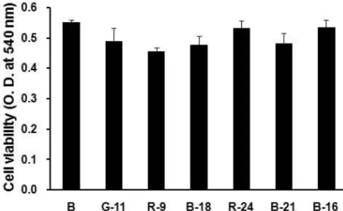
in normal human dermal cells and human fibrosarcoma cells. First of all, the cytotoxic effects of seaweeds methanolic extracts were examined by MTT assay. As shown in Fig.
1, even though all data were not shown here, all seaweeds methanolic extracts did not exert any cytotoxic effect on HDFs at the 1 μg/ml. Therefore, all seaweeds methanolic extracts with no cytotoxity could be focused for screening of therapeutic candidates in this study.
Inhibitory effect of seaweeds methanol extracts on MMPs activity in human dermal fibroblasts
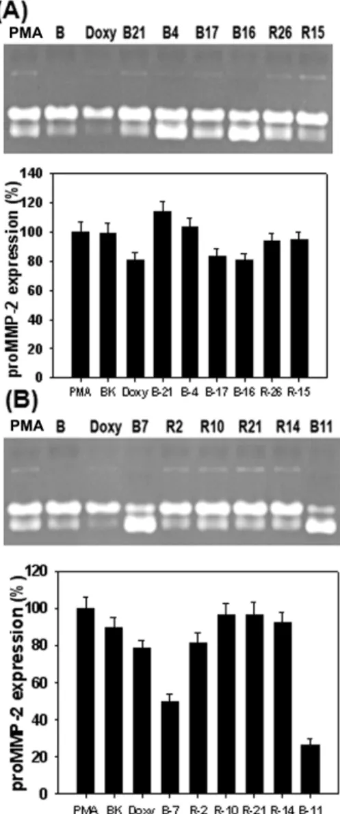
In order to investigate whether seaweeds methanolic ex- tracts inhibits MMPs secreted from HDFs cultured primarily from human fetal skin, conditioned medium of HDFs treated with seaweeds methanolic extracts at 1 μg/ml for 6 day after treated with PMA to induce MMPs expression was subjected to gelatin zymography. As shown in Fig. 2A, HDFs secreted mainly proMMP-2 and a little amount of proMMP-9. It also observed that MMP-2 was activated by treatment with dox- ycycline at 1 μg/ml, a positive control. In this experiment the M. myagroides methanolic extract greatly increased MMP-2 activity. In contrast, G. lanceolate methanolic extract inhibited MMP-2 activity. It showed higher inhibition of MMP-2 activity compared to that of doxycycline at same concentration. As shown in Fig. 2B, in this study the E. linza methanolic extract remarkably activated pro-MMP-2 com- pared with PMA treatment group. On the other hand, P.
Fig. 1. Effect of seaweeds methanolic extracts on viability of human dermal fibroblasts. The cells were treated with 1 μg/ml of seaweeds methanolic extracts and cell via- bility was determined by MTT assay after 24 hr. Data are given as means of values±SD from three in- dependent experiments. The seaweeds used in this ex- periment were as follows: G11 (
M. nitidum
), R9 (L. oka- murae
), B18 (M. myagroides
), R24 (P. japonica
), B21 (P.
coriaceum
) and B16 (S. coreanum
).Fig. 2. Effects of seaweeds methanolic extracts on activities of MMP-2 and MMP-9 from HDFs. The cells were treated with 10 ng/ml of PMA under serum-free conditions to induce MMP expressions. After 6 days of incubation, conditioned media were reacted with various concen- trations of seaweeds methanolic extracts for 1 hr, and then gelatin zymography was performed. Doxycycline (Doxy) was used as the positive control. (A) The sea- weeds used in this experiement were as follows: R13 (
P. capillacea
), R19 (S. okamura
e), G2 (C. contractum
), R9 (L. okamurae
), B18 (M. myagroides
) and R4 (G. lanceolate
).(B) The seaweeds used in this experiement were as fol- lows: B15 (
S. horneri
), G9 (S. okamura
e), R25 (C. un- dulates
), R1 (G. textorii
), B5 (I. sinicola
) and R24 (P. japon- ica
). Lower panel represents respective relative enzyme activities as percent of blank group.japonica methanolic extract inhibited MMP-2 activation by more 30 % than doxycycline treatment group. In next experi- ment, both I. okamurai and S. coreanum methanolic extracts
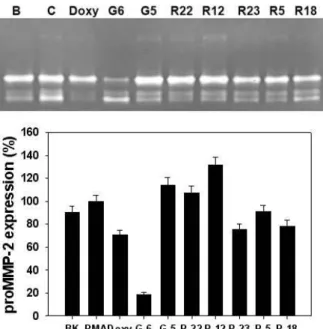
Fig. 3. Effects of seaweeds methanolic extracts on activities of MMP-2 and MMP-9 from HDFs. The cells were treated with 10 ng/ml of PMA under serum-free conditions to induce MMP expressions. After 6 days of incubation, conditioned media were reacted with various concen- trations of seaweeds methanolic extracts for 1 hr, and then gelatin zymography was performed. Doxycycline (Doxy) was used as the positive control. (A) The sea- weeds used in this experiement were as follows: B21 (
P.
coriaceum
), B4 (I. okamurai
), B17 (S. siliquastrum
), B16 (S.
coreanum
), R26 (H. dilatata
) and R15 (G. furcata
). (B) The seaweeds used in this experiement were as follows: B7 (P. bighamiae
), R2 (G. verrucosa
), R10 (C. cssicaulis
), R21 (C. crispus
), R14 (P. cornea
) and B11 (S. thunbergii
). Lower panel represents respective relative enzyme activities as percent of blank group.showed MMP-2 inhibition by 50% compared with Blank
group. However, it was found that G. furcata methanolic ex-
tract inhibited MMP-2 activation compared with PMA treat-
Fig. 4. Effects of seaweeds methanolic extract on activities of MMP-2 and MMP-9 from HDFs. The cells were treated with 10 ng/ml of PMA under serum-free conditions to induce MMP expressions. After 6 days of incubation, conditioned media were reacted with various concen- trations of seaweeds methanolic extracts for 1 hr, and then gelatin zymography was performed. Doxycycline (Doxy) was used as the positive control. The seaweeds used in this experiement were as follows: G6 (
E. com- pressa
), G5 (U. pertusa
), R22 (M. denticulate
), R12 (L. cate- nata
), R23 (A. flabellatum
), R5 (S. lancifolia
) and R18 (G.
amansii). Lower panel represents respective relative en- zyme activities as percent of blank group.
ment group, as shown in Fig. 3A. In contrast, both P. bigha- miae and S. thunbergii methanolic extracts greatly enhanced MMP-2 activity compared to PMA treatment group, as shown in Fig. 3B. They exhibited 3 times more inhibition of MMP-2 activity than PMA treatment group. The excellent inhibitory effect of E. compressa methanolic extract on MMP-2 activity was observed in Fig. 4. Furthermore, 50% more in- hibition of MMP-2 activity was observed at 1 μg/ml com- pared with doxycycline treatment group.
Inhibitory effect of seaweeds methanol extracts on MMPs activity in human fibrosarcoma cells
In this experiment, the effect of seaweeds methanolic ex- tracts on MMPs was examined in human fibrosarcoma cells, HT1080 cell line that has widely used to study metastasis.
In a similar method to normal human dermal fibroblasts, the conditioned medium of HT1080 cells treated with sea- weeds methanolic extracts at 1 μg/ml for 3 d after treated
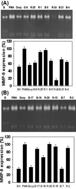
Fig. 5. Effects of seaweeds methanolic extracts on expression and activity of MMP-2 and MMP-9 in HT1080 cells. The cells stimulated with 10 ng/ml of PMA to induce MMP expression were treated with various concentrations of seaweeds methanolic extracts under serum-free con- ditions for 3 days. MMP activities in conditioned media were determined by zymography as described in the text. Doxycycline (Doxy) was used as the positive control. (A) The seaweeds used in this experiement were as follows: R10 (
C. cssicaulis
), R21 (C. crispu
), R14 (P.
cornea
), B22 (E. cava
), B11 (S. thunbergii
), G6 (E. com- pressa
) and G5 (U. pertusa
). (B) The seaweeds used in this experiement were as follows: R22 (M. denticulate
), R12 (L. catenata
), R23 (A. flabellatum
), R5 (S. lancifolia
), R18 (G. amansii
), B9 (H. fusiform
) and G4 (U. conglobata
).Lower panel represents respective relative enzyme ac- tivities as percent of blank group.
Fig. 6. Effects of seaweeds methanolic extracts on expression and activity of MMP-2 and MMP-9 in HT1080 cells. The cells stimulated with 10 ng/ml of PMA to induce MMP expression were treated with various concentrations of seaweeds methanolic extracts under serum-free con- ditions for 3 days. MMP activities in conditioned media were determined by zymography as described in the text. Doxycycline (Doxy) was used as the positive control. (A) The seaweeds used in this experiement were as follows: R17 (
S. dubyi
), B19 (P. arborescen
s), R3 (G.
elliptica
), B13 (S. fulvellum
), R8 (C. affinis
), R7 (G. filicina
) and R13 (P. capillacea
). (B) The seaweeds used in this experiement were as follows: R19 (S. okamura
e), G2 (C.
contractum
), G11 (M. nitidum)
, R9 (L. okamurae
), B18 (M.
myagroides
), R4 (G. lanceolate
) and B15 (S. horneri
). Lower panel represents respective relative enzyme activities as percent of blank group.with PMA was analyzed to analyzed using gelatin zymography. As shown in Fig. 5A, the gelatinolytic activity
Fig. 7. Effects of seaweeds methanolic extracts on expression and activity of MMP-2 and MMP-9 in HT1080 cells. The cells stimulated with 10 ng/ml of PMA to induce MMP expression were treated with various concentrations of seaweeds methanolic extracts under serum-free con- ditions for 3 days. MMP activities in conditioned media were determined by zymography as described in the text. Doxycycline (Doxy) was used as the positive control. (A) The seaweeds used in this experiement were as follows: G9 (
E. linza
), R25 (C. undulates
), R1 (G. textor- ii)
, R24 (P. japonica
), B21 (P. coriaceum
) and B4 (I. okamur- ai
). (B) The seaweeds used in this experiement were as follows: B17 (S. siliquastrum
), B16 (S. coreanum
), R26 (H.
dilatata)
, R15 (G. furcata
), B7 (P. bighamiae
) and R2 (G.
verrucosa
). Lower panel represents respective relative en- zyme activities as percent of blank group.at 92 kDa, which corresponds to the molecular mass of
pro-MMP-9, was more clearly detected in the conditioned
medium from PMA-stimulated HT1080 cells than HDFs. The inhibitory effect of doxycycline at 1 μg/ml on MMP-9 activ- ity was clearly observed in zymogram. Among seaweeds methanolic extracts in this experiment, S. thunbergii and E.
compressa methanolic extracts greatly reduced MMP-9 activ- ity as well as MMP-activity. However, there was no sig- nificant difference in inhibitory effect on MMP-9 activity be- tween other seaweeds and PMA treated group. Fig. 5B illus- trated that L. catenata methanolic extract greatly enhanced MMP-9 activation compared to PMA treated group. In con- trast, A. flabellatum showed the highest inhibitory effect on MMP-9 activity in other seaweeds. It was not observed that there was any effective algae with inhibition or activation on MMP-2 and MMP-9 in Fig. 6A. In the next experiment, as shown in Fig. 6B, several seaweeds exhibited an effective inhibition on MMP-2 and MMP-9 activities. In particular, both L. okamurae and G. amansii methanolic extracts exhibited a significant inhibitory effect on MMP-9 activity. As shown
Fig. 8. Effects of seaweeds methanolic extracts on expression and activity of MMP-2 and MMP-9 in HT1080 cells. The cells stimulated with 10 ng/ml of PMA to induce MMP expression were treated with various concentrations of
S. thunbergii
andE. linza
methanolic extracts under se- rum-free conditions for 3 days. MMP activities in con- ditioned media were determined by zymography as de- scribed in the text. Doxycycline (Doxy) was used as the positive control. Lower panel represents respective rela- tive enzyme activities as percent of blank group.in Fig. 7A. two seaweeds were found to be effective in in- hibiting MMP-2 and MMP-9 activities. E. linza methanolic extract showed a similar inhibitory effect to doxycycline treatment group. The other seaweed, P. japonica methanolic extract completely inhibited MMP-2 and MMP-9 activities.
In results of Fig. 7B, it was found that Peltaronia bighamiaeme- thanolic extract has a inhibitory effect on MMP-9 activity.
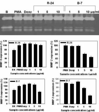
Inhibitory effect of 4 seaweeds methanol extracts in a dose dependent manner on MMP-2 and -9 activies in human fibrosarcoma cells
In order further confirm whether the seaweeds selected from above screening assay exert an inhibitory effect on MMP-2 or MMP-9 activity, 4 kinds of seaweeds methanolic extracts with increasing concentration were treated to HT1080 cells. As shown in Fig. 8, S. thunbergii methanolic extract did not show any clear inhibitory effect on MMP-9 activity, wheras it at 10 μg/ml exhibited an inhibitory effect
Fig. 9. Effects of seaweeds methanolic extracts on expression and activity of MMP-2 and MMP-9 in HT1080 cells. The cells stimulated with 10 ng/ml of PMA to induce MMP expression were treated with various concentrations of R24 (
P. japonica
) and B7 (P. bighamiae
) methanolic extracts under serum-free conditions for 3 days. MMP activities in conditioned media were determined by zymography as described in the text. Doxycycline (Doxy) was used as the positive control. Lower panel represents respective relative enzyme activities as percent of blank group.on MMP-2 activity. In contrast, E. linza methanolic extract not only revealed an inhibitory effect on MMP-9 activity at a concentration of 10 μg/ml but also exerted a dose depend- ent inhibitory effect on MMP-2 activity. Other two seaweeds, P. japonica and P. bighamiae, were investigated for the effect on MMPs activity. As shown in Fig. 9, it was observed that while P. japonica methanolic extract did not exhibit any in- hibitory effect on MMP-9 activity, it showed a clear in- hibitory effect on MMP-9 activity at a concentration of 10 μ g/ml. However, P. bighamiae methanolic extract exerted an inhibitory effect on both MMP-9 and MMP-2 activities in a dose dependent manner, indicating that it may be a poten- tial candidate for chemoprevention of metastasis
Discussion
In our study several marine seaweeds including green al- gae, brown algae and red algae collected from the southern coast of Korea was used to screen novel MMP inhibitors from marine natural resources. Methanol was applied to the seaweeds to extract as much ingredients as possible.
ethanol. In order to study the inhibitory effects of these ex- tracts on MMPs activity, gelatin zymography was utilized as an assay because of its accuracy and reproducibility even though there are many ways for study of MMPs. Using this assay, we investigated inhibitory effect of the seaweeds methanolic extracts on MMPs released from the cultured cells corresponding to normal human fibroblasts and human fibrosarcoma cells. For reliability of this experiment, doxycy- cline was used as a positive control to compare their effects.
Doxycycline is a synthetic tetracycline derivative that can inhibit MMP activity and potentially an effective therapeutic agent [1]. Longitudinal double-blind studies on humans with adult periodontitis have demonstrated that a sub-anti- microbial dose of doxycycline, previously reported to sup- press collagenase activity in the periodontal pocket, is safe and effective and has recently been approved by the FDA as an adjunct to scaling and root planning [7]. Furthermore, Periostat
®(CollaGenex Pharmaceuticals Inc.), a tetracycline analog containing doxycycline has been reported to inhibit collagenase activity but failed to act as an antibiotic [8].
We wondered whether a pothetial candidate could be screen from seaweeds and what is difference in effect on MMPs activity between normal cells and cancer cells. Our results demonstrate that same seaweeds are able to exert different effects on MMPs activity. These results suggest a possibility
that a therapeutic agent can act an efficacy in normal in- dividual and patient with cancer diffentially. In our study especially P. bighamiae , a brown algae, and E. linza, a green algae, revealed any clear inhibitory effect on both MMP-9 and MMP-2 activities in human fibrosarcoma cells, indicat- ing that they can possibly be a candidate to further study a concrete mechanism and single compound from them.
Previous study on Peltaronia bighamiae have reported that high content of active compounds such as polysaccharides
>30 kDa pholorotannins is responsible for anticoagulant ac- tivitiy [26]. Enteromorpha linza was reported to have an an- ti-inflammatory activity [16]. Furthermore, its inhibitory ef- fects on MMP-2 and MMP-9 activity at the same concen- tration were more effective and that of doxycycline. In addi- tion, our results demonstrated that both P. japonica, a red algae, and P. bighamiae, a brown alga, methanolic extracts revealed an excellent inhibitory effect on MMP-2 activity.
Our findings are consistent with previous study in detail on P. japonica that it attenuates Wnt/beta-catenin signaling via activation of NF-kappaB and can potentially be used as a chemopreventive agent against colon cancer [9]. On the other hand, recent study has shown that a water-soluble ex- tract of P. binghamiae inhibits the expression of adipogenic regulators in 3T3-L1 preadipocytes and reduces adiposity and weight gain in rats fed a high-fat diet [15]. Moreover, the inhibitory effects on MMP-2 were observed in L. okamur- ae, P japonica, G.. lanceolate and S. lancifolia of only red algae.
In contrast, the positive effects on MMP-2 activity were found in E. compressa and E. linza of green algae, and P. bigha- miae and S. thunbergii of brown algae. In human fibrosarcoma cells, the inhibitory effects on MMP-9 were observed in S.
thunbergii of brown algae, P. japonica in red algae and E. com- pressa and E. linza of green algae. The positive effects on MMP-9 activity were not observed in any algae used in this study. HT1080 cells mainly release proMMP-9 and a little amount of proMMP-2. In contrast, proMMP-2 is mainly ex- pressed in HDFs. The interesting finding is that E. compressa and E. linza of green algae, and S. thunbergii of brown algae exhibited a positive effect on MMP-2 in normal cells, but a negative effect on MMP-9 in cancer cell line. Previously it has been reported that fibrosarcoma HT1080 cells secrete type IV collagenase, MMP-2 and MMP-9, and that these en- zymes play a major role in cancer metastasis [27].
Furthermore, MMP-2 and MMP-9 can degrade type IV colla-
gen of base membranes and known to play a crucial role
in cancer invasion such as ovarian carcinoma [22].
Moreover, above results confirmed that the innibitory effect on MMP activity exert depending on cell types. Therefore, it can be presumed that E. compressa and E. linza of green algae, and S. thunbergii of brown algae contain potential ther- apeutic ingredients for cancer treatment. In particular, S.
thunbergii that inhibits MMP-9 in this study was reported to contain not only (2S)-1-O-(5Z,8Z,11Z,14Z,17Z-eicosa- pentaenoyl)-2-O-(9Z,12Z,15Z-octadeca trienoyl)-3-O-β-d-gal- actopyranosyl-sn-glycerol and (2S)-1-O-(9Z,12Z,15Z-octade- catri enoyl)-2-O-(6Z,9Z,12Z,15Z-octadecatetraenoyl)-3-O-β -d-galactopyranosyl-sn-glycerol [17] but also 9-(3,4-dihydro- 2,8-dimethyl-6-hydroxy-2H-1-benzopyran-2-yl)-6-methyl-2- (4-methyl-3-pent enyl)-(2E,6E)-nonadienoic (hunbergols A) acid, 10-(2,3-dihydro-5-hydroxy-7-methyl-1-benzofuran-2- yl)-10-hydroxy-6-methyl-2-(4-methyl-3-pentenyl)-(2E,6E)- undecadienoic acid (hunbergols B) and tetraprenyltoluqui- nols [23]. Therefore, these results suggest that the activity which S. thunbergii exerts MMP-9 inhibition as well as ROS scavenging effect may be caused by hunbergols and tetraprenyltoluquinols.
In conclusion, although our results show in vitro effects of the seaweeds methanolic extracts on MMP-2 and MMP-9 activities, it provides the first experimental evidence that some seaweeds can be a potential candidate to develop ac- tive compounds capable of inhibiting MMP activity for che- moprevention of metastasis.
Acknowledgement
This work was supported by Dong-Eui University (2011AA100).
References
1. Ashley, R. A. 1999. Clinical trials of a matrix metal- loproteinase inhibitor in human periodontal disease.
Ann.
N.Y. Acad
.Sci
. 878, 335-346.2. Callow, M. E. 1996. Ship-fouling: the problem and method of control.
Biodeterioration Abstr
. 10, 411-421.3. Chakrabarti, S. and K. D. Patel. 2005. Matrix metal- loproteinase-2 (MMP-2) and MMP-9 in pulmonary pathology.
Exp. Lung Res.
31, 599-621.4. Chang, Y. H., I. L. Lin, G. J. Tsay, S. C. Yang, T. P. Yang, K. T. Ho, T. C. Hsu, and M. Y. Shiau. 2008. Elevated circu- latory MMP-2 and MMP-9 levels and activities in patients with rheumatoid arthritis and systemic lupus erythematosus.
Clin. Biochem.
41, 955-959.5. Donguibogam Committee. 1999. Translated Donguibogam.
Bubinmunwha Press, Seoul, pp. 2198.
6. Foronjy, R., T. Nkyimbeng, A. Wallace, J. Thankachen, Y.
Okada, V. Lemaitre, and J. D’Armiento. 2008. Transgenic expression of matrix metalloproteinase-9 causes adult-onset emphysema in mice associated with the loss of alveolar elastin.
Am. J. Physiol. Lung Cell Mol. Physiol.
294, L1149- L1157.7. Golub, L. M., S. Ciancio, N. S. Ramamamurthy, M. Leung, and T. F. McNamara. 1990. Low-dose doxycycline therapy:
Effect on gingival and crevicular fluid collagenase activity in humans.
J. Periodontal Res.
25, 321-330.8. Golub, L. M, H. M. Lee, M. E. Ryan, W. V. Giannobile, J.
Payne, and T. Sorsa. 1998. Tetracyclines inhibit connective tissue breakdown by multiple non-antimicrobial mechanisms.
Advances Dent. Res.
12, 12-26.9. Gwak, J., S. Park, M. Cho, T. Song, S. H. Cha, D. E. Kim, Y. J. Jeon, J. G. Shin, and S. Oh. 2006. Polysiphonia japonica extract suppresses the Wnt/beta-catenin pathway in colon cancer cells by activation of NF-kappaB.
Int. J. Mol. Med.
17, 1005-1010.
10. Higashi-Okai, K., S. Otani, Y. Okai, and K. Hiqashi-Okai.
1999. Potent suppressive effect of a Japanese edible seaweed, Enteromorpha prolifera (Sujiao-nori) on initiation and pro- motion phases of chemically induced mouse skin tumorigenesis.
Cancer Lett
. 140, 21-25.11. Hrabec, E., M. Strek, D. Nowak, J. Greger, M. Suwalski, and Z. Hrabec. 2002. Activity of type IV collagenases (MMP-2 and MMP-9) in primary pulmonary carcinomas: a quantita- tive analysis.
J. Cancer Rre. Clin.
128, 197-204.12. Inomata, S., Y. Matsunaga, S. Amano, K. Takada, K.
Kobayashi, M. Tsunenaga, T. Nishiyama, Y. Kohno, and M.
Fukuda. 2003. Possible involvement of gelatinases in base- ment membranedamage and wrinkle formation in chroni- cally ultraviolet B-exposed hairless mouse.
J. Invest.
Dermatol.
120, 128-134.13. Itoh, T., M. Tanioka, H. Yoshida, T. Yoshioka, H. Nishimoto, and S. Itohara. 1998. Reduced angiogenesis and tumor pro- gression in gelatinase A-deficient mice.
Cancer Res
. 58, 1048-1051.14. Kang, J. W. 1968. Illustrated Encyclopedia of Fauna and Flora of Korea: Marine Algae. pp. 465, Samhwa Press, Seoul.
15. Kang, S. I., M. H. Kim, H. S. Shin, H. M. Kim, Y. S. Hong, J. G. Park, H. C. Ko, N. H. Lee, W. S. Chung, and S. J.
Kim. 2010. A water-soluble extract of Petalonia binghamiae inhibits the expression of adipogenic regulators in 3T3-L1 preadipocytes and reduces adiposity and weight gain in rats fed a high-fat diet.
J. Nutr. Biochem
. 21, 1251-1257.16. Khan, M. N., J. S. Choi, M. C. Lee, E. Kim, T. J. Nam, H.
Fujii, and Y. K. Hong. 2008. Anti-inflammatory activities of methanol extracts from various seaweed species.
Environ.
Biol.
29, 465-469.17. Kim, Y. H., E. H. Kim, C. H. Lee, M. H. Kim, and J. R.
Rho. 2007. Two new monogalactosyl diacylglycerols from brown Alga
Sargassum thunbergii
.Lipids
42, 395-399.18. Nelson, A. R., B. Fingleton, M. L. Rotherberg, and L. M.
Matrisian. 2000. Matrix metalloproteinases:biologic activity and clinical implications.
J. Clin.Oncol.
18, 1135-1149.초록:사람피부섬유아세포 및 섬유아육종세포로부터 유래된 기질금속단백질효소에 대한 해조류의 효능 박인환
1․이상훈
2․김세권
3․Dai-Nghiep Ngo
4․전유진
5․김문무
1*
(
1동의대학교 화학과,
2한국식품연구원,
3부경대학교 화학과,
4베트남국립대학 생화학과,
5제주대학교 해양의 생명과학부)
최근에 해양자원에 있는 동물, 해조류 곰팡이 세균에서 신규 잠재적인 후보약효제가 조사되어 왔다. 본 연구에 서는 치료제를 탐색하기 위하여 암전이, 관절염, 만성염증 및 주름형성에 주요한 역할을 하는 기질금속단백질분 해효소(s) (MMPs)를 목적효소로 이용하였다. 5종의 녹조류, 18종의 홍조류, 4종의 갈조류를 포함한 다양한 해조 류가 사람피부섬유아세포 및 섬유아육종세포로부터 유래된 기질금속단백질효소에 미치는 영향을 gelatin zy- mography를 이용하여 조사하였다. 사람피부섬유아세포에서는 홍조류중에서 Laurencia okamurae, Polysiphonia ja- ponica, Grateloupia lanceolate 및 Sinkoraena lancifolia에서 MMP-2 억제효과가 관찰되었다. 반면에 녹조류의 Enteromorpha compressa와 Enteromorpha linza, 갈조류의 Peltaronia bighamiae and Sargassum thunbergii에서는 MMP-2 활성화가 관찰되었다. 사람섬유아육종세포에서는 MMP-9 활성화가 갈조류인 Sargassum thunbergii, 홍조 류의 Polysiphonia japonica, 녹조류의 Enteromorpha compressa와 Enteromorpha linza의 존재 하에서는 감소되었다.
본 연구에서 흥미로운 발견은 녹조류의 E. compressa와 E. linza 및 갈조류의 S. thunbergii는 정상세포에서는 MMP-2에 대하여 활성화 효과를 나타내었으나, 암세포에서는 MMP-9응 억제하는 효과를 나타낸 것이다. 이러한 결과는 녹조류의 E. compressa와 E. linza 및 갈조류의 S. thunbergii는 항암 효능을 발휘할 수 있는 성분을 함유하 고 있다는 것을 암시하고 있다.
19. Okai, Y. and K. Higashi-Okai. 1997. Potent anti-in- flammatory activity of pheophytin-a derived from edible green algae enteromorpha prolifera (sujiao-nori).
Int. J.
Immunopharmacol.
19, 355-358.20. Pageon, H., H. Bakala, V. M. Monnier, and D. Asselineau.
2007. Collagen glycation triggers the formation of aged skin
in vitro
.Eur. J. Dermatol.
17, 12-20.21. Russell, R. E., S. V. Culpitt, C. DeMatos, L. Donnelly, M.
Smith, J. Wiggins, and P. J. Barnes. 2002. Release and activity of matrix metalloproteinase-9 and tissue inhibitor of metal- loproteinase-1 by alveolar macrophages from patients with chronicobstructive pulmonary disease.
Am. J. Respir. Cell Mol. Biol.
26, 602-609.22. Schmalfeldt, B., D. Prechtel, K. Harting, K. Spathe, S. Rutke, E. Konik, R. Fridman, U. Berger, M. Schmitt, W. Kuhn, and E. Lengyel. 2001. Increased expression of matrix metal- loproteinases (mmp)-2, mmp-9 and the urokinase-type plas- minogen activator is associated with progression from be- nign to advanced ovarian cancer.
Clin. Cancer Res.
7, 2396- 2404.23. Seo, Y, K. E. Park, Y. A. Kim, H. J. Lee, J. S. Yoo, J. W.
Ahn, and B. J. Lee. 2006. Isolation of tetraprenyltoluquinols from the brown alga Sargassum thunbergii.
Chem. Pharm.
Bull.
54, 1730-1733.24. Van Kempen, L. C. and L. M. Coussens. 2002 MMP9 poten- tiates pulmonary metastasis formation.
Cancer Cell
2, 251- 25. Venkata-Raman, B., D. N. Rao, and T. M. Radhakrishnan.252.2004. Enteromorpha compressa (L.) greville an edible green alga as a source of antiallergic principle.
Indian J. Clin.
Biochem.
19, 105-109.26. Yasantha, A., K. W. Lee, S. K. Kim, and Y. J. Jeon. 2007.
Anticoagulant activity of marine green and brown algae col- lected from Jeju Island in Korea.
Bioresource Technol.
98, 1711-1716.27. Yoon, S. O., M. M. Kim, and A. S. Chung. 2001. Inhibitory effect of selenite on invasion of HT1080 tumor cells.