Anti-inflammatory and Cellular Proliferation Effects of Ethanol Extracts from 5 Kinds of Oriental Medical Plants
Min-Hwa Jung*
Department of Cosmetology, Daedong College, 88, Dongbugok-ro 27 beon-gil, Geumjeong-gu, Busan 46270, Korea Received June 25, 2018 /Revised August 31, 2018 /Accepted September 19, 2018
This study was carried out to search for the anti-inflammatory activities of ethanol extracts obtained from 5 kinds of oriental medical plant; Pleuropterus multiflorus extract (PME), Acorus calamus L. extract (ACE), Lithospermum erythrorhizon Siebold & Zucc. extract (LEE), Xanthium strumarium L. extract (XSE), Lonicera japonica extract (LJE), which have traditionally been used as a drug in oriental medical plants in Korea. XSE showed cytotoxicity at 100, 200 μg/ml concentration in RAW264.7 cells (p<0.05) and ACE showed cytotoxicity at 200 μg/ml concentration in RAW264.7 cells (p<0.05). But other oriental medical plants did not showed cytotoxicity was observed in RAW264.7 cells below 200 μg/ml concentration. These extracts at non-toxic concentrations showed anti-inflammatory effects. PME, ACE, XSE and LJE showed a concentration-dependent inhibitory effect on NO production and PGE
2pro- duction in LPS-induced RAW264.7 cells. In particular, XSE showed the highest NO production in- hibition (52.9 μg/ml, IC
50) as well as the highest PGE
2production inhibition at 50 μg/ml (73.6%). ACE and LEE showed cell proliferation effects on HaCaT keratinocyte cells. Especially, LEE showed 21.1, 53.5 and 99.6% proliferative activity by incubation for 1, 3, 5days at 100 μg/ml concentration. ACE also showed 11.2, 26.0% proliferative activity for 1day and 3days at 10 μg/ml concentration. As a re- sult of this study, ethanol extracts obtained from 5 kinds of oriental medical plant showed anti-in- flammatory activity and HaCaT cell regeneration effect.
Key words : Anti-inflammatory, cell proliferation, cytotoxicity, ethanol extracts, oriental medical
plants
*Corresponding author
*Tel : +82-51-510-4936, Fax : +82-51-510-4939
*E-mail : [email protected]
This is an Open-Access article distributed under the terms of the Creative Commons Attribution Non-Commercial License (http://creativecommons.org/licenses/by-nc/3.0) which permits unrestricted non-commercial use, distribution, and reproduction in any medium, provided the original work is properly cited.
Journal of Life Science 2018 Vol. 28. No. 9. 1022~1029 DOI : https://doi.org/10.5352/JLS.2018.28.9.1022
서 론
전 세계적으로 삶의 질적 향상과 함께 건강과 장수에 대해 관심이 높아지고 있으며 또한 그와 더불어 사람들의 관심이 건강하게 오래 살며 아름답게 늙어가는 것으로 집중이 되고 있다[24]. 이러한 추세에 따라 현대인들은 신체적으로 가장 많 이 노출되고 있는 피부에 대하여 관리를 많이 쓰고 있으나.
피부는 외부환경에의 노출과 내재적 인자에 의한 자연노화에 의해서 항상 활성산소종의 생성이 왕성한 상태에 놓여지게 된다. 활성산소가 원인이 되는 여러 질환 중 염증성 반응이 지속적으로 또는 과도하게 일어나면 과민성 알러지 질환이나 만성 염증질환, 순환기 장애, 암 등과 같은 다양한 질환의 원인 을 제공하게 된다[9, 20]. 이와 같이 염증성 질환에 대한 치료 및 개선제로서 최근에는 부작용이 없으며 친환경적인 천연물 과 한방제제 추출 성분 중 생리활성이 뛰어난 성분들을 이용
한 천연추출물질이 함유된 천연 화장품 연구 분야가 급부상 하고 있으며[15], 유효성과 안전성이 뛰어난 새로운 기능성 화 장품, 특히 피부의 주름 개선을 포함한 노화방지용 화장품에 대한 연구개발이 활발하게 진행되고 있다[16, 32]. 우리나라에 서 대표적으로 널리 애용하는 하수오(何首烏: Pleuropterus multiflorus)는 마디풀과에 속하는 덩굴성 초본으로 대표적인 지표물질로서의 안트라퀴논 화합물인 에모딘은 항균, 항염증, 항산화작용, 항암작용 등이 있는 것으로 보고되어져 있으며 [12], 지치(芝草: Lithospermum erythrorhizon Sieb. et Zucc.)는 allantoin cyanoglucoside, fumaric acid, succinic acid anhy- dride, shikonin, acetylshikonin과 같은 다양한 성분들이 함유 되어 있어 항균, 항바이러스, 항종양, 착색 등 다양한 효능, 효과가 있는 것으로 알려져 있다[37, 38]. 또한, 창이자(蒼耳子:
xanthium strumarium L.)는 중이염, 알레르기 비염 또는 취비증
같은 염증질환에 치료제로 사용되어온 한국의 전통 약재로서
[19] 애용되어 왔으며, 창포(菖蒲: Acorus calamus L.)의 근경은
백창(白菖)이라 하여 민간에서는 이것을 분말로 만들어 기름
에 개어 종기에 발라서 농즙을 제거하는 약용으로 사용하였다
[18, 38]. 그리고 인동초(忍冬草: Lonicera japonica Thunberg, 일
명: 금은화)는 플라보노이드, 탄닌, 알칼로이드, 루테루닌, 이
노시톨, 사포닌, 로니세린 성분을 함유하고 있으며, 폐경, 비
경, 해열, 해독 효과가 있다고 알려져 있다[35, 36].
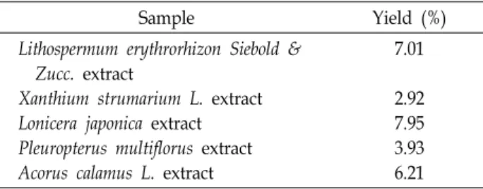
Table 1. Yields of 70% ethanol extracts from medicinal plant
Sample Yield (%)
Lithospermum erythrorhizon Siebold &
Zucc. extract
Xanthium strumarium L. extract Lonicera japonica extract Pleuropterus multiflorus extract Acorus calamus L. extract
7.01
2.92 7.95 3.93 6.21
상기의 한약재 5종은 페놀화합물의 함량이 높고 뛰어난 항 산화활성을 가진 것으로 이전 연구에서 밝혀졌다[5]. 실제로 항산화 효과가 있는 물질들은 동시에 높은 항염증 작용을 수 반하는 경우가 많다[2]. 이는 면역세포의 nitiric oxide syn- thase (NOS)에서 생성되며, 외부의 자극에 의해 발현되는 NO 의 경우 세포 활성화 물질 및 reactive oxygen intermediates (ROI)에 의해 유발된 세포독성을 최소화시킴으로써 항산화와 더불어 면역활성 발현에도 관여함이 알려져 있다[4].
이에 본 연구에서는 항산화 활성이 높은 물질이 산화적 손 상과 관련이 높은 만성염증성 질환에 대한 치료 및 개선제로 서의 개발 가능성을 구명할 목적으로 다양한 유용성분과 항산 화 활성을 가지고 있는 5종의 한약재(하수오, 창포, 창이자, 인동초, 지치)들을 에탄올로 추출하여 RAW264.7 대식세포로 부터 LPS로 유발된 염증반응에 대한 염증억제효과와 HaCaT 세포의 증식에 대한 효과에 대해서 확인하고자 하였다.
재료 및 방법
한약재 추출물
본 실험에 사용한 5종의 한약재인 하수오(Pleuropterus mul- tiflorus), 창포(Acorus calamus L.), 지치(Lithospermum erythro- rhizon Siebold & Zucc.), 창이자(Xanthium strumarium L.), 인동 초(Lonicera japonica)는 (주)화림한약(Busan)에서 건조된 상태 로 구입하여 사용하였다. 시료로 사용된 한약재 건조물 무게 를 재어 10배에 해당하는 70% 에탄올을 가하여 실온에서 7일 동안 유효 성분을 추출하였다. 이를 여과한 다음 회전식 진공 농축기(Tokyo Rikakikai Co., LTD, Bunkyo-ku, Tokyo, Japan) 로 감압 하에 농축한 후 동결건조하여 -20℃에 보관하면서 실 험을 위한 시료로 사용하였다. 이들 5종의 한약재 추출물들의 수율은 Table 1에 나타내었다.
세포 배양
실험에 사용한 RAW264.7 세포주는 한국세포주은행(KCLB, Seoul, Korea)에서 분양을 받아 사용하였다. 각 세포주를 cul- ture plate에서 배양하여 1주일에 2~3회 계대하였으며, 배지는 10% Fetal Bovine Serum (FBS, Lonza, Valais, Switzerland)을 함유한 성장배지를 이용하였다. 세포주는 습도 95%, 5% CO
2, 37℃로 조절된 배양기에서 배양하였으며, 배지는 2~3일에 한
번씩 교환하였다. 이때 미생물의 오염이나 증식을 억제하기 위해 배지용 항생제(Penicillin streptomycin, Gibco, CA, USA)를 사용하였다.
RAW264.7세포에서의 세포독성
한약재 추출물의 RAW264.7 세포에 대한 독성을 확인하였 다. 즉 세포 배양용 플라스크에 배양한 RAW264.7 세포를 10%
FBS (Lonza, Valais, Switzerland)가 함유된 Dulbecco's modi- fied Eagle's Medium (Hyclone)으로 배양하였다. 1×10
5cells/
well의 농도로 96well plate에 분주하고, 24시간 동안 37℃, 습 도 95%, 5%로 조절된 CO
2배양기에서 배양하였다. 새로운 배 지에 시료의 최종 농도가 1, 10, 50, 100 및 200 μg/ml이 되도록 녹여 세포주에 처리한 후 24시간 동안 세포를 배양하였다. 세 포주의 생존율은 Cell Counting Kit-8 (CCK-8, Dojindo, Kumamoto, Japan)을 이용하여 측정하였다. 시약을 처리한 후 microplate reader (Perkin Elmer 1420, VICTORTM X Multilabel Plate Readers, Waltham, MA, USA)로 450 nm에서 흡광도를 측정하였다. 세포의 생존율은 시료를 처리하지 않은 대조군에 대비한 시료 처리군의 흡광도로 표시하였다.
NO 생성량
RAW 264.7 세포를 3×10
5cells/well 농도로 96well에 분주 한 후 세포가 바닥의 약 70~80% 정도를 덮을 때까지 배양하였 다. 대조군으로는 배지만을 배양하였고, 음성 대조군에는 LPS 만을 1 μg/ml의 농도로 처리하였다. 시험군에는 LPS 1 μg/ml 과 함께 시료의 최종 농도가 1, 10, 50, 100, 200 μg/ml가 되도 록 배지에 처리하였고, 시료 처리 후 24시간 동안 배양하였다.
NO 생성량은 Griess 시약으로 측정하였다. Griess 시약은 2.5%의 phosphate 용액에 1% sulfanilamide와 0.1% naph- thylethylene-diamine dihydrochloride를 섞어 만들었다. 위에 배양한 세포의 상등액 100 μl를 96well plate에 취하고 여기에 Griess 시약 100 μl를 가해 15분 동안 실온에서 반응시킨 후 microplate reader (Molecular Devices, VersaMax ELISA Microplate Reader, USA)를 이용하여 540 nm에서 흡광도를 측정하였다. NO 생성량은 LPS만을 처리한 음성 대조군의 생 성량에 대해 LPS와 함께 시료를 처리한 시험군의 NO 생성량 을 비교하여 측정하였다.
Prostaglandin E
2생성에 대한 효과
Prostaglandin E
2(PGE
2)의 측정은 Prostaglandin E
2EIA
Kit를 Cayman Chemical Company (Ann Arbor, Michigan,
USA)에서 구매하여 측정하였다. 즉, NO측정과 같은 방법으
로 RAW264.7 세포에 한약재 추출물을 24시간 전처리 후 세포
배양 상층액을 취해 PGE
2생성량을 측정하였다. 배양액을
goat anti-mouse로 coating된 96well plate에 각각 배양액을
50 μl씩 loading 하고, primary antibody solution 50 μl와 PGE
2conjugate 50 μl씩 첨가하여 4℃에서 18시간 동안 반응시킨 후, Washing buffer로 5회 세척한다. Ellman's reagent을 200 μl씩 처리하여 60~90분간 교반하면서 반응 시킨 후, 405~420 nm에서 흡광도를 측정하여 PGE
2생성량을 계산하였다.
HaCaT 세포에서의 세포독성
세포 배양용 플라스크에 배양한 HaCaT 세포는 FBS가 함유 되지 않은 Dulbecco's modified Eagle's Medium (Hyclone)로 배양하였다. HaCaT 세포주를 1×10
5cells/well의 농도로 96 well plate에 분주하고, 24시간 동안 37℃, 습도 95%, CO
25%
로 조절된 CO
2배양기에서 배양하였다. 새로운 배지에 시료를 최종 농도가 1, 10, 100 및 200 μg/ml이 되도록 녹여 세포주에 처리한 후 24시간 동안 배양하였다. 세포주의 생존률을 측정 하기 위해 Cell Counting Kit-8 (Dojindo, Kumamoto, Japan) 을 이용하여 세포의 생존률을 측정하였다. 시약을 처리한 후 microplate reader (Perkin Elmer 1420, VICTORTM X Multi- label Plate Readers, Waltham, MA, USA)로 450 nm에서 흡광 도를 측정하였다. 세포의 생존율은 시료를 처리하지 않은 대 조군에 대비한 시료 처리군의 흡광도로 표시하였다.
HaCaT 세포 증식능
피부에 대한 세포 증식능은 인간 표피세포인 HaCaT 세포 로 확인하였다. 즉, 세포 배양용 플라스크에 배양한 HaCaT 세포는 FBS가 함유된 Dulbecco's modified Eagle's Medium (Hyclone)로 배양하였다. HaCaT 세포를 2×10
3cells/well의 농도로 96well plate에 분주하고, 24시간 동안 37℃, 습도 95%, CO
25%로 조절된 CO
2배양기에서 배양하였다. 새로운 배지에 2%의 FBS와 함께 시료를 최종 농도가 0.1, 1, 10 및 50 μg/ml이 되도록 조제하고, 세포에 처리한 후 각각 1일, 3일, 5일 동안 배양하였다. 세포주의 증식능은 Cell Counting Kit-8 (Dojin- do, Kumamoto, Japan)을 이용하여 확인하였다. 시약을 처리 한 후 microplate reader (Perkin Elmer 1420, VICTORTM X Multilabel Plate Readers, Waltham, MA, USA)로 450 nm에서 흡광도를 측정하였다. 세포의 생존율은 시료를 처리하지 않은 대조군에 대비한 시료 처리군의 흡광도로 표시하였다.
통계 분석
본 연구의 모든 실험 결과는 3회 이상 반복으로 평균값으로 나타내었다. 통계분석은 Student's t-test를 이용하였으며, 각 각의 시료에 대해 평균±S.D.으로 나타내었다. 각 시료군에 대 한 유의성 검증은 분산분석을 한 후 p<0.05 수준에서 각 실험 군 평균값 간의 유의성을 검증하였다.
결과 및 고찰
RAW264.7세포독성에 미치는 영향
5종의 한약재의 에탄올 추출물이 세포에 미치는 독성을 확 인하고, 세포실험에 대한 시료의 적정처리 농도를 확인하기 위하여 대식세포주인 RAW 264.7 세포에 한약재 에탄올 추출 물을 1, 10, 50, 100 및 200 μg/ml로 24시간 동안 처리하였다.
하수오, 지치, 인동초는 상기의 농도 구간에서 세포에 대한 독성은 보이지 않았다(Fig. 1). 반면, 창이자는 100~200 μg/ml 의 농도 구간에서 세포의 생존률이 감소하여 독성을 확인하였 고, 창표는 200 μg/ml 농도에서 유의적인 세포독성을 확인하 였다. 따라서 Nitric oxide (NO)와 PGE
2생성 억제력은 세포 독성이 나타나지 않는 농도구간에서 실시하였다.
Nitric oxide (NO) 생성 억제력의 평가
NO는 주로 LPS 또는 TNF-α 등의 염증 유발물질에 의해 자극된 세포가 염증반응을 유발할 때 분비되는 염증반응 매개 물질로 알려져 있다. 따라서 염증반응이 진행되는 동안 유의 적으로 증가하는 NO를 효과적으로 억제하는 것은 염증질환 치료에서 유용한 방법으로 여겨지고 있다[10, 26, 27]. LPS는 그람 음성 세균의 세포벽에서 분리된 생물학적 독소로서[30], 염증 매개 인자들을 분비하도록 대식세포를 자극하는 대표적 성분 중 하나이다[6, 22, 29]. 항염증 생리활성 평가를 위해 LPS 로 활성화된 RAW264.7 대식세포가 널리 쓰이고 있다[25, 33, 40].
5종의 한약재(하수오, 창포, 지치, 창이자, 인동초) 에탄올 추출물들이 RAW264.7 세포에 대한 NO 생성량의 변화를 알 아보기 위하여 추출물들의 독성이 없는 농도로 처리한 뒤, LPS 및 시료를 처리하지 않은 대조군의 NO 생성량과 비교하 였다(Fig. 2). LPS 1 μg/ml 처리 후 NO의 생성량은 정상세포 에 비하여 약 6배 가까이 증가되었다. 이에 반해 하수오, 지치, 인동초, 창이자를 처리한 군에서는 모든 농도 구간에서 농도 의존적으로 NO 생성량이 유의적으로 감소되었다. 특히 하수 오는 다른 시료들에 비해 다소 높은 농도인 200 μg/ml의 농도 구간에서 NO 생성량이 81.1% 감소되었다. 하수오, 지치, 인동 초는 RAW 264.7 세포 독성실험에서 독성을 나타나지 않은 것으로 보아 이들 한약재들의 NO 생성억제는 세포 독성 작용 에 의한 것이 아닌 것으로 판단하였다. NO 생성량이 50% 저해 되는 IC
50은 창이자가 52.9 μg/ml에서 확인되어 가장 높은 NO 생성 억제력을 보였으며 다음으로 하수오, 창포, 인동초의 순 으로 나타났다. 지치의 경우 농도 의존적으로 NO 생성억제 활성을 보이기는 하나 실험에 사용된 농도에서는 가장 낮은 저해활성을 보였다. 이러한 결과로 보아 이들 한약재 추출물 들은 NO의 생산을 억제함으로써 염증반응에 의한 조직손상 을 감소시킬 수 있을 것으로 생각된다.
PGE
2생성에 대한 효과
COX는 arachidonic acid을 prostaglandin으로 전환시키는
데 중요한 역할을 하는 효소로 염증반응 매개물질인 PGE
2를
Fig. 1. The effects of 5 kinds of oriental medical plants on the RAW 264.7 cell viability. RAW 264.7 cells were treated with various concentrations (from 1 to 200 μg/ml) of 5 kinds of oriental medical plants and then incubated for 24 hr. RAW 264.7 cell viabilities were assessed using Cell Counting Kit-8 (CCK-8). Values are means ± S.D. of three independent experiments.
*p<0.05 vs. control; significant differences between treated groups were determined Student's t-test. PME; Pleuropterus multi- florus extract, ACE; Acorus calamus L. extract, LEE; Lithospermum erythrorhizon Siebold & Zucc. Extract, XSE; Xanthium strumarium L. extract, LJE; Lonicera japonica extract
Fig. 2. The effects of 5 kinds of oriental medical plant ethanol extracts on nitrite oxide (NO) production in LPS-stimulated RAW 264.7 macrophages. NO production was measured by the Griess reaction assay and expressed as a percentage of the control (LPS alone). Values are the mean ± S.D. of the three independent experiments. *p<0.05 vs. (-) control; LPS (1 μg/ml) alone;
significant differences between treated groups were determined Student's t-test. PME; Pleuropterus multiflorus extract, ACE;
Acorus calamus L. extract, LEE; Lithospermum erythrorhizon Siebold & Zucc. Extract, XSE; Xanthium strumarium L. extract, LJE;
Lonicera japonica extract.
생성하는 효소이다[1, 13]. COX는 COX-1, COX-2 두 종류가 존재하며, COX-1은 생체 내의 대부분의 조직에서 발현하는 반면 COX-2는 growth factor, mitogenes, cytokines 등과 같은
요인에 의해 발현이 증가되어 염증반응을 조절하는데 중요한
역할을 한다[17, 34]. COX-2에 의하여 생성된 PGE
2는 염증반
응에 동반되는 홍반, 부종 및 통증 등과 같은 증상의 원인이다
Table 2. NO production inhibition activities by treatment of 5 kinds of oriental medical plant extracts for 24 hr
Sample IC50 (μg/ml)
PME ACE LEE XSE LJE
86.4 144.7 - 52.9 163.6
*p<0.05; compared with control group.
PME; Pleuropterus multiflorus extract.
ACE; Acorus calamus L. extract.
LEE; Lithospermum erythrorhizon Siebold & Zucc. extract.
XSE; Xanthium strumarium L. extract.
LJE; Lonicera japonica extract.
LPS; lipopolysaccharide.
Fig. 3. The effects of 5 kinds of oriental medical plant ethanol extracts on PGE2 (Prostaglandin E2) production in LPS-stimulated RAW 264.7 macrophages. PGE2 production expressed as a percentage of the control (LPS alone). Values are the mean ± S.D of the three independent experiments. *p<0.05 vs. (-) control; LPS (1 μg/ml) alone; significant differences between treated groups were determined Student's t-test. PME; Pleuropterus multiflorus extract, ACE; Acorus calamus L. extract, LEE;
Lithospermum erythrorhizon Siebold & Zucc. Extract, XSE; Xanthium strumarium L. extract, LJE; Lonicera japonica extract.
[7, 8]. 뿐만 아니라 PGE
2는 염증성 질환을 포함한 다양한 생체 반응에 있어서도 세포분열이나 증식에 영향을 줌으로서 각종 질병의 유발과 진행에 관여한다[23].
LPS에 의해 RAW264.7 대식세포로부터 생성되는 PGE
2에 대한 억제효과를 측정하기 위해 5종의 한약재 추출물들을 세 포에 처리하여 LPS 및 시료를 처리하지 않은 대조군의 PGE
2생성량과 비교하였다(Fig. 3). 하수오, 창포, 창이자, 인동초 추 출물들의 경우 생성억제 효과가 농도 의존적으로 나타났으며, 창포는 100 μg/ml의 농도구간에서 79.5% 감소하여 가장 높은 생성 억제효과를 보여주었다. 하수오는 NO 생성 억제활성은 매우 높았으나 PGE
2생성 억제활성은 다른 한약재보다 높지 않았다. 반면, 지치의 경우 농도가 증가함에 따라 PGE
2가 증가
함을 보였다. PGE
2는 피부 상처 치유의 중요한 매개체이며, PGE
2가 EP
2수용체 경로를 통한 섬유아세포의 근섬유모세포 로의 분화를 억제할 수 있다고 보고하였다[21].
HaCaT 세포에서의 세포독성
LPS로 활성화된 RAW 264.7 세포에 대해 항염증 활성을 보였던 5종의 한약재 추출물들이 피부의 1차 면역방어체로 작용하는 표피세포의 증식에 미치는 영향을 알아보기 위해 먼저 한약재 추출물들이 인간 표피세포인 HaCaT 세포에서의 세포독성을 측정하였다. 한약재 추출물들을 0.01, 0.1, 1, 10 및 100 μg/ml로 처리한 결과 지치의 경우 모든 농도 구간에서 독성을 보이지 않았으며, 하수오, 창포, 창이자, 인동초의 경우 100 μg/ml 농도에서 세포생존률이 감소하여 세포독성을 확인 하였다(Fig. 4). HaCaT 세포에서의 세포독성이 없는 농도에서 한약재 추출물들의 세포 증식능에 미치는 영향을 측정하였다.
HaCaT 세포의 증식능에 미치는 영향
피부의 가장 표면층인 표피는 각질세포, 모낭, 멜라닌 세포,
랑겔한스세포로 이루어져 있으며 외부의 자극으로부터 신체
를 보호하는 역할을 한다[14]. 그 중 각질형성세포는 표피세포
의 대부분을 구성하는 성분으로서 각질을 형성하고 여러 cyto-
kines를 생산하여 다양한 염증반응과 면역반응에 관여한다
[3]. 인체와 외부 환경사이의 투과막인 피부각질장벽이 손상되
면 경표피수분손실(trans epidermal water loss)이 증가되고
수분결합력(water-binding capacity)이 감소되어 소양증이 심
해지고 피부가 건조해진다[31]. 그러므로 이러한 각질형성세
포의 증식은 알러지성 염증질환을 완화시킬 수 있다.
Fig. 4. The effects of 5 kinds of oriental medical plant ethanol extract on the HaCaT cell viability. HaCaT cells were treated with various concentrations (from 0.01 to 100 μg/ml) of 5 kinds of oriental medical plants and then incubated for 24 hr. HaCaT cell viabilities were assessed using Cell Counting Kit-8 (CCK-8). Values are means ± S.D. of three independent experiments.
*p<0.05 vs. control; significant differences between treated groups were determined Student's t-test. PME; Pleuropterus multi- florus extract, ACE; Acorus calamus L. extract, LEE; Lithospermum erythrorhizon Siebold & Zucc. Extract, XSE; Xanthium strumarium L. extract, LJE; Lonicera japonica extract.
피부재생 효과를 측정하기 위해 독성을 나타내지 않은 농도 범위내에서 HaCaT 세포에 대한 세포의 증식능을 측정하였다.
그 결과 지치와 창포는 농도가 증가할수록 높은 증식능을 확 인할 수 있었으며, 나머지 3종의 한약재는 세포 증식활성이 없었다. 창포는 10 μg/ml 농도구간에서 1일, 3일 배양 시 11.2, 26.0%의 증식능을 확인하였으나, 5일 배양 시에는 오히려 감 소하였다(Fig. 5). 지치의 경우 100 μg/ml 농도구간에서 1, 3, 5일간 배양 시 21.1, 53.5, 99.6%의 증식능을 보였다(Fig. 6).
특히 지치는 PGE
2생성 증가활성을 보여주었고, 피부재생효 과는 매우 뛰어남을 알 수 있었다. 이것은 앞서 Kolodsick 외 [21] 가 PGE
2를 피부 상처 치유의 중요한 매개체로 보고한 내 용와 유사한 결과를 보여주었다.
이들 5종의 한약재 에탄올 추출물의 함염증 활성 효과를 위해 LPS로 자극된 마우스 대식세포 RAW264.7 세포에 대한 세포 독성 실험으로 세포 독성이 나타나지 않는 농도에서 실 시하여 기본적으로 세포의 생존율에 영향을 주지 않는다는 사실을 알 수 있었다. 따라서 이들 한약재의 항염증 효과가 세포 생존율 감소에 의한 것이 아니라 이들 한약재 고유의 특성임을 보여주었다.
RAW264.7 세포에 한약재 추출물을 전처리 한 후 LPS를 처리하여 24시간 염증반응을 유도한 결과 한약재 추출물들의 처리에 의해 LPS로 유도되었어야 할 NO, PGE
2의 생성량이 농도의존적으로 크게 감소되었다. 창이자는 NO 생성 저해활 성과 PGE
2생성 저해 활성이 뛰어났으며, 창포의 경우 NO
생성 억제활성에 비해 PGE
2생성 억제활성이 더 뛰어났다.
피부세포 재생 효과를 조사하기 위해 인간 표피세포인 HaCaT 세포에 대한 추출물들의 독성과 세포 증식률을 측정한 결과 항염증 활성이 비교적 약했던 지치와 창포가 세포 증식 률이 높게 나타나 우수한 피부재생 효과를 보였다.
이상의 결과들로부터 한약재로 널리 이용되고 있는 5종의 약용식물(하수오, 창포, 지치, 창이자, 인동초)의 항염증 효능 과 피부세포 재생 활성 및 기능적 활성을 확인하였고, 화장품 원료로의 활용 가능성을 확인하였다.
Gu 외[11]의 논문에 의하면 항산화 및 항염증 활성이 알려 진 3종의 한약재 추출물의 혼합비를 달리하였을 때, 혼합비에 따라 항산화 및 항염증 활성 차이를 비교하였다. Oh 외[28]는 한약재 단독 추출물과 혼합액의 비율을 달리한 실험에서 한약 재 단독 추출물보다 혼합액의 활성이 더 높았고, 혼합비에 따 라 활성이 달라짐을 보고하였다.
본 연구에서는 5종의 한약재 추출물의 항염증 활성 및 HaCaT 세포재생 활성을 확인하였으며, 5종의 한약재 추출물 의 최적 혼합비 활성실험을 추가적으로 실시한다면, 한약재 혼합액 피부 미용액 소재 개발이 가능할 것으로 사료된다.
References
1. Aktan, F. 2003. iNOS-mediated nitric oxide production and its regulation. J. Life Sci. 75, 639-653.
2. Ames, B. N., Shienaga, M. K. and Hagen, T. M. 1993.
Oxidants, antioxidants and the degenerative diseases of aging. Proc. Natl. Acad. Sci. USA. 90, 7915-7922.
3. Beissert, S., Cavazzana, I., Mascia, F., Meroni, P., Pastore, S., Tessari, G. and Girolomoni, G. 2006. Mechanisms of im- mune-mediated skin diseases: an overview. Clin. Exp. Rheu- matol. 24, S1-6.
4. Cho, E. K., Song, H. J., Cho, H. E., Choi, I. S. and Choi, Y. J. 2010. Development of functional beverage (Sanya) from fermented medicinal plants and evaluation of its physio- logical activities. J. Life Sci. 20, 82-89.
5. Chung, B. S. and Shin, M. K. 1990. Encylopedia of local med- ical hurbs. pp. 278-279, Younglim Press, Seoul, Korea.
6. Fujiwara, N. and Kobayashi, K. 2005. Macrophages in inflammation. Curr. Drug Targets Inflamm. Allergy 4, 281-286.
7. Funk, C. D. 2001. Prostaglandins and leukotrienes: advances in eicosanoid biology. Science 294, 1871-1875.
8. Giercksky, K. E. 2001. COX-2 inhibition and prevention of cancer. Best Pract. Res. Clin. Gastroenterol. 15, 821-833.
9. Gracie, J. A., Forsey, R. J., Chan, W. L., Gilmour, A., Leung, B. P., Greer, M. R., Kennedy, K., Carter, R., Wei, X. Q., Xu, D., Field, M. Foulis, A., Liew, F. Y. and McInnes, I. B. 1999.
A proinflammatory role for IL-18 in rheumatoid arthritis.
J. Clin. Invest. 104, 1393-1401.
10. Grisham, M. B., Jourd'Heuil, D. and Wink, D. A. 1999. Nitric Oxide I. Physiological chemistry of nitric oxide and its me- tabolites : implications in inflammation. Am. J. Physiol. Gas- trointest. Liver Physiol. 276, G315.
11. Gu, Y. R. and Hong, J. H. 2017. Physicochemical character- istics and physiological activities of mixture extracts from Liriope platyphylla, Schizandra chinensis, and Panax gin- seng C.A. Meyer. Kor. Soci. Food Pre. 24, 431-439.
12. Hatano, T., Uebayashi, H., Ito, H., Shiota, S., Tsuchiya, T.
and Yoshida, T. 1999. Phenolic constituents of Cassia seeds and antibacterial effect of some naphthalenes and an- thraquinones on methicillin-resistant Staphylococcus aureus.
Chem. Pharm. Bull. 47, 1121-1128.
13. Hur, S., Lee, Y. S., Yoo, H., Yang, J. H. and Kim, T. Y. 2010.
Homoisoflavanone inhibits UVB-induced skin inflammation through reduced cyclooxygenase-2 expression and NF-kappaB nuclear localization. J. Dermatol. Sci. 59, 163-169.
14. Ishida, H., Ray, R. and Ray, P. 2008. Sulfur mustard down- regulates iNOS expression to inhibit wound healing in a human keratinocyte model. J. Dermatol. Sci. 49, 207-216.
15. Jeon, S. B. Jeon, J. A. and Jeong, B. G. 2010. Anti-oxidative activities and tyrosinase inhibition ability of rice bran etha- nol extract. J. Kor. Soc. Cosm. 16, 602-606.
16. Kang, H. H. 1997. Anti-aging in cosmetics. J. Cosmet. Sci.
23, 57-61.
17. Kean, W. F. and Buchanan, W. W. 2005. The use of NSAIDs in rheumatic disorders 2005: a global perspective. Inflammo- pharmacology 13, 343-370.
18. Kim, C. M., Shin, M. K., Lee, K. S. and Ahn, D. K. 1998.
Dictionary of Chinese pharmacy. pp. 78-79, Joungdam pub- lisher, Seoul, Korea.
19. Kim, H. M., Yi, J. M. and Lim, K. S. 1999 Magnoliae flos inhibits mast cell-dependent immediate-type allergic reactions.
Pharmacol. Res. 39, 107-111.
20. Kinne, R. W., Bräuer, R., Stuhlmüller, B., Palombo-Kinne, E. and Burmester, G. R. 2000. Macrophages in rheumatoid arthritis. Arthritis Res. Ther. 2, 189-202.
21. Kolodsick, J. E., Peters-Golden, M., Larios, J., Toews, G. B., Thannickal, V. J. and Moore, B. B. 2003. Prostaglandin E2 inhibits fibroblast to myofibroblast transition via E. prosta- noid receptor 2 signaling and cyclic adenosine mono- phosphate elevation. Am. J. Respir. Cell Mol. Biol. 29, 537-544.
22. Kundu, J. K. and Surh, Y. J. 2005. Breaking the relay in de- regulated cellular signal transduction as a rationale for che- moprevention with anti-inflammatory phytochemicals. Mutat.
Res. 591, 123-146.
23. Lee, E., Choi, M. K., Lee, Y. J., Ku, J. L., Kim, K. H., Choi, J. S. and Lim, S. J. 2006. α-Tocopheryl succinate, in contrast to α-tocopherol and α-tocopheryl acetate, inhibits prosta- glandin E2 production in human lung epithelial cells. Carci- nogenesis 27, 2308-2315.
24. Lee, K. S., Ahn, D. K., Shin, M. K. and Kim, C. M. 1998.
The encyclopedia of oriental herbal medicine. pp. 4657-4663, Jungdam Pubishing Co., Seoul, Korea.
25. Lee, Y. G. and Cho, J. Y. 2007. Inhibitory effect of curcumin on nitric oxide production in lipopolysaccharide-stimulated RAW 264.7 cells and its suppressive mechanism. Hanguk Yakyong Changmul Hakhoe Chi 15, 451-456.
26. Min, J. Y. and Park, Y. K. 2009. Effect of dipsaci radix water extract on LPS-induced inflammatory response in RAW 264.7 mouse macrophages. Kor. J. Herbology 24, 189.
27. Nathan, C. 1992. Nitric oxide as a secretory product of mam- malian cells. FASEB J. 6, 3051-3064.
28. Oh, H., S. and Kim, J. H. Physiological functionalities of hot water extract of Codonopsis lanceolata and some medici- nal materials, and their mixtures. Kor. J. Comm. Living Sci.
18, 407-415
29. Paul, A., Cuenda, A., Bryant, C. E., Murray, J., Chilvers, E. R., Cohen, P., Gould, G. W. and Plevin, R. 1999. Involve- ment of mitogen-activated protein kinase homologues in the regulation of lipopolysaccharide-mediated induction of cy- clo-oxygenase-2 but not nitric oxide synthase in RAW 264.7 macrophages. Cell. Signal. 11, 491-497.
30. Roth, R. A., Harkema, J. R., Pestka, J. P. and Ganey, P. E.
1997. Is exposure to bacterial endotoxin a determinant of susceptibility to intoxication from xenobiotic agents?. Toxicol.
Appl. Pharmacol. 147, 300-311.
31. Schäfer, L. and Kragballe, K. 1991. Abnormalities in epi- dermal lipid metabolism in patients with atopic dermatitis.
J. Invest. Dermatol. 96, 10-15.
32. Shao, L. X. 2003. Effects of the extract from bergamot and boxthorn on the delay of skin aging and hair growth in mice. Zhongguo Zhong Yao Za Zhi 28, 766-772.
33. Shen, T., Lee, Y. J. and Cho, J. Y. 2008. Effect of hot water extract from Scutellaria barbata on the macrophages acti- vated by lipopolysaccharide. Hanguk Yakyong Changmul Hakhoe Chi 16, 313-319.
34. Sirsjö, A., Karlsson, M., Gidöf, A., Rollman, O. and Törmä, H. 1996. Increased expression of inducible nitric oxide syn-
초록:5종의 한약재 에탄올 추출물의 항염증 및 표피세포 증식 활성
정민화*
(
대동대학교 헤어디자인과
)이 연구는 한국에서 전통적으로 사용되어 온 5종의 한약재인 하수오(Pleuropterus multiflorus), 창포(Acorus cala- mus L.), 지치(Lithospermum erythrorhizon Siebold & Zucc.), 창이자(Xanthium strumarium L.), 인동초(Lonicera japon- ica) 에탄올 추출물의 항염증에 미치는 효과를 탐색하였다. 창이자는 RAW264.7 세포에 100, 200 μg/ml 농도에서 세포 독성을 나타냈고(p<0.05), 창포는 RAW264.7 세포에 200 μg/ml 농도에서 세포 독성을 나타냈다(p<0.05). 다 른 한약재들은 RAW264.7 세포에 200 μg/ml 농도 이하에서 세포 독성이 나타나지 않았다. 독성이 없는 농도에서 5종의 한약재 추출물들의 항염증 효과를 확인하였다. 하수오, 창포, 창이자, 인동초는 LPS 유도된 RAW264.7세포 에서 NO 생산과 PGE
2생산에 대해 농도 의존적으로 억제 효과를 보였다. 특히, 창이자는 52.9 μg/ml (IC
50)로 가장 뛰어난 NO 생성 저해효과를 나타냈을 뿐만 아니라 50 μg/ml 농도 구간에서 가장 뛰어난 PGE
2생성 저해능을 나타냈다(73.6%). 창포와 지치는 HaCaT 세포에 대하여 세포가 증식하는 효과를 나타냈다. 특히 지치는 100 μg/
ml 농도구간에서 1, 3, 5일 배양 시 21.1, 53.5, 99.6%의 증식능을 보였다. 창포 또한 10 μg/ml 농도구간에서 1, 3일 배양 시 11.2, 26.0%의 증식능을 보였다. 따라서 본 연구를 통해 5종의 한약재 에탄올 추출물의 항염증 활성 및 HaCaT 세포 재생에 미치는 효과를 확인하였다.
thase in psoriatic skin and cytokine-stimulated cultured keratinocytes. Br. J. Dermatol. 134, 643-648.
35. Son, K. H., Park, J. O., Chung, K. C., Chang, H. W., Kim, H. P., Kim, J. S. and Kang, S. S. 1992. Flavonoids from the aerial parts of Lonicera japonica. Arch. Pharm. Res. 15, 365- 370.
36. Son, K. H., Kim, J. S., Kang, S. S., Kim, H. P. and Chang, H. W. 1994. Isolation of flavonoids from Lonicera japonica.
Kor. J. Pharmacogn. 25, 24-27.
37. Tabata, M., Tsukada, M. and Fukui, H. 1987. Antimicrobial activity of quinone derivatives from Echium lysopsis callus cultures. Planta Med. 44, 234-236.
38. Taga, M. S., Miller, E. E. and Pratt, D. E. 1984. Chia seeds as a source of natural lipid antioxidants. J. Am. Oil Chem.
Soc. 61, 928-993.
39. Yoon, K. B. and Chang, J. L. 1989. Useful plants in good health. pp. 129, Seok-O Publishers, Seoul, Korea.
40. Yun, J. Y., Choi, S. Y., Park, P. J., Chung, H. G., Shin, H.
M., Suk, K. H. and Lim, B. O. 2008. Extract of Artemisia princeps Pampanini inhibit lipopolysaccharide-induced ni- tric oxide, cyclooxygenase-2, prostaglandin E2, and tumor necrosis factor-a production from murine macrophages RAW 264.7 cells. Hanguk Yakyong Changmul Hakhoe Chi 16, 326-331.