돼지 단위 발생 난자의 체외 발달에 있어서 피라칸타 추출액의 처리 효과
민성훈, 연지영, 김진우, 박수용, 이용희, 강선철, 구덕본*
대구대학교 생명공학과
Pyracantha Extract Acts as an Antioxidant Agent to Support Porcine Parthenogenetic Embryo Development In Vitro
Sung-Hun Min, Ji-Yeong Yeon, Jin-Woo Kim, Soo-Yong Park, Yong-Hee Lee, Sun-Chul Kang and Deog-Bon Koo*
Department of Biotechnology, Daegu University, Gyeongsan, 712-714, Korea
Abstract
Pyracantha is a genus of thorny evergreen large shrubs in the family of Rosaceae, with common names Firethorn or Pyracantha. It's extract has also been used in cosmetics as a skin-whitening agent and functioning through tyrosinase inhibition. Recent studies have shown that pyracantha extract possesses antioxidant activities and may significantly improve lipoprotein metabolism in rats. Although the mode of action of Pyracantha extract is not fully understood, a strong relationship was observed between antioxidant and apoptosis in some types of cells. Thus, the aim of this study was to evaluated the effect of pyracantha extract on blastocysts formation and their quality of the porcine parthenogenetic embryos. After parthenogenetic activation by chemicals, presumptive porcine parthenogenetic embryos were cultured in PZM-3 medium supplemented with extracts of pyracantha leaf, stalk and root for 6 day (1, 5 and 10 µg/ml, respectively). In our results, the frequency of blastocyst formation in pyracantha root extract (5 µg/ml) treated group had increased that of other groups. Furthermore, blastocysts derived from pyracantha root extract (5 µg/ml) treated group had increased the total cell numbers and reduced apoptotic index. Blastocyst development was significantly improved in the pyracantha root extract (5 µg/ml) treated group when compared with the H2O2 treated group (p<0.05). Subsequent evaluation of the intracellular levels of ROS in pyracantha root extract (5 µg/ml) treated groups under H2O2 induced oxidative stress were decreased (p<0.05). In conclusion, our results indicate that treatment of pyracantha root extract may improve in vitro development of porcine parthenogenetic embryos through its antioxi- dative and antiapoptotic effects.
(Key words : parthenogenetic embryos, pyracantha extract, antioxidant, apoptosis, porcine)
†본 연구는 농촌진흥청 차세대바이오그린21 사업(PJ009530012013) 지원에 의해 수행되었음.
*Correspondence : E-mail : [email protected]
서 론
단위 발생 유도 기술은 수정 과정에서 번식 기능과 유전적 각인 그리고 핵이식과 정자 주입술과 같은 다양한 초기배의 발생 생물학적 분야에 있어서 매우 중요한 수단으로 이용되 고 있다(Kim 등, 2007). 또한, 단위 발생은 조직 적합 배아 유 래의 줄기세포를 유도할 수 있으며, 조직 적합성 기관이나 세 포를 이용하여 손상된 조직이나 질환을 겪는 환자들에게 이 식할 수 있기 때문에 재생의학 분야에서도 유용하게 이용되 어질 수가 있을 것으로 판단하고 있다(Yuh 등, 2010). 특히, 단위발생 난자를 이용하여 체외 발달 과정에서 악영향을 미
치는 활성 산소종의 수준을 제어할 수 있는 다양한 실험적 접 근은 돼지 초기배 발생 연구에 효과적으로 이용될 수 있다.
최근에 항산화 효과가 뛰어난 식물 추출물들이 보고되고 있 어 이러한 추출물을 이용하여 포유동물 초기배의 발생 효율 을 효과적으로 유도할 수 있을 것으로 기대하고 있다.
일반적으로 페놀계 합성 항산화제로 널리 사용되고 있는 butylated hydroxy anisol과 butylated hydroxy toluene는 그 효 과와 경제성 그리고 안정성 때문에 많이 사용해 왔지만, 합성 식품 첨가물의 일반적인 기피 현상뿐만 아니라 과량 섭취 시 간, 위장 점막, 폐, 신장, 순환계 등에 심각한 독성 작용을 일 으키는 것으로 알려져 안전한 대체 항산화제의 개발이 요구
되었다(Choe 와 Yang, 1982). 따라서 인체에 무해하고 항산화 력이 우수한 천연 항산화제에 관한 연구가 오래전부터 진행되 어 왔으며, 지금까지 보고된 대부분의 천연 항산화제는 식물 유래이다. 대부분의 식물들의 항산화능 화합물은 주로 폴리페 놀 물질들이며, 천연 항산화제로서의 기능이 잘 알려져 있다 (Huang 등, 1992). 식물로부터 유래된 페놀 물질의 항산화제는 일부가 금속 복합체를 형성하는 작용을 하나, 주요 기능은 이 들의 항산화 활성에 있다. 따라서 식물 추출물로부터 radical 제거 기능을 탐색함으로써 천연 항산화제를 개발할 수 있다.
본 연구에서 이용하고자 하는 피라칸타는 장미과(薔薇科 Rosaceae) 피라칸타속(Pyracantha 屬)에 속하는 가시가 달린 상록관목으로써 유럽 남동부와 아시아가 원산지로 알려져 있 다. 이것은 중국의 남부와 북서부 지역에 널리 분포되어 있으 며, 이것의 열매 추출물은 소화 불량 치료제로써 중국의 전통 적인 의약품으로 널리 사용되어져 왔다(Deng 등, 1990). 게다 가 일본에서는 tyrosinase 억제 기능으로 인해 미백제로써 화장 품에 이용되어져 왔다(Van Gelder 등, 1997). 최근 연구에서 피라칸타는 항산화 활성을 가지고 있으며, 랫에서 lipoprotein 대사 활성을 유의하게 향상시킨다고 보고하였다(Hou 등, 2002).
또한, 이전의 몇몇 보고들에서 다양한 생리학적 과정에서 산 화적 스트레스의 해로운 효과를 검증하였고(Hou 등, 2003), free radical의 생성과 제거에 있어서 불균형은 노화와 관련되 어 있으며, free radical은 세포막의 lipid peroxidation, 지질과 단백질 사이에 상호작용 변경 및 불완전한 효소들의 발생을 일으킨다고 보고하였다(Huang 등, 2007).
호기성 기관에서 활성산소는 산소의 정상적인 대사 과정에 서 자연적인 부산물이며, 세포의 신호전달에 중요한 역할을 하지만, 활성산소의 농도가 증가하면 세포 구조에 영향을 주 어 신호전달에 악영향을 미치게 된다(Agarwal 등, 2005). 특 히, 증가된 활성산소는 수정란 발달과 질적 수준에 밀접하게 관련되어 있으며, 체외에서 미세조작 처리 환경에서 활성산소 가 다량 생성되어 체세포 복제 배아의 미토콘드리아, DNA 그 리고 reprogamming에 영향을 준다고 보고되었다(Guerin 등, 2001). 또한, 체외 배양 과정에서 발생하는 활성산소종의 수 준은 체내 환경과 비교하여 매우 높아, 체외 수정란의 경우 상당한 산화적 스트레스에 노출된다고 할 수 있다. 이러한 활 성 산소종은 세포막을 쉽게 통과하여 산화적 스트레스를 유 도할 수 있기 때문에 체외 수정란 발달에 악 영향을 미친다.
따라서 높은 농도의 활성 산소종은 지질, 단백질 그리고 핵산 같은 세포의 분자 수준을 변형시킴으로 인하여 미토콘드리아 손상, 수정란 발달 장애, ATP 결손 그리고 세포 사멸을 일으 킨다(Takahashi, 2012). 특히, 돼지 초기배의 경우 산화적 스트 레스의 완화를 통해 착상 전 단계의 배반포의 질적 수준의 향 상시킬 수 있는 다양한 연구적 접근이 요구된다.
본 연구에서는 체외배양 배지에 피라칸타의 잎, 줄기 및 뿌 리 추출액을 첨가하여 돼지 단위발생 수정란이 배반포로의 발달 양상과 배반포에서의 질적 측면을 세포 사멸과 활성산 소종의 발현 수준을 통해 비교 분석하였다.
재료 및 방법
1. 배양액
본 논문에서 언급하지 않은 모든 시약은 Sigma Aldrich Korea(Yongin, Korea)에서 구입하였다. 돼지 난소로부터 미성 숙 난포란 회수 및 세척용 배지는 25 mM N-2-hydroxyenthyl- piperazine-N’-2-ethanesulfoic acid(Hepes)와 3 mg/ml BSA가 첨가된 Tyrode’s lactate(TL)-Hepes 용액(Prather 등, 1995)을 사용하였다. 돼지 미성숙 난포란의 체외 성숙 배지는 North Carolina State University(NCSU) 23 용액(Petters와 Wells, 1993) 에 0.6 mM cysteine, 10 ng/ml EGF, 10 IU/ml PMSG, 10 IU/ml hCG와 10% 난포액(Funahashi 등, 1993; Niwa, 1993)을 첨가 하여 사용하였다. 돼지 난포액은 난소에서 직경이 3∼6 mm 정도의 난포로부터 채취하여 4℃에서 30분간 1,600 × g로 원 심 분리 후 상층액을 0.45 µm filters로 여과하여 -20℃에 보 관 후 사용하였다. 단위 발생 유도를 위한 조작용 용액은 0.3 M mannitol 용액에 100 µM MgSO4, 100 µM CaCl2와 0.01%
BSA가 첨가된 용액을 사용하였다. 단위발생 난자의 체외배양 용 배지는 0.3% BSA가 첨가된 porcine zygotes medium(PZM) 3 용액(Yoshioka 등, 2002)을 사용하였다.
2. 체외 성숙, 단위 발생 및 체외 배양
도축장에서 난소를 회수하여 75 µg/ml penicillin G와 50 µg/ml streptomycin이 첨가된 생리식염수 용액에 옮겨 25∼30℃
의 온도를 유지하여 실험실로 운반하였다. 난포란은 직경 3∼6 mm의 난포로부터 18 게이지 주사바늘이 부착된 주사기로 흡 입한 후 실체 현미경 하에서 회수하였다. 회수한 난포란은 TL- Hepes 용액으로 3번 세척한 뒤 NCSU-23에서 3회 세척하였 다. 난포란은 체외성숙용 배지에서 39℃, 5% CO2 배양기에서 22시간 동안 배양(IVM I)한 후, 다시 PMSG와 hCG가 첨가되 지 않은 배지에서 추가적으로 22시간 배양(IVM II)하여 체외 성숙을 유도하였다. 체외성숙이 완료된 난포란은 0.1% hyalu- ronidase가 첨가된 TL-Hepes 용액에 2분간 침지하여 pipetting 을 통해 난구 세포를 제거하고, 단위발생 유도를 위한 조작용 용액에 옮겨 cell fusion generator(LF201, NepaGene, Chiba, Japan)를 이용하여 직류(DC) 24 volt에서 40 µsec, 1 pulse 조 건으로 전기 자극 후 2 mM 6-dimethlyaminopurin이 첨가된 PZM-3 용액에서 4시간 배양하여 단위발생을 유도하였다. 이 후 난자는 PZM-3 용액에서 3회 세척하여 50 µl의 소적에 10
∼30개씩 넣어 39℃, 5% CO2 배양기에서 배양하였다.
3. 피라칸타 제조
본 실험에 사용된 피라칸타는 대구대학교 생명공학과에서 검증(No. DU-PR95)된 것을 이용하였으며, 간단하게 제조 방 법을 서술하면 다음과 같다. 시료는 멸균수로 세척하고, 40℃
에서 48시간 건조한 후, 100% methanol과 함께 환류 냉각 장 치가 된 추출기에서 3회 반복하여 추출물을 얻었다. 추출물은 Whatman filter paper를 이용하여 필터하였고, rotary vacuum evaporator(EYELA N1000, SB-1000, Tokyo, Japan)을 이용하 여 농축되었다. 메탄올 추출물은 dimethyl sulfoxide(DMSO)에 녹여 사용하기 전까지 -20℃에서 보관하였다.
4. TUNEL 분석
TUNEL kit는 In Situ Cell Death Detection Kit, Fluore- scent(Roche Diagnostics GmbH, Mannheim, Germany) 1번과 2번을 사용하였으며, 혼합은 1:9의 비율로 희석하였다. 단위 발생 유도 후 배양 6일째 생산된 배반포를 0.1% polyvinyl- pyrrolidone(PVA)이 첨가된 dPBS(dPBS-PVA) 용액으로 3회 세척한 후 4% paraformaldehyde 가 첨가된 dPBS 용액에 침지 시켜 4℃에서 1시간 고정시켰다. 고정된 배반포를 dPBS-PVA 용액으로 3회 세척하고, 0.1% Triton X-100이 첨가된 dPBS 용액에 다시 침지시켜 4℃에서 30분간 보관한다. 그리고 dPBS- PVA 용액으로 3회 세척하고, TUNEL kit에서 혼합한 용액에 침지시켜 39℃, 5% CO2배양기에서 1시간 배양한 후, dPBS- PVA 용액으로 3회 세척한 후 DAPI가 첨가된 10 µl
mounting 용액으로 고정된 배반포를 옮겨 형광현미경
(Olympus, Tokyo, Japan)하에서 배반포의 전체 세포 수와 세 포 사멸 수를 조사하였다.
5. ROS 수준 분석
단위 발생 유도 후 배양 6일째 생산된 배반포를 10 mM dichlorohydrofluorescein diacetate(DCHFDA)가 첨가된 체외 배 양 용액에 침지시킨다. 배양 20분 후 수정란은 0.1% PVA이 첨가된 dPBS 용액으로 3회 세척한 후 형광현미경(Olympus, Tokyo, Japan)으로 조사하였다. 기록된 형광 이미지는 Image J software(National Institutes of Health, Bethesda, MD)로 분 석하였다.
6. 실험 설계
1) 실험 1
돼지 단위 발생란의 체외배양 시기에 H2O2 농도를 각각 50, 100 그리고 200 µM로 보정하여 처리하였다. 단위 발생란 은 배양 2일째에 난할율을 확인하였으며, 배양 6일째 배반포
형성을 관찰하였다.
2) 실험 2
돼지 단위 발생란의 체외 배양 시기에 피라칸타 잎, 줄기와 뿌리 추출액 각각을 1, 5 그리고 10 µg/ml의 농도로 처리하였 다. 단위 발생란은 배양 2일째에 난할율을 확인하였으며, 배 양 6일째 배반포 형성을 관찰하였다.
3) 실험 3
돼지 단위 발생란의 체외 배양 시기에 각각의 처리군에 비 해 배반포 형성율, 전체 세포 수에서 다소 증가하고, 세포 사 멸 지수에서는 각각의 처리군보다 다소 줄어드는 경향을 보 이는 피라칸타 뿌리 추출액을 5 µg/ml의 농도로, H2O2는 200 µM의 농도로 단독 또는 병행 처리하여 6일간 배양 후 배반포 발달율과 질적 수준을 확인하였다. 처리시간과 농도는 실험 1 과 실험 2의 결과를 토대로 하였다.
7. 통계 처리
본 실험에서 수행된 각각의 실험은 최소한 4회 이상 반복 실시하였다. 모든 퍼센트 데이터는 mean ± SD로 나타내었으 며, 배반포의 발달율, 배반포에서 전체 세포수와 세포 사멸 수 그리고 배반포에서 ROS의 발현 수준과 관련된 데이터는 SAS 프로그램의 GLM 과정을 이용한 Duncan’s multiple range tests를 이용하여 유의차를 검증하였다. p<0.05일 때 통계적 유의차를 인정하였다.
결 과
1. 돼지 단위 발생란에서 체외 배양 기간 동안 H2O2와 피라 칸타 추출액 처리 효과
첫 번째로 돼지 단위 발생 난자의 체외 배양 시 H2O2 처리 효과는 Table 1과 같다. 50 µM과 100 µM 농도 처리군의 경우,
Table 1. Effect of various concentrations of H2O2 during in vitro culture on developmental competence of porcine parthenogenetic embryos
Group (µM)
No. of embryos examined
No. (%)of embryos cleaved
No. (%)of blasto- cysts produced 0 149 138(92.1 ± 2.8)a 51(34.2 ± 3.5)a 50 151 135(90.8 ± 3.4)a 48(31.9 ± 3.1)a 100 150 132(89.3 ± 3.6)a 44(30.4 ± 3.3)a 200 152 119(78.1 ± 3.2)b 29(20.4 ± 2.5)b
a,b Data are the mean ± SD. Values with different superscripts within a column differ significantly(p<0.05).
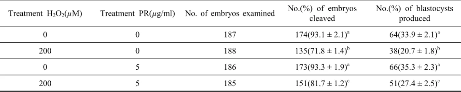
Table 5. Effect of PR extract on development of porcine parthenogenetic embryos cultured under oxidative stress conditions
Treatment H2O2(µM) Treatment PR(µg/ml) No. of embryos examined No.(%) of embryos cleaved
No.(%) of blastocysts produced
0 0 187 174(93.1 ± 2.1)a 64(33.9 ± 2.1)a
200 0 188 135(71.8 ± 1.4)b 38(20.7 ± 1.8)b
0 5 186 173(93.3 ± 1.9)a 66(35.3 ± 2.3)a
200 5 185 151(81.7 ± 1.2)c 51(27.4 ± 2.5)c
a,b Data are the mean ± SD. Values with different superscripts within a column differ significantly(p<0.05).
난할율 및 배반포로의 발달율은 대조군과 유사하게 나타났다.
그러나 200 µM 처리군에서의 난할율(78.1 ± 3.2%)과 배반포 발달율(20.4 ± 2.5%)은 대조군(92.1 ± 2.8%, 34.2 ± 3.5%)에 비 해 유의하게 낮게 나타났다(p<0.05).
둘째로 돼지 단위 발생 난자의 체외 배양 시 피라칸타 잎, 줄기와 뿌리 추출액 각각의 처리효과는 Table 2, 3 및 4에 제 시한 결과와 같다. 피라칸타 잎, 줄기와 뿌리 추출액의 경우, 1과 5 µg/ml 농도의 처리군에서 난할율 및 배반포로의 발달 율은 대조군과 유사한 양상을 보였으며, 피라칸타 뿌리 추출 액 경우, 5 µg/ml 처리군에서의 배반포 발달율이 36.4 ± 2.3%
로서 대조군의 34.2 ± 2.5%보다 다소 높게 나타나는 경향을 확인할 수 있었다. 그러나 10 µg/ml 농도의 각 처리군에서 배 반포로의 발달율은 대조군보다 유의하게 낮게 나타남을 알 수 있었다(p<0.05).
마지막으로 Table 1∼4에서 나타난 결과를 종합하여, 돼지 단위 발생 난자의 체외 배양 시 효과적이라고 판단되는 피라 칸타 뿌리 추출액 5 µg/ml와 산화적 스트레스를 유도하는 H2O2 200 µM을 병행 처리하여 배발달 양상을 확인하였다 (Table 5). 피라칸타 뿌리 추출액 5 µg/ml를 단독 처리군에서 의 배반포 발달율은 대조군과 비교하여 다소 높게 나타남을 다시 확인할 수 있었다(35.3 ± 2.3% vs. 33.9 ± 2.1%). 한편, H2O2 200 µM 처리군에서의 배반포로의 발달율은 대조군과
Table 2. Effect of various concentrations of pyracantha leaf(PL) extract during in vitro culture on development of porcine parthenogenetic embryos
Group (µg/ml)
No. of embryos examined
No. (%)of embryos cleaved
No. (%)of blasto- cysts produced 0 132 120(90.4 ± 2.0) 45(34.0 ± 2.8)a 1 138 126(91.1 ± 2.1) 46(33.1 ± 2.6)a 5 120 111(91.7 ± 1.9) 39(32.7 ± 2.9)a 10 123 110(90.7 ± 2.2) 30(24.0 ± 3.1)b
a,b Data are the mean ± SD. Values with different superscripts within a column differ significantly(p<0.05).
Table 3. Effect of various concentrations of pyracantha stalk(PS) extract during in vitro culture on development of porcine parthenogenetic embryos
Group (µg/ml)
No. of embryos examined
No. (%)of embryos cleaved
No. (%)of blasto- cysts produced 0 162 150(92.0 ± 1.9) 57(35.5 ± 3.3)a 1 150 141(93.9 ± 1.8) 51(33.8 ± 3.7)a 5 155 142(91.3 ± 2.1) 52(33.2 ± 2.5)a 10 145 132(90.9 ± 2.2) 38(26.3 ± 2.3)b
a,b Data are the mean ± SD. Values with different superscripts within a column differ significantly(p<0.05).
Table 4. Effect of various concentrations of pyracantha root(PR) extract during in vitro culture on development of porcine parthenogenetic embryos
Group (µg/ml)
No. of embryos examined
No. (%)of embryos cleaved
No. (%)of blasto- cysts produced 0 167 153(92.1 ± 1.8) 57(34.2 ± 2.5)a 1 162 148(91.8 ± 1.4) 56(35.9 ± 2.1)a 5 163 151(92.3 ± 1.6) 59(36.4 ± 2.3)a 10 162 146(90.1 ± 1.2) 48(29.4 ± 2.2)b
a,b Data are the mean ± SD. Values with different superscripts within a column differ significantly(p<0.05).
비교하여 유의하게 낮게 나타났다(p<0.05). 반면, H2O2 200 µM와 피라칸타 뿌리 추출액 5 µg/ml을 병행 처리군에서 배 반포 발달율은 27.4 ± 2.5%로서 200 µM 농도의 H2O2 단독 처 리군(20.7 ± 1.8%)의 배반포 발달율보다 유의하게 높게 나타 남을 확인할 수 있었다(p<0.05).
2. 피라칸타 추출액 처리에 의해 생산된 단위 발생 유래 배 반포의 질적 분석
돼지 단위 발생 난자에 피라칸타 잎, 줄기 또는 뿌리 추출액
처리에 의해 생산된 배반포의 질적 수준을 조사하였다(Fig. 1).
피라칸타 잎과 줄기 추출액 처리군에서 생산된 배반포의 총 세포 수는 대조군에서 생산된 배반포의 그것과 유사함을 확 인할 수 있었다. 또한, 피라칸타 잎과 줄기 추출액 처리군에서 생산된 배반포의 세포 사멸 지수가 대조군에서 생산된 배반 포의 그것과 유사하였다. 반면, 뿌리 추출액 5 µg/ml 처리군 에서 생산된 배반포의 총 세포 수가 다소 증가하였으며, 세포 사멸 지수 또한 다소 감소하는 경향을 보여 주었다.
피라칸타 뿌리 추출액 5 µg/ml와 H2O2 200 µM을 돼지 단 위 발생 난자에 단독 또는 병행 처리하여 생산된 배반포에서 ROS의 수준을 비교 분석하였다(Fig. 2). 피라칸타 뿌리 추출 액 처리군에서 생산된 배반포의 ROS 수준은 대조군에서 생 산된 배반포에서 보다 유의하게 낮았다(p<0.05). 한편, H2O2
단독으로 처리하여 생산된 배반포에서 ROS의 수준은 피라칸 타 뿌리 추출액과 병행 처리한 군보다 유의하게 높게 나타남 을 확인할 수 있었다(p<0.05).
고 찰
체외배양 과정에서 수정란은 체내 환경과 다르게 산화적 스트레스에 많이 노출되어 수정란 내에 ROS의 농도가 증가
Fig. 1. Comparison of epifluorescent images on apoptotic index in porcine parthenogenetic blastocysts derived from PL, PS and PR extracts treated groups. Total cells number and apoptotic index in porcine parthenogenetic blastocyst deri- ved from PL(A), PS(B) and PR(C) extracts treatment. Data are the mean ± SD. The chromatin contentis stained by DAPI (blue), fragmented DNA is labeled by the TUNEL reaction (green), and colocalization with DAPI appears sky-blue. Scale bars=
100 μm.
A
B
Fig. 2. Comparison of antioxidant characteristics in porcine partheno- genetic blastocysts derived from H2O2 and/or PR extract treatment groups. Fluorescence microscopy imaging of intra- cellular ROS expression(A) and ROS relative intensity(B) in porcine parthenogenetic blastocysts derived from H2O2 and/
or PR extract treatment. Data are the mean ± SD. Statisti- cally significant differences are indicated by asterisks (p<0.05).
Scale bars=100 μm.
한다(Kitagawa 등, 2004). 활성산소는 free radical을 가진 산소를 의미하며, 광범위하게는 lipid peroxide, lipid peroxy radical, peroxynitrite 등이 포함된다. 활성산소는 불안정한 특성을 지 니고 있으며, 주위의 물질과 반응성이 아주 강해 세포내 단백 질이나 지질 분자는 물론이고, 유전 정보를 함유한 DNA에도 산화적 손상을 입히며, 결과적으로 미토콘드리아 fission/fu- sion dynamic에 영향을 주어 수정란 발달 정지, ATP 결핍 그 리고 세포 사멸을 일으킨다(Guerin 등, 2001). 특히, 산화적 스 트레스는 착상 전 체외수정란에 해로운 영향을 미치고, 체외 배양 환경에서 산화적 스트레스를 줄이는 것이 체외수정란의
생산에 있어서 중요한 요인으로 인식되어지고 있다(Olson과 Seidel, 2000; Oris와 Leese, 2001). 기존의 연구에서 피라칸타 는 tannins, ascorbic acid, β-carotene 그리고 lycopene을 함유 하고 있다고 보고하였다(Pal 등, 2013). 이러한 식물 기원의 물 질들은 활성 산소종을 제거하는 데 효과적인 항산화제(Benzie 등, 1996)이며, 세포배양과정에서 산화적 스트레스로부터 DNA 손상을 막을 수 있다고 보고하였다(Rice-Evans 등, 1996). 식 물성 천연 페놀성 물질은 산화 환원의 특성 때문에 식물에서 발견되는 항산화제로서, 항균, 항염증 및 암 예방 물질로도 알 려져 있다(Jiang, 2000; Gomes 등, 2003). 본 연구에서 피라칸 타 추출액은 페놀 함량과 1,1-diphenyl-2-picrylhydrazyl (DPPH), [2,2’-azinobis(3-ethylbenzothiazoline-6-sulfonic acid)] diammo- nium salt(ABTS) 및 ferric reducing antioxidant powder(FRAP) 와 같은 3가지 다른 방법의 항산화 결과에 있어서 유의한 정 의 상관관계가 있었다(미발표). 이는 페놀성 물질과 항산화 활 성간의 높은 상관관계가 있다고 보고한 기존의 논문과도 비 슷한 경향을 보여주었다(Pulido 등, 2000). 따라서 본 연구에 서는 돼지 단위발생 난자의 체외 발달에 있어서 피라칸타의 효과와 항산화 능력을 확인하였다.
일반적으로 항산화제로 vitamin E와 C의 경우 체외배양 기 간 동안 첨가함으로써 배반포 발달율을 향상시킬 수 있다고 보고하였다(Hossein 등, 2007; Hu 등, 2012). 그러나 본 연구 에서 피라칸타 추출액은 배반포 발달율에는 특별한 효과가 없는 것으로 나타났다. 이전의 연구들에서 ROS는 착상 전 체 외 수정란의 발달에 해로운 영향을 미치기 때문에, 체외 배양 환경에서 ROS를 제거하는 것이 수정란의 발달 능력을 향상시 킨다고 보고하였다(Deleuze와 Goudet, 2010; Takahashi, 2012).
과산화수소(H2O2)는 세포와 화학적 반응을 일으켜 산화적 스 트레스를 유도하기 위하여 사용되었으며, 수정란 발달 과정에 서 과산화수소에 의한 손상 기능은 ROS(O2-)가 세포막을 통 하여 이동할 수 있다. 그래서 지질, 단백질, 그리고 핵산과 같 은 세포의 분자를 변형시킴으로 인해 미토콘드리아 손상, 수 정란 발달 정지 및 ATP 결손을 일으킨다(Guerin 등, 2001).
또한, 배양 과정 동안 높은 농도의 과산화수소 처리는 수정란 발달상에 독성 작용을 일으켜 결국 배반포 생성율에 영향을 미친다고 보고되었다(Morales 등, 1999). 따라서 본 연구에서 는 단위 발생 난자의 발달 과정 동안 과산화수소를 50, 100 그리고 200 µM 농도를 처리하였고, 과산화수소 200 µM 농도 에 노출된 단위발생 난자는 배반포로의 발달 능력과 질적 수 준이 현저히 감소하였다(Table 1, Fig. 2). 반면에 과산화수소 를 이용하여 산화적 스트레스를 유도한 배양 환경에서 피라 칸타 뿌리 추출액 5 µg/ml의 첨가는 단위 발생 난자의 발달 능력을 향상시켰고, 배반포의 질적 수준 또한 향상됨을 확인 할 수 있었다(Table 5, Fig. 2). 따라서 피라칸타 뿌리 추출액 은 ROS를 제거하는 항산화 기능을 가지고 있으며, 이로 인하
여 돼지 단위 발생 난자의 발달 능력과 배반포에서의 질적 수 준을 향상시켰다고 판단된다.
포유동물 배반포에서 전체 세포 수는 배반포의 질적 수준 과 생존 능력을 판단할 수 있는 지표이며, 수정란 발달 동안 세포 사멸은 핵과 염색체 이상으로 인해 수정란의 질적 수준 을 저하시킬 수 있고(Metwee 등, 2000), 수정란은 체외배양 동안 ROS 발생으로 인하여 세포 사멸이 유도될 수 있다고 보 고되었다(Tatemoto 등, 2000). 특히, ROS는 세포 외부 요소들 과 반응하여 미토콘드리아 의존적 세포 사멸을 매개할 수 있 다고 보고되었다(Herrera 등, 2001). 이와는 대조적으로, 체외 배양 배지에 항산화제 첨가는 산화적 스트레스를 완화시킴으 로써 생산된 수정란의 세포 사멸을 감소시킬 수 있다고 보고 되고 있다(Liu 등, 2003; Uhm 등, 2007). 따라서 본 연구에서 는 단위 발생 난자의 배양 과정 동안 피라칸타 추출액을 첨가 하여 생산된 배반포를 대상으로 DAPI/TUNEL 분석을 통하여 전체 세포 수와 세포 사멸 지수를 확인하였다(Fig. 1). 피라칸 타 잎, 줄기 및 뿌리 추출액 첨가군에서 전체 세포 수와 세포 사멸 지수는 대조군과 유사한 양상을 확인할 수 있었다. 한편, 인위적으로 유도된 산화적 스트레스 환경에서 피라칸타 뿌리 추출액을 첨가한 결과, 발달한 배반포에서 ROS의 수준이 감 소되는 것을 확인할 수 있었다(Fig. 2). 이러한 결과는 피라칸 타 뿌리 추출액에 존재하는 식물성 항산화제인 페놀 성분 등 이 free radical의 제거제로 작용을 함으로써 돼지 단위 발생 난자의 발달 과정 동안 산화적 스트레스가 완화되어 생산된 단위 발생 배반포의 질적 수준이 향상될 수 있었던 것으로 생 각된다.
결론적으로, 활성 산소는 배양 과정 동안 산화적 스트레스 를 일으켜 포유동물 수정란의 배반포 발달율과 질적 수준을 감소시킨다는 것을 확인하였다. 이러한 산화적 스트레스를 감 소시키기 위해 피라칸타 뿌리 추출액을 체외 배양 용액에 첨 가함으로써 돼지 단위 발생 난자의 발달 능력과 배반포에서 의 질적 수준이 향상됨을 확인할 수 있었다. 따라서 포유동물 수정란의 체외 생산에 있어서 피라칸타 뿌리 추출액은 항산 화제로써의 효능이 있다고 판단된다.
인 용 문 헌
Agarwal A, Allamaneni SS, Nallella KP, George AT and Mascha E. 2005. Correlation of reactive oxygen species levels with the fertilization rate after in vitro fertilization: a qua- lified meta-analysis. Fertil. Steril. 84: 228-231.
Benzie I and Strain J. 1996. The ferric reducing ability of pla- sma (FRAP) as a measure of antioxidant power. Anal. Bio- chem. 239: 70-76.
Choe SY and Yang KH. 1982. Toxicological studies of antio-
xidants butylated hydroxytoluene(BHT) and butylated hyd- roxy anisol(BHA). Kor. J. Food. Sci. Technol. 14: 283-288.
Deleuze S and Goudet G. 2010. Cysteamine supplementation of in vitro maturation media: a review. Reprod. Domest.
Anim. 45: 476-482.
Deng RF, Wang SG and Li GR. 1990. Studies on the nutri- tional components of the fruit of the wild plant-firethorn.
Acta. Nutrimenta. Sinica. 12: 79-84.
Funahashi H and Day BN. 1993. Effects of the duration of ex- posure to hormone supplements on cytoplasmic maturation of pig oocytes in vitro. J. Reprod. Fertil. 98: 179-185.
Gomes CA, Cruz TG, Andrade JL, Milhazes N, Borges F and Marques MPM. 2003. Anticancer activity of phenolic acids of natural or synthetic origin: A structure-activity Study. J.
Med. Chem. 46: 5395-5401.
Guerin P, El Mouatassim S and Menezo Y. 2001. Oxidative stress and protection against reactive oxygen species in the pre-implantation embryo and its surroundings. Hum. Reprod.
Update. 7: 175-89.
Herrera B, Alvarez AM, Sánchez A, Fernández M, Roncero C, Benito M and Fabregat I. 2001. Reactive oxygen species (ROS) mediates the mitochondrial-dependent apoptosis in- duced by transforming growth factor (beta) in fetal hepato- cytes. FASEB. J. 15: 741-751.
Hossein MS, Hashem MA, Jeong YW, Lee MS, Kim S, Kim JH, Koo OJ, Park SM, Lee EG, Park SW, Kang SK, Lee BC and Hwang WS. 2007. Temporal effects of alpha- tocopherol and L-ascorbic acid on in vitro fertilized porcine embryo development. Anim. Reprod. Sci. 100: 107-117.
Hou JJ, Xue H, Li YS, Liu XL and Wei WK. 2002. Effects of Pyracantha on plasma lipid and blood rheology of rats fed a high fat diet. China Public Health 18: 1059-1060.
Hou JJ, Wei WK, Huang H and Wu MG. 2003. Antioxidation effects of Pyracantha on aging mice model induced by over- dose of d-galactuse. China J. Pub. Health. 19: 944-945.
Hu J, Cheng D, Gao X, Bao J, Ma X and Wang H. 2012.
Vitamin C enhances the in vitro development of porcine pre-implantation embryos by reducing oxidative stress. Rep- rod. Domest. Anim. 47: 873-879.
Huang MT, Ho CT and Lee CY. 1992. Phenolic Compounds in Food and Their Effects on Health(II). Antioxidants and Cancer Prevention. American Chem. Soci. Pulication. Washington.
DC. p. 54-71.
Huang YC, Yang F and Duan YF. 2007. Study on physicoche- mical property of PP-A3 from water-soluble Pyracantha
fortuneana polysaccharides. Food. Res. Dev. 28: 75-79.
Jiang YM. 2000. Role of anthocyanins, polyphenol oxidase and phenols in Lychee pericarp browning. J. Sci. Food.
Agri. 80: 305-310.
Kim K, Lerou P, Yabuuchi A, Lengerke C, Ng K, West J, Kirby A, Daly MJ and Daley GQ. 2007. Histocompatible embryonic stem cells by parthenogenesis. Science 315: 482- 486.
Kitagawa Y, Suzuki K, Yoneda A and Watanabe T. 2004.
Effects of oxygen concentration and antioxidants on the in vitro developmental ability, production of reactive oxygen species (ROS), and DNA fragmentation in porcine embryos.
Theriogenology 62: 1186-1197.
Liu L, Trimarchi JR, Navarro P, Blasco MA and Keefe DL.
2003. Oxidative stress contributes to arsenic-induced telo- mere attrition, chromosome instability, and apoptosis. J. Biol.
Chem. 278: 31998-32004.
Matwee C, Betts DH and King WA. 2000. Apoptosis in the early bovine embryo. Zygote 8(1): 57-68.
Morales H, Tilquin P, Rees JF, Massip A, Dessy F and Van Langendonckt A. 1999. Pyruvate prevents peroxide-induced injury of in vitro preimplantation bovine embryos. Mol.
Reprod. Dev. 52: 149-157.
Niwa K. 1993. Effectiveness of in vitro maturation and in vitro fertilization techniques in pigs. J. Reprod. Fertil. Suppl. 48:
49-59.
Olson SE and Seidel GE Jr. 2000. Culture of in vitro-produced bovine embryos with vitamin E improves development in vitro and after transfer to recipients. Biol. Reprod. 62: 248- 52.
Orsi NM and Leese HJ. 2001. Protection against reactive oxygen species during mouse preimplantation embryo development:
role of EDTA, oxygen tension, catalase, superoxide dismu- tase and pyruvate. Mol. Reprod. Dev. 59: 44-53.
Pal RS, Arun Kumar R, Agrawal PK and Bhatt JC. 2013. Anti- oxidant capacity and related phytochemicals analysis of methanilic extract of two wild edible fruits from north wes- tern Indian Himalaya. Int. J. Pharm. Bio. Sci. 4: 113-123.
Petters RM and Wells KD. 1993. Culture of pig embryos. J.
Reprod. Fertil. Suppl. 48: 61-73.
Prather RS, Boice ML, Gibson J, Hoffman KE and Parry TW.
1995. In vitro development of embryos from sinclair minia- ture pigs: A preliminary report. Theriogenology 43: 1001- 1007.
Pulido R, Bravo L and Saura-Calixto F. 2000. Antioxidant
activity of dietary polyphenols as determined by a modi- fied ferric reducing/antioxidant power assay. J. Agricul.
Food. Chem. 48: 3396-3402.
Rice-Evans CA, Miller NJ and Paganga G. 1996. Structure anti- oxidant activity relationship of flavonoids and phenolic acids.
Free Radical Bio. Med. 20: 933-956.
Song K, Hyun SH, Shin T and Lee E. 2009. Post-activation treatment with demecolcine improves development of so- matic cell nuclear transfer embryos in pigs by modifying the remodeling of donor nuclei. Mol. Reprod. Dev. 76:
611-619.
Takahashi M. 2012. Oxidative stress and Redox regulation on in vitro development of mammalian embryos. J. Reprod.
Dev. 58: 1-9.
Tatemoto H, Sakurai N and Muto N. 2000. Protection of porcine oocytes against apoptotic cell death caused by oxidative stress during in vitro maturation: role of cumulus cells. Biol. Reprod. 63: 805-810.
Uhm SJ, Gupta MK, Yang JH, Lee SH and Lee HT. 2007.
Selenium improves the developmental ability and reduces the apoptosis in porcine parthenotes. Mol. Reprod. Dev.
74: 1386-1394.
Van Gelder CWG, Flurkey WH and Wichers HJ. 1997. Se- quence and structural features of plant and fungal tyrosina- ses. J. Phytochem. 45: 1309-1323.
Yoshioka K, Suzuki C, Tanaka A, Anas IM and Iwamura S.
2002. Birth of piglets derived from porcine zygotes cultu- red in a chemically defined medium. Biol. Reprod. 66:
112-119.
Yuh HS, Yu DH, Shin MJ, Kim HJ, Bae KB, Lee DS, Lee HC, Chang WK, Park SB, Lee SG, Park HD, Ha JH, Hyun BH and Ryoo ZY. 2010. The effects of various antioxida- nts on the development of parthenogenetic porcine embryos.
In Vitro. Cell. Dev. Biol. Ani. 46: 148-154.
(접수: 2013. 08. 15/ 심사: 2013. 08. 16/ 채택: 2013. 08. 29)