Effect of Oocyte Maturation Medium, Cytochalasin Treatment and Electric Activation on Embryonic Development after
Intracytoplasmic Sperm Injection in Pigs
Joohyeong Lee1, Jung Hoon Choi1,2, Seung Tae Lee3, Sang-Hwan Hyun4 and Eunsong Lee1,2,*
1College of Veterinary Medicine, Kangwon National University, Chuncheon 200-701, Korea
2Institute of Veterinary Science, Kangwon National University, Chuncheon 200-701, Korea
3Department of Animal Biotechnology, College of Animal Life Sciences, Kangwon National University, Chuncheon 200-701, Korea
4College of Veterinary Medicine, Chungbuk National University, Cheongju 361-763, Korea
ABSTRACT
The objective of this study was to examine the effect of in vitro maturation (IVM) medium, cytochalasin B (CB) treatment during intracytoplasmic sperm injection (ICSI), and electric activation on in vitro development ICSI-derived embryos in pigs. Immature pig oocytes were matured in vitro in medium 199 (M199) or porcine zygote medium (PZM)-3 that were supplemented with porcine follicular fluid, cysteine, pyruvate, EGF, insulin, and hormones for the first 22 h and then further cultured in hormone-free medium for an additional 21~22 h. ICSI embryos were produced by injecting single sperm directly into the cytoplasm of IVM oocytes. The oocytes matured in PZM-3 with 61.6 mM NaCl (low-NaCl PZM-3) tended to decrease (0.05<P<0.1) nuclear maturation when compared with oocytes matured in M199 (76.9% vs. 83.8%) but no significant differences were found in embryo cleavage, blastocyst formation, and mean number of cells in blastocyst (73.8% vs. 74.6%, 11.1% vs. 12.1%, and 28.4 cells vs. 30.1 cells, respectively).
The oocyte degeneration was not reduced by CB treatment during ICSI (11.9%) when compared with no treatment control (11.3%) while the treatment showed detrimental effects (P<0.05) on embryonic cleavage (40.0%) and blastocyst formation (1.8%) rates when compared with control (60.0% and 11.5%, respectively). For activation of ICSI oocytes, additional electric stimulus has no positive or negative effect on in vitro development of preimplantation stage ICSI porcine embryos. Our results demonstrate that CB treatment during ICSI inhibits embryonic development of ICSI oocytes and additional electric activation after ICSI has no effect in improving ICSI embryonic development in pigs.
Further studies are needed to improve ICSI efficiency by investigating factors influencing embryonic development after ICSI in pigs.
(Key words : cytochalasin B, electric activation, embryonic development, ICSI, pig)
†This research was supported by the Bio-Industry Technology Development Program (IPET112015-4), Ministry for Food, Agriculture Forestry and Fisheries, Republic of Korea.
*Correspondence : E-mail : [email protected]
INTRODUCTION
Intracytoplasmic sperm injection (ICSI) is a useful techni- que and can be used to maximize the utilization of male gene- tic resources including spermatozoa and round spermatids.
This technique was developed in hamsters in the 1970s (Uehara and Yanagimachi, 1976, 1977) and has been used subsequently to achieve fertilization and live births in rabbits, cattle, and humans (Iritani et al., 1988; Lanzendorf et al., 1988; Goto et al., 1990; Palermo et al., 1992). Presently, ICSI is an impor-
tant tool for investigating and understanding the early events of fertilization, and is used as an assisted reproductive tech- nology (ART) in humans. For decades, a variety of studies has been performed for the efficient production of mammalian embryos by using ARTs like ICSI and in vitro fertilization (IVF). It is well known that embryos derived from in vitro maturation (IVM), IVF and ICSI show lower developmental capacity than embryos derived in vivo. Even though many species have been successfully generated as live ICSI-derived offspring, the efficiency of live ICSI-derived piglets production
has only been reported as 4~6% in pigs (Katayama et al., 2007; Watanabe et al., 2012), which means the current pig ICSI-in vitro production (IVP) system is suboptimal and needs to be improved.
Developmental competence of IVP pig embryos is influ- enced by various factors including oocytes maturation, culture media, and other culture environments (Park et al., 2005; Viet Linh et al., 2009; Kim et al., 2010; Lee et al., 2013). Of those factors, the quality of oocytes may be one of the most critical factors in determining the success of IVP of pig embryos by IVF, ICSI, and somatic cell nuclear transfer (SCNT) (Yoon et al., 2000; Kim et al., 2010). Recently, it was found that lowering NaCl concentration in IVM medium could improve the cytoplasmic maturation and subsequent development of embryos derived from parthenogenetic activation and SCNT (Lee et al., 2013) but no report is available on the effect of IVM medium with low concentration of NaCl on the ICSI embryonic development.
In nuclear transfer manipulation, cytochalasin B (CB) re- laxes the cytoskeleton and enhances the flexibility of the oocyte. Treating oocytes of various species with CB during nuclear transfer prevented damage to the oolemma, allowed the insertion of a glass pipette through the ZP without lysing the oocyte (Baguisi et al., 1999; Polejaeva et al., 2000), and resulted in an improved success rate of embryonic develop- ment during the cloning of rabbits and sheep (Smith and Wil- mut, 1989; Collas and Robl, 1990).
ICSI also needs mechanical manipulation of oocytes to in- ject sperm into the oocyte cytoplasm. Presently, studies descri- bing the effects of CB-supplemented manipulation media on oocyte and embryonic development following ICSI are rarely reported. Spermatozoa derived from several mammalian spe- cies are also unable to activate oocytes, mainly due to lack of oocyte activation factors. Recent studies have shown that spe- cies differences exist in oocyte activation after ICSI. An addi- tional activation procedure is required to allow oocyte ferti- lization following spermatozoa injection in cattle (Rho et al., 1998) and pigs (Lee et al., 2003), whereas injection alone is sufficient to activate oocytes in mice and humans (Tesarik, 1996). Although mechanical stimulation by the injection pipe- tte occasionally results in activation, further stimulation may be needed for porcine oocytes.
The objective of this study was to examine the effect of IVM medium (M199 and low-NaCl PZM-3), CB treatment during ICSI, and additional electric activation after ICSI on in
vitro development ICSI-derived embryos in pigs.
MATERIALS AND METHODS
1. Culture Media
Unless otherwise stated, all chemicals were purchased from Sigma-Aldrich (St. Louis, USA). The base medium for IVM of oocytes was medium199 (M199) (Invitrogen, Grand Island, USA) or low-NaCl PZM-3 (61.6 mM) supplemented with 0.6 mM cysteine, 0.91 mM pyruvate, 10 ng/ml epidermal growth factor, 75 μg/ml kanamycin, 1 μg/ml insulin, and 10 % (v/v) porcine follicular fluid. Two media, PZM-3 (Yoshioka et al., 2002) and mPZM-3 (Lee et al., 2012) were used for in vitro culture (IVC) of ICSI embryos. PZM-3 was modified by supplementation with 2.77 mM myo-inositol, 0.34 mM triso- dium citrate, and 10 μM β-mercaltoehtanol (this medium was designated mPZM-3) according to the experimental design.
2. Oocyte Collection and IVM
Porcine ovaries were obtained from prepubertal gilts at a local abattoir and transported to the laboratory. Cumulus-oocyte complexes (COCs) were aspirated from superficial follicles (3~8 mm in diameter) in the ovaries using an 18-gauge needle and a 10 ml syringe. COCs that had multiple layers of compacted cumulus cells were selected and washed three times in HEPES-buffered Tyrode’s medium containing 0.05% (w/v) polyvinyl alcohol (TLH-PVA). COCs were placed into each well of a four-well multi-dish (Nunc, Roskilde, Denmark) con- taining 500 μl of IVM medium with 80 μg/ml FSH (Antrin R-10, Kyoritsu Seiyaku, Japan) and 10 IU/ml hCG (Intervet International BV, Holland). The COCs were cultured at 39℃ with 5% CO2 at maximum humidity. After 22 h in the ma- turation culture, the COCs were washed three times in fresh hormone-free IVM medium and then cultured in hormone-free IVM medium for an additional 21~22 h for ICSI.
3. ICSI
After IVM, COCs with expanded cumulus cells were treated with 0.1 % (w/v) hyaluronidase in IVM medium, and the cu- mulus cells were removed by repeated gentle pipetting. De- nuded oocytes were placed in droplets of TLH containing 0.4
% BSA, and oocytes with a uniform cytoplasm were chosen under the microscope. Spermatozoa for ICSI were prepared from extended liquid boar semen commercially available. Sper- matozoa were washed two times with D-PBS containing 0.1%
PVA and centrifuged at 700 g for 3 min. The sperm pellet was resuspended in D-PBS containing 5% polyvinyl pyrrolidone (PVP). Microinjection of whole spermatozoa into oocytes was performed at 39℃ using a 5.0~5.7 μm pipette (Origio Huma- gen, Charlottesville, USA). A sperm was aspirated into the injection pipette with a minimal amount of medium and intro- duced into the cytoplasm. ICSI was performed in the presence or absence of 5 μg/ml CB in manipulation medium. ICSI was completed within 2 h after collection of IVM oocytes.
4. Electric Activation of ICSI Oocytes and Embryo Culture All oocytes used for ICSI were washed three times with IVC medium. After washing, ICSI embryos were activated with two pulses of 120 V/mm DC for 60 μsec in a 280 mM mannitol solution containing 0.01 mM CaCl2 and 0.05 mM MgCl2
according to the experimental design. Activated or non- activated ICSI embryos were washed three times in fresh cul- ture medium, transferred into 30-μl droplets of IVC medium under mineral oil, and then cultured at 39℃ in a humidified atmosphere of 5 % CO2, 5% O2, and 90% N2 for 7 days.
5. Experimental Design
In Experiment 1, the effects of two maturation media, M199 and low-NaCl PZM-3 on nuclear maturation and embryonic development of pig oocytes after ICSI were compared. It was examined in Experiment 2 whether CB supplementation to the manipulation medium during ICSI would prevent oocyte dege- neration and improve embryonic development of ICSI oocytes.
In addition, effect of additional activation of ICSI oocytes by electric stimulus on embryonic development was examined in Experiment 3. Degeneration of ICSI oocytes was examined 2 h after sperm injection. Embryo cleavage and blastocyst for- mation were evaluated on Days 2 and 7, respectively (the day of ICSI was designated as Day 0). The mean cell number in blastocysts developed on Day 7 was examined using Hoechst
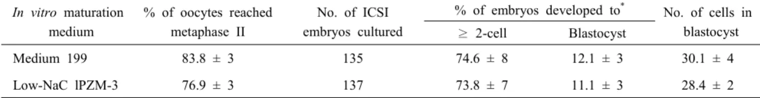
Table 1. In vitro development of intracytoplasmic sperm injection (ICSI)-derived pig embryos that were matured in vitro in medium 199 and PZM-3 with 61.6 mM NaCl (Low-NaCl PZM-3)
In vitro maturation medium
% of oocytes reached metaphase II
No. of ICSI embryos cultured
% of embryos developed to* No. of cells in blastocyst
≥ 2-cell Blastocyst
Medium 199 83.8 ± 3 135 74.6 ± 8 12.1 ± 3 30.1 ± 4
Low-NaC lPZM-3 76.9 ± 3 137 73.8 ± 7 11.1 ± 3 28.4 ± 2
Six replicates.
* Percentage of the number of ICSI embryos cultured.
33342 staining using an epifluorescence microscope.
6. Statistical Analysis
All statistical analyses were performed using the Statistical Analysis System (version 9.3; SAS Institute, USA). Data were analyzed using a general linear model followed by the least square method when the treatments differed at P<0.05. Per- centage data were arcsine-transformed prior to analysis to main- tain the homogeneity of variances. The results are expressed as the mean ± standard error of the mean (SEM).
RESULTS
1. Experiment 1: Preimplantation Development of ICSI Emb- ryos that were Matured in Two Different Culture Media: M199 and low-NaCl PZM-3
Maturation culture of oocytes in PZM-3 with 61.6 mM Na- Cl (low-NaCl PZM-3) tended to decrease (0.05<P<0.1) nuclear maturation compared to maturation in M199 (76.9% vs. 83.8%).
No significant differences were found in embryo cleavage, bla- stocyst formation, and mean number of cells in blastocyst (73.8% vs. 74.6%, 11.1% vs. 12.1%, and 28.4 cells vs. 30.1 cells, respectively) (Table 1).
2. Experiment 2: Effect of CB Treatment during ICSI on Oocyte Degeneration and Subsequent Embryonic Development
CB treatment during ICSI did not reduce oocyte degene- ration (11.9%) compared to no treatment (11.3%). Instead, CB significantly reduced embryo cleavage (40.0% vs. 60.0%) and blastocyst formation (1.8% vs. 11.5%) compared to control (Table 2).
3. Experiment 3: Effect of Additional Activation of ICSI Oocytes by Electric Stimulus on Preimplantation Development of ICSI Embryos
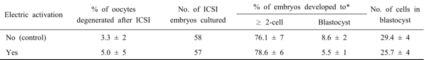
Activation of ICSI oocytes by electric stimulus did not alter embryonic development to the cleavage (78.6% vs. 76.1%) and
Table 2. Effect of cytochalasin B (CB) treatment during intracytoplasmic sperm injection (ICSI) on degeneration of oocytes and subsequent embryonic development after ICSI
CB treatment during ICSI
% of oocytes degenerated after ICSI
No. of ICSI embryos cultured
% of embryos developed to* No. of cells in blastocyst
≥ 2-cell Blastocyst
No (control) 11.3 ± 4 112 60.0 ± 7a 11.5 ± 2a 27.2 ± 4
Yes 11.9 ± 3 112 40.0 ± 4b 1.8 ± 1b 24.0 ± 6
Six replicates.
* Percentage of the number of ICSI embryos cultured.
a,b Different superscript letters indicates a significant difference within a column (P<0.05).
Table 3. Effect of electric activation on embryonic development of pig oocytes after ICSI
Electric activation % of oocytes degenerated after ICSI
No. of ICSI embryos cultured
% of embryos developed to* No. of cells in blastocyst
≥ 2-cell Blastocyst
No (control) 3.3 ± 2 58 76.1 ± 7 8.6 ± 2 29.4 ± 4
Yes 5.0 ± 5 57 78.6 ± 6 5.5 ± 1 25.7 ± 4
Three replicates.
* Percentage of the number of ICSI embryos cultured.
the blastocyst stages (5.5% vs. 8.6%) compared to control (Table 3).
DISCUSSION
Developmental competence of IVP embryos by IVF, ICSI, or SCNT is influenced by various factors. In this study, we examined the effects of several factors such as oocyte matu- ration medium, CB treatment during ICSI, and electrical acti- vation of ICSI oocytes on subsequent embryonic development.
It was demonstrated that CB treatment during ICSI inhibits embryonic development of ICSI oocytes while maturation of oocytes in low-NaCl PZM-3 and additional electric activation after ICSI has no effect in improving ICSI embryonic deve- lopment in pigs.
The quality of oocytes is one of the most important factors determining the success of ART in various species (Wang et al., 1998). Recently, it has been reported that oocytes having a wide perivitelline space show lower incidence of poly- spermic fertilization after IVF (Funahashi et al., 1994; Wang et al., 1998) and higher developmental competence after par- thenogenetic activation (PA) and somatic cell nuclear transfer (SCNT) (Lee et al., 2013). In this study, embryonic develop- ment of ICSI oocytes was not improved by the maturation of
oocytes in low-NaCl PZM-3. This result was not consistent with the previous result that embryonic development of PA and SCNT embryos was increased by the culture of oocytes in low- NaCl PZM-3 (Lee et al., 2013). The rate of embryos developed to the blastocyst state was around 10% that was lower than that of PA or SCNT embryos in our previous study. It was not clear the reason why no improving effect was shown in ICSI embryonic development. Relatively low developmental compe- tence of ICSI oocytes might be too low to be improved by the beneficial effect of IVM in low-NaCl PZM-3.
ICSI is a manipulative technique in which a single sperm is injected directly into the ooplasm of a mature oocyte using a micropipette. When sperm is injected into the oocyte cyto- plasm, oocytes can be ruptured or degenerated by mechanical damage to the ooplasmic membrane or cytoplasmic structures.
During the manipulation of oocytes for the enucleation or donor cell injection in SCNT, CB is commonly used to pre- vent oocytes from degenerating by stabilizing oocyte cyto- skeletons (Sugimura et al., 2008; Song et al., 2009). Treatment of oocytes with CB during sperm injection had no effect in preventing oocyte degeneration and was detrimental to emb- ryonic development of ICSI oocytes. It was considered that diameter of ICSI pipette was too small to result in mechanical damage to the oocytes. CB inhibits cytoplasmic division by
blocking the formation of contractile microfilaments. Although nuclear remodeling of ICSI oocytes was not examined in this study, it was probable that CB treatment during sperm in- jection affected nuclear remodeling such as inhibition of polar body extrusion in ICSI oocytes and resultantly reduced emb- ryonic development. This result indicates that exposure of ICSI oocytes to CB for short time (within 15 min) is detri- mental to embryonic development.
In the normal fertilization process, sperm has to be capa- citated and acrosome reaction is necessary before penetration (Matas et al., 2010; Tsai et al., 2010). Once sperm has pene- trated into the oocytes, oocytes are activated by the signal from the penetrated sperm (Wang et al., 1998). In ICSI, sperm is directly injected into oocytes without either acrosome reac- tion or capacitation processes. This is not physiological pro- cess and thus it is suspected whether the same pattern of oocyte activation will occur in ICSI oocytes as in normal ferti- lized oocytes. Electric activation has been widely used for activation of ICSI or SCNT oocytes (Prather et al., 1985; Jol- liff et al., 1997; Wang et al., 1998; Kure-bayashi et al., 2000).
Oocytes activation by electric stimulus in this study showed neither beneficial nor detrimental effects on embryonic deve- lopment of ICSI oocytes. Our result was not consistent with previous reports in which electric activation of ICSI oocytes increased embryo cleavage and blastocyst formation (Lee et al., 2003; Yoo et al., 2011) but was similar with the result that electrical activation was not necessary in the modified porcine ICSI (Yong et al., 2005). From present and previous results, it is considered that the effect of electric activation on ICSI emb- ryonic development can be various depending on the method of sperm injection and culture environments of ICSI oocytes.
In summary, our results demonstrate that CB treatment during ICSI inhibits embryonic development of ICSI oocytes and additional electric activation after ICSI has no effect in impro- ving ICSI embryonic development in pigs. Further studies are needed to improve ICSI efficiency by investigating factors influencing embryonic development after ICSI in pigs.
ACKNOWLEDGEMENT
We thank Gangwon Veterinary Service for the help in col- lecting pig ovaries.
REFERENCES
Baguisi A, Behboodi E, Melican DT, Pollock JS, Destrempes MM, Cammuso C, Williams JL, Nims SD, Porter CA, Midura P, Palacios MJ, Ayres SL, Denniston RS, Hayes ML, Ziomek CA, Meade HM, Godke RA, Gavin WG, Over- strom EW and Echelard Y. 1999. Production of goats by somatic cell nuclear transfer. Nat. Biotechnol. 17:456-461.
Collas P and Robl JM. 1990. Factors affecting the efficiency of nuclear transplantation in the rabbit embryo. Biol. Rep- rod. 43:877-884.
Funahashi H, Cantley TC, Stumpf TT, Terlouw SL and Day BN. 1994. In vitro development of in vitro-matured porcine oocytes following chemical activation or in vitro fertiliza- tion. Biol. Reprod. 50:1072-1077.
Goto K, Kinoshita A, Takuma Y and Ogawa K. 1990. Ferti- lisation of bovine oocytes by the injection of immobilised, killed spermatozoa. Vet. Rec. 127:517-520.
Iritani A, Utsumi K, Miyake M, Hosoi Y and Saeki K. 1988.
In vitro fertilization by a routine method and by micro- manipulation. Ann. N. Y. Acad. Sci. 541:583-590.
Jolliff WJ and Prather RS. 1997. Parthenogenic development of in vitro-matured, in vivo-cultured porcine oocytes beyond blastocyst. Biol. Reprod. 56:544-548.
Katayama M, Rieke A, Cantley T, Murphy C, Dowell L, Sutovsky P, Day BN. 2007. Improved fertilization and embryo development resulting in birth of live piglets after intracytoplasmic sperm injection and in vitro culture in a cysteine-supplemented medium. Theriogenology 67:835-847.
Kim J, You J, Hyun SH, Lee G, Lim J and Lee E. 2010.
Developmental competence of morphologically poor oocytes in relation to follicular size and oocyte diameter in the pig.
Mol. Reprod. Dev. 77:330-339.
Kure-bayashi S, Miyake M, Okada K and Kato S. 2000.
Successful implantation of in vitro-matured, electro-activated oocytes in the pig. Theriogenology 53:1105-1119.
Lee J, Hyun S and Lee E. 2012. A comparative study on the parthenogenetic development of pig oocytes cultured in North Carolina State University-23 and porcine zygote me- dium-3. J. Emb. Trans. 27:121-126.
Lee J, You J, Lee G-S, Hyun S-H, Lee E. 2013. Pig oocytes with a large perivitelline space matured in vitro show grea- ter developmental competence after parthenogenesis and so- matic cell nuclear transfer. Mol. Reprod. Dev. In press.
Lee, JW, Tian XC and Yang X. 2003. Failure of male pro- nucleus formation is the major cause of lack of fertilization and embryo development in pig oocytes subjected to intra-
cytoplasmic sperm injection. Biol. Reprod. 68:1341-1347.
Matás C, Sansegundo M, Ruiz S, García-Vázquez FA, Gadea J, Romar R and Coy P. 2010. Sperm treatment affects capacitation parameters and penetration ability of ejaculated and epididymal boar spermatozoa. Theriogenology 74:1327- 1340.
Palermo G, Joris H, Devroey P and Van Steirteghem AC.
1992. Pregnancies after intracytoplasmic injection of single spermatozoon into an oocyte. Lancet 340:17-18.
Park Y, Hong J, Yong H, Lim J and Lee E. 2005. Effect of exogenous carbohydrates in a serum-free culture medium on the development of in vitro matured and fertilized porcine embryos. Zygote 13:269-275.
Polejaeva IA, Chen SH, Vaught TD, Page RL, Mullins J, Ball S, Dai Y, Boone, J, Walker S, Ayares DL, Colman A and Campbell KH. 2000. Cloned pigs produced by nuclear transfer from adult somatic cells. Nature 407:86-90.
Prather RS, Sims MM and First NL. 1989. Nuclear trans- plantation in early pig embryos. Biol. Reprod. 41:414-418.
Rho GJ, Kawarsky S, Johnson WH, Kochhar K and Betteridge KJ. 1998. Sperm and oocyte treatments to improve the for- mation of male and female pronuclei and subsequent deve- lopment following intracytoplasmic sperm injection into bovine oocytes. Biol. Reprod. 59:918-924.
Smith LC and Wilmut I. 1989. Influence of nuclear and cytoplasmic activity on the development in vivo of sheep embryos after nuclear transplantation. Biol. Reprod. 40:
1027-1035.
Song K, Hyun SH, Shin T and Lee E. 2009. Post-activation treatment with demecolcine improves development of so- matic cell nuclear transfer embryos in pigs by modifying the remodeling of donor nuclei. Mol. Reprod. Dev. 76:611- 619.
Sugimura S, Kawahara M, Wakai T, Yamanaka K, Sasada H and Sato E. 2008. Effect of cytochalasins B and D on the developmental competence of somatic cell nuclear transfer embryos in miniature pigs. Zygote 16:153-159.
Tesarik J. 1996. Fertilization of oocytes by injecting sperma- tozoa, spermatids and spermatocytes. Rev. Reprod. 1:149- 152.
Tsai PS, Garcia-Gil N, van Haeften T and Gadella BM. 2010.
How pig sperm prepares to fertilize: stable acrosome dock- ing to the plasma membrane. PLoS One 18:e11204.
Uehara T and Yanagimachi R. 1976. Microsurgical injection of spermatozoa into hamster eggs with subsequent trans- formation of sperm nuclei into male pronuclei. Biol. Re- prod. 15:467-470.
Uehara T, Yanagimachi R. 1977. Behavior of nuclei of testi- cular, caput and caudapididymal spermatozoa injected into hamster eggs. Biol. Reprod.16:315-321.
Viet Linh N, Dang-Nguyen TQ, Nguyen BX, Manabe N and Nagai T. 2009. Effects of cysteine during in vitro matu- ration of porcine oocytes under low oxygen tension on their subsequent in vitro fertilization and development. J.
Reprod. Dev. 55:594-598.
Wang WH, Abeydeera LR, Prather RS and Day BN. 1998.
Functional analysis of activation of porcine oocytes by spermatozoa, calcium ionophore, and electrical pulse. Mol.
Reprod. Dev. 51:346-353.
Watanabe M, Kurome M, Matsunari H, Nakano K, Umeyema K, Shiota A, Nakauchi H and Nagashima H. 2012. The creation of transgenic pigs expressing human proteins using BAC-derived, full-length genes and intracytoplasmic sperm injection-mediated gene transfer. Transgenic Res. 21:605- 618.
Yong HY, Hong JY, Pak SI, Kang SK, Lee BC, Lee ES and Hwang WS. 2005. Effect of centrifugation and electrical activation on male pronucleus formation and embryonic development of porcine oocytes reconstructed with intra- zcytoplasmic sperm injection. Reprod. Fertil. Dev. 17:557- 563.
Yoo J, Hur C, Park M, Park J, Hwang K, Kim J, Kim J and Cho S. 2011. Electrical activation enhances pre-implanta- tion embryo development following sperm injection into in vitro matured pig oocytes. J. Vet. Med. Sci. 74:429-434.
Yoon KW, Shin TY, Park JI, Roh S, Lim JM, Lee BC, Hwang WS and Lee ES. 2000. Development of porcine oocytes from preovulatory follicles of different sizes after matu- ration in media supplemented with follicular fluids. Reprod.
Fertil. Dev. 12:133-139.
(접수: 2013. 4. 29 / 심사: 2013. 4. 29 / 채택: 2013. 5. 17)