Tuberc Respir Dis 2012;72:452-456
CopyrightⒸ2012. The Korean Academy of Tuberculosis and Respiratory Diseases. All rights reserved.
Disseminated Mycobacterium intracellulare Infection in an Immuno- competent Host
Won-Young Kim, M.D., Sun-Joo Jang, M.D., Taejin Ok, M.D., Gwang Un Kim, M.D., Han-Seung Park, M.D., Jaechan Leem, M.D., Bo Hyoung Kang, M.D., Se Jeong Park, M.D., Dong Kyu Oh, M.D., Byung Ju Kang, M.D., Bo Young Lee, M.D., Won-Jun Ji, M.D., Tae Sun Shim, M.D.
Department of Pulmonary and Critical Care Medicine, Asan Medical Center, University of Ulsan College of Medicine, Seoul, Korea
Disseminated Mycobacterium avium complex (MAC) infection can occur in immunocompromised patients, and rarely in immunocompetent subjects. Due to the extensive distribution of the disease, clinical presentation of disseminated MAC may mimic malignancies, and thorough examinations are required in order to make accurate diagnosis. We report a case of disseminated Mycobacterium intracellulare disease in an immunocompetent patient, which involved the lung, lymph nodes, spleen, and multiple bones. F-18 fluorodeoxyglucose positron-emission tomography imaging showed multiple hypermetabolic lesions, which are suggestive of typical hematogenous metastasis. However, there was no evidence of malignancy in serial biopsies, and M. intracellulare was repeatedly cultured from respiratory specimens and bones. Herein, we should know that disseminated infection can occur in the immunocompetent subjects, and it can mimic malignancies.
Key Words: Nontuberculous Mycobacteria; Immunocompetence; Positron-Emission Tomography; Hybridization, Genetic
Address for correspondence: Tae Sun Shim, M.D.
Department of Pulmonary and Critical Care Medicine, Asan Medical Center, University of Ulsan College of Medicine, 88, Olympic-ro 43-gil, Songpa-gu, Seoul 138-736, Korea Phone: 82-2-3010-3892, Fax: 82-2-3010-6968
E-mail: [email protected] Received: Sep. 23, 2011 Revised: Oct. 4, 2011 Accepted: Nov. 16, 2011
Introduction
Mycobacterium intracellulare is one of the Mycobac- terium avium complex (MAC) and is the most frequently identified pathogen in nontuberculous mycobacterial (NTM) lung disease in the United States1. The infection is usually confined to the lung, but extrapulmonary or disseminated forms can occur, especially in immuno- compromised patients but rarely in immunocompetent subjects2-4.
In case of disseminated MAC disease, in which clin- ical presentations could often mimic those of malig- nancies, the confirmative diagnosis should be made
with a careful history taking and thorough examina- tions. Isolation of causative organisms is the first step to the correct diagnosis and F-18 fluorodeoxyglucose positron-emission tomography (FDG PET) can be useful to evaluate the extent of disease even though the main use of PET is to evaluate the extents of various malig- nancies5,6.
In this report, we describe a case of disseminated M.
intracellulare infection in an immunocompetent male.
The PET finding was so typical of hematogenous meta- stasis of malignancies, repeated histologic examinations were performed to exclude the possibility of malig- nancy.
Case Report
A 71-year-old man was referred to our hospital for the evaluation of recurrent fever, cough, sputum, short- ness of breath, and recent weight loss of 3 kg (5%
weight loss). Seven months ago, the patient was admit-
Figure 1. Chest X-ray showed the ill-defined patchy mass opacity with surrounding ground-glass opacity in left mid-lung field.
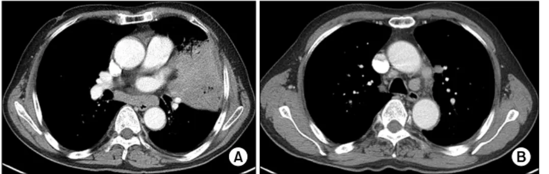
Figure 2. Contrast enhan- ced computed tomogra- phy showed consolidation in the lingual segment of left upper lobe (A) and bi- lateral meditational lymph nodes swelling (B).
ted to another hospital, and chest computed tomog- raphy (CT) revealed a 14-mm nodule and loculated effu- sion in left lower lung field. Under the diagnosis of em- pyema, drainage of pleural fluid concomitant with anti- biotics therapy was performed and left pleural effusion and a lung nodule disappeared. However, the symp- toms had recurred three months ago and persisted de- spite antibiotic treatment, and the patient was referred to our hospital for further evaluation. The patient had previously been healthy with no significant history of lung disease, other than a previously documented em- pyema and a lung nodule, and has smoked 20 cigarettes a day for 40 years. He was in mild respiratory distress, respiratory rate 24/min, temperature 37.6oC, and pulse 82/min. The lung sound was slightly diminished over
left lower lung field.
Findings of laboratory studies showed a white-blood- cell count of 18,000/mm3 with 76.6% neutrophils, he- moglobin 10.8 g/dL, and platelet count 462,000/mm3. Electrolytes, measures of renal function, and liver en- zymes were within normal limits. C-reactive protein (CRP) was elevated to 15.34 mg/dL. Serologic test for human immunodeficiency virus gave negative result.
Initial blood and sputum cultures remained negative, but the patient was started on piperacillin/tazobactam (18 g per day) and ciprofloxacin (800 mg per day) as an empirical antibacterial therapy. Chest X-ray showed the ill-defined patchy mass opacity with surrounding ground-glass opacity in left mid-lung field (Figure 1).
Chest CT showed huge mass-like consolidation in the lingular segment of left upper lobe and enlarged bi- lateral supraclavicular and mediastinal lymph nodes, suggestive of malignancy or inflammatory disease (Figure 2).
During five days of antibiotic treatment, neither clin- ical nor radiologic improvement was observed. Brocho- alveolar lavage (BAL) and endobronchial ultrasound- guided transbronchial needle aspiration (EBUS-TBNA) for paratracheal and subcarinal lymph nodes were per- formed, and the findings were unremarkable. The pa- tient remained febrile after seven days of initial anti- biotic therapy, and the regimen was changed to mer- openem (3 g per day) and vancomycin (2 g per day).
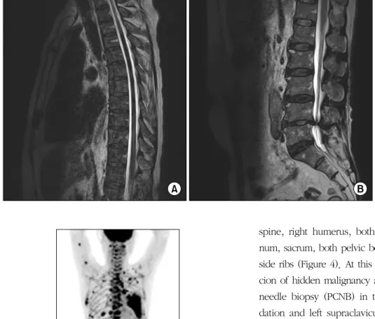
In addition to respiratory symptoms, the patient com- plained of back pain. Magnetic resonance imaging (MRI) of the thoracic and lumbar spine was performed and showed diffuse heterogenous low signal change with heterogenous enhancement (Figure 3). From this
Figure 3. Magnetic reso- nance imaging of the thor- acic (A) and lumbar (B) spine showed diffuse heter- ogenous low signal change with heterogenous enhan- cement.
Figure 4. Maximum intensity projection image of F-18 flu- orodeoxyglucose positron-emission tomography on ad- mission showed multifocal hypermetabolic lesions in the left upper lung, lymph nodes, spleen, and multiple bones.
MRI finding, marrow infiltrative diseases such as multi- ple myeloma, malignant lymphoma, and multiple meta- stasis of unknown origin were considered. There was no evidence of monoclonal gammopathy based on se- rum and urine electrophoresis. FDG PET was performed and maximum intensity projection image showed multi- focal hypermetabolic lesion in the lingular segment of left upper lung, bilateral supraclavicular and mediastinal lymph nodes, spleen, and multiple bones, including
spine, right humerus, both scapula, left clavicle, ster- num, sacrum, both pelvic bones, both femurs, and both side ribs (Figure 4). At this point, we had a high suspi- cion of hidden malignancy and performed percutaneous needle biopsy (PCNB) in the left upper lobe consoli- dation and left supraclavicular lymph node. However, both pathologies revealed nonspecific chronic inflam- mation and interstitial fibrosis and there was no evi- dence of malignancy or granulomas. Then, CT-guided bone biopsy was performed in the first lumbar verte- brae, and the pathology showed myeloid hyperplasia with marked plasmacytosis, without the evidence of ma- lignant cell infiltration.
From the day of admission, serial sputum specimens were collected and cultured for mycobacteria using both mycobacteria growth indicator tuve (MGIT; Becton Dickinson Diagnostic Systems, Sparks, MD, USA) and Ogawa (Korean Institute of Tuberculosis, Seoul, Korea) media. On the day 30 of admission, acid-fast bacilli were repeatedly cultured in MGIT media and then in Ogawa media, which were identified as nontuberculous mycobacteria. Subsequent cultures from previous BAL and PCNB of left upper lobe consolidation specimens also were identified as nontuberculous mycobacterium.
On the day 37 of admission, M. intracellulare was iden- tified, and anti-MAC treatment was initiated with clari- thromycin (1 g per day), rifampicin (600 mg per day),
ethambutol (800 mg per day), and streptomycin (1 g per day). Even though the bone biopsy specimen did not reveal acid-fast bacilli, the reverse blot hybridization assay (REBA Myco-IDⓇ; Molecules and Diagnostics, Wonju, Korea) using bone biopsy specimen revealed M.
intracellulare. Later, M. intracellulare was also identified from bone biopsy specimen.
From these radiological and microbiological findings, the diagnosis of disseminated M. intracellulare infection was made. Anti-MAC therapy was continued and the patient was discharged with improved symptoms on the day 63 of admission. The drug sensitivity test revealed that the organism was susceptible to clarithromycin.
The minimum inhibitory concentration of clarithromycin was 1 mg/L (susceptible). Unfortunately, additional in- formation after discharge was not available due to trans- fer to another hospital.
Discussion
Disseminated MAC disease primarily occurs in im- munocompromised patients, like those with acquired immunodeficiency syndrome, renal or cardiac trans- plantation, leukemia, or history of immunosuppressive therapy7. Recently, numerous disseminated MAC in- fection cases have been reported in immunocompetent hosts2,8,9.
The clinical symptoms of disseminated NTM infection include fever (>80%), night sweats (>35%), and weight loss (25%)10. The most commonly identified lab- oratory abnormalities include anemia and elevated lev- els of alkaline phosphatase, aspartate aminotransferase, lactate dehydrogenase, and CRP. In our case, mild ane- mia and CRP elevation were observed. The most com- monly involved organs are the spleen, lymph nodes, liv- er, intestines, and bone marrow; the lungs are less com- monly involved11. However, in a recent Taiwanese study by Chou et al.12, many patients (42.5%) with dis- seminated NTM infection had lung involvement and they explained that increased suspicion for pulmonary NTM infection and improved culture techniques may be the reason of increasing incidence. In our case, we also
had positive cultures of M. intracellulare in sputum, BAL fluid, and lung parenchyma.
CT and MRI can well demonstrate multiple lesions of disseminated disease such as lymph nodes enlargement, splenomegaly, and bone marrow involvement. On the other hand, FDG PET can recognize whole body dis- tribution more easily and importantly, can recognize le- sions which show as normal in other imaging modali- ties. In our case of disseminated disease, CT finding of spleen was unremarkable, although FDG PET revealed a focal hypermetabolic lesion. FDG uptake increases when metabolic activity is increased by the presence of inflammatory cells, and there are several reports related to FDG accumulation in MAC infection13,14. Moreover, FDG PET can well demonstrate the activity of dis- seminated MAC, assess the treatment response after an- ti-MAC therapy, and provide suitable biopsy sites15. The distribution of FDG accumulation is similar be- tween disseminated MAC and other systemic diseases, such as malignancies and other granulomatous diseases.
Therefore, histopathologic diagnosis is essential for fur- ther treatment. We performed four core needle biopsies in lung parenchyma, supraclavicular lymph node, and spines and EBUS-TBNA in enlarged mediastinal lymph nodes and there was no evidence of malignancy in any of these specimens.
In the initial period of evaluation, cultures in respira- tory specimens were positive for NTM but bone biopsy specimen did not reveal NTM. Instead, molecular tech- nique using REBA method detected M. intracellulare DNA from the bone biopsy specimen and later M. intra- cellulare was isolated from bone biopsy specimen.
Hence, molecular technique may permit rapid diagnosis of disseminated NTM disease. In our case, REBA techni- que was used and rapid identification among NTM spe- cies was also possible by using this kit.
In conclusion, our report indicates that the clinical presentations of disseminated MAC infection can mimic those of malignancies. Thorough examinations, includ- ing histopathologic and microbiologic diagnosis, are required. FDG PET is a useful diagnostic tool to assess the exact extent of disease and provide suitable biopsy
sites. Along with culture method, REBA is an improved rapid molecular diagnostic tool in identification of mycobacterium.
References
1. Griffith DE, Aksamit T, Brown-Elliott BA, Catanzaro A, Daley C, Gordin F, et al. An official ATS/IDSA state- ment: diagnosis, treatment, and prevention of non- tuberculous mycobacterial diseases. Am J Respir Crit Care Med 2007;175:367-416.
2. Song JY, Park CW, Kee SY, Choi WS, Kang EY, Sohn JW, et al. Disseminated Mycobacterium avium complex infection in an immunocompetent pregnant woman.
BMC Infect Dis 2006;6:154.
3. Kim SY, Oh DW, Yu JH, Kim D, Noh S, Roh J, et al.
A case of disseminated Mycobacterium intracellulare infection in an immunocompromised host. Tuberc Respir Dis 2009;67:32-6.
4. Chung JW, Cha YJ, Oh DJ, Nam WJ, Kim SH, Lee MK, et al. Disseminated Mycobacterium avium complex in- fection in a non-HIV-infected patient undergoing con- tinuous ambulatory peritoneal dialysis. Korean J Lab Med 2010;30:166-70.
5. Lowe VJ, Naunheim KS. Positron emission tomography in lung cancer. Ann Thorac Surg 1998;65:1821-9.
6. Meller J, Sahlmann CO, Scheel AK. 18F-FDG PET and PET/CT in fever of unknown origin. J Nucl Med 2007;
48:35-45.
7. Diagnosis and treatment of disease caused by non- tuberculous mycobacteria. This official statement of the American Thoracic Society was approved by the Board of Directors, March 1997. Medical Section of the
American Lung Association. Am J Respir Crit Care Med 1997;156(2 Pt 2):S1-25.
8. Okada H, Yoshioka K. Acute disseminated encephalo- myelitis associated with meningitis due to Mycobacte- rium intracellulare. Intern Med 2010;49:2113-6.
9. Azzam HC, Gahunia MK, Sae-Tia S, Santoro J. Myco- bacterium avium-avium-associated typhlitis mimicking appendicitis in an immunocompetent host. Am J Med Sci 2009;337:218-20.
10. Nightingale SD, Byrd LT, Southern PM, Jockusch JD, Cal SX, Wynne BA. Incidence of Mycobacterium avium-intracellulare complex bacteremia in human im- munodeficiency virus-positive patients. J Infect Dis 1992;165:1082-5.
11. Kasperbauer SH, Daley CL. Diagnosis and treatment of infections due to Mycobacterium avium complex.
Semin Respir Crit Care Med 2008;29:569-76.
12. Chou CH, Chen HY, Chen CY, Huang CT, Lai CC, Hsueh PR. Clinical features and outcomes of dis- seminated infections caused by non-tuberculous myco- bacteria in a university hospital in Taiwan, 2004-2008.
Scand J Infect Dis 2011;43:8-14.
13. Zhuang H, Pourdehnad M, Yamamoto AJ, Rossman MD, Alavi A. Intense F-18 fluorodeoxyglucose uptake caused by Mycobacterium avium intracellulare in- fection. Clin Nucl Med 2001;26:458.
14. Bandoh S, Fujita J, Ueda Y, Tojo Y, Ishii T, Kubo A, et al. Uptake of fluorine-18-fluorodeoxyglucose in pul- monary Mycobacterium avium complex infection. In- tern Med 2003;42:726-9.
15. Sato M, Hiyama T, Kaito K, Hayashi Y, Okumura T.
Usefulness of F-18 FDG PET/CT in the assessment of disseminated Mycobacterium avium complex infection.
Ann Nucl Med 2009;23:757-62.