• 검색 결과가 없습니다.

The Comparison of Muscle Activities in the Right Leg during Squatting According to Various Ankle Angles

N/A
N/A
Protected

Academic year: 2021

Share "The Comparison of Muscle Activities in the Right Leg during Squatting According to Various Ankle Angles"

Copied!
8
0
0

로드 중.... (전체 텍스트 보기)

전체 글

(1)

| Abstract |

Purpose: This study aimed to compare muscle activities in the right leg during squatting on an angle-adjustable inclined wooden plate at three different angles.

Methods: The subjects were 19 healthy adult men and women. An angle-adjustable inclined wooden plate was used for the experiment, and the subjects performed squatting at three adjusted angles of 0° ankle angle, 10° ankle flexion, and 10° plantar flexion. Squatting was randomly performed without a sequence. The knee angle was set at 45°, and a goniometer was used to measure the angles accurately. Electromyography was employed to measure and compare muscle activity in the right leg in each condition. The measured data were converted to root mean square values to calculate the muscle activities.

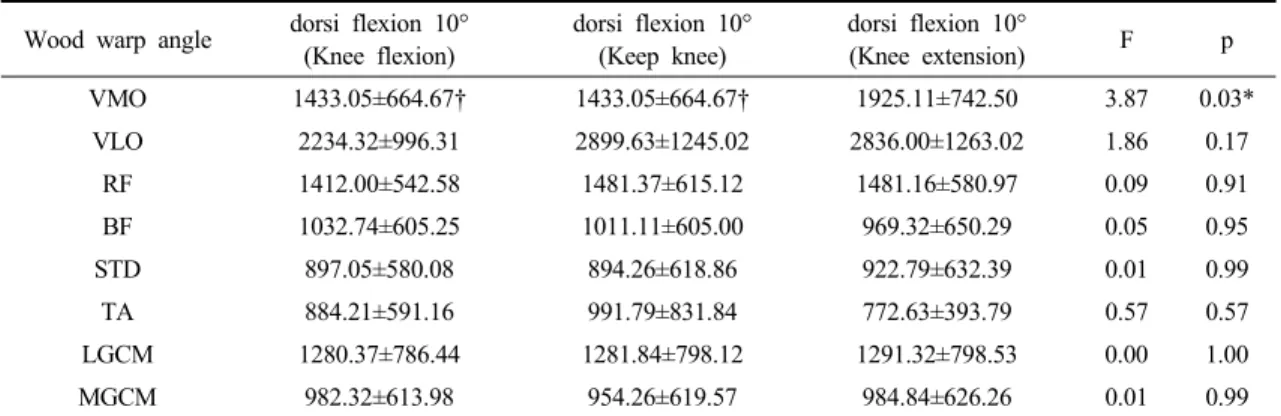
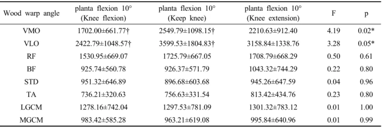
Results: This study showed no statistically significant difference at a 0° ankle angle, but a statistically significant difference was found in the vastus medialis at 10° of ankle flexion. Moreover, statistically significant differences were observed in the vastus medialis and lateralis at 10° of plantar flexion.

Conclusion: This study showed a statistically significant difference in the vastus medialis at 10° of ankle flexion and statistically significant differences in the vastus medialis and lateralis at 10° of plantar flexion. Therefore, it may be effective to perform squatting at 10° of ankle flexion when intending to selectively strengthen the vastus medialis and at 10° of plantar flexion when intending to strengthen both the vastus medialis and lateralis.

Key Words: Squart, Wood warp, Goniometer, Electromyography

†Corresponding Author : Su-Hong Ahn ([email protected])

Original article Open Access

다양한 발목각도에 따른 스쿼트 시 오른쪽 다리의 근 활성도 비교

안수홍⋅이수경

1†

⋅이광준⋅박진성⋅황제웅

1)

동의대학교 보건의과학대학원,

1

동의대학교 물리치료학과

The Comparison of Muscle Activities in the Right Leg during Squatting According to Various Ankle Angles

Su-Hong Ahn⋅Su-Kyoung Lee

1†

⋅Kwang-Jun Lee⋅Jin-Seong Park⋅Jea-Woong Hwang Department of Biomedical Health Science, Dong-Eui University

1

Department of Physical Therapy, College of Nersing and Healthcare Sciences, Dong-Eui University

Received: August 14, 2017 / Revised: September 28, 2017 / Accepted: September 29, 2017

ⓒ 2017 Journal of Korea Proprioceptive Neuromuscular Facilitation Association

This is an Open Access article distributed under the terms of the Creative Commons Attribution Non-Commercial License

(http://creativecommons.org/licenses/by-nc/3.0) which permits unrestricted non-commercial use, distribution, and reproduction

in any medium, provided the original work is properly cited.

(2)

Ⅰ. 서 론

최근 대부분의 사람들은 생활의 여유를 자신의 건 강을 위하여 운동과 식생활 개선 등과 같이 여러 방면 으로 관심을 많이 가지며 특히 운동 및 여가 스포츠 활동에 대한 관심과 실천의 비중이 커지고 있다(Jeong, 2006). 건강한 삶을 위한 대표적인 사례인 체력 증진은 근력 , 근지구력, 심폐지구력과 같은 요소들을 포함하 는데, 근력과 지구력을 증가시킬 수 있는 웨이트 트레 이닝은 전반적으로 체력증진에 주요한 역할을 한다 (Lee et al., 2004). 웨이트 트레이닝의 기본적인 운동에 포함되는 저항운동은 근육이 약한 성인을 대상으로 근력을 증가시킬 수 있는 안정적이고 효과적인 운동 방법이며(Peterson et al., 2010), 자신의 체중을 이용한 운동 방법과 중량기구를 사용하여 다양한 형태로의 운동이 가능하다고 하였다(Kwon et al., 2012). 특히 닫힌 사슬 운동은 체중 부하를 이용하여 몸의 먼 쪽은 고정시킨 상태에서 몸 쪽 부위와 먼 쪽 부위에 동시에 저항을 적용하여 일어나는 운동이며 (Prince, 1999), 열 린 사슬 운동보다 관절 내 구조물이나 주위 조직들이 특정한 기계적 수용기를 효율적으로 자극하고, 관절 에 대한 안정성과 일치성을 증가시켜 체중지지를 하 는 동안 부하에 대한 축을 생성하는 동시에 체중지지 자세에서 수행을 하기 때문에 주동근과 길항근에 대 한 협력 수축을 촉진시키고 , 동적 안정성을 증진시킨 다(Kisner & Colby, 2007). 또한 몸통 안정성과 같이 신체분절에 대한 움직임 조절을 할 수 있는 다리 근력 은 몸통을 지지하면서 보행에서 주된 능동적인 역할 을 한다고 하였고(Bohannon, 1995), Sharp 와 Brouwer (1997)는 다리의 근력 강화 운동을 통하여 무릎관절에 대한 신전력이 증가한다고 주장하였다. 이러한 다리 근력을 증가시킬 수 있는 대표적인 방법으로는 스쿼 트 운동이 있으며 물리치료나 스포츠 환경에서 훈련 프로그램의 중요한 요소이다 (Dionisio et al., 2008). 스 쿼트 운동은 대 근육 운동이며 , 여러 다리 부위의 근육 들이 참여하는 복합적인 운동이다(Na, 2013). 스쿼트 운동은 체중지지를 통해 보다 많은 관절을 움직임으

로써 넙다리네갈근이 근력강화가 되며(Selseth et al., 2000), 관절주변의 협력수축과 압박력, 고유수용성 감 각의 입력, 다 분절 관절과 근육군의 동원을 동반하는 원심성과 구심성의 근육 작용효과를 볼 수 있다(Lee, 2012). 스쿼트를 통한 주요 활성화가 되는 운동부위는 안쪽넓은근, 가쪽넓은근, 넙다리곧은근, 넙다리두갈 래근 등이 근력에 영향을 미치며 (Consitt et al., 2002), 넙다리근육군 형성에 중요한 역할을 한다(Selseth et al., 2000). 특히 넙다리네갈근은 무릎을 정상적으로 유지하는데 가장 필요한 근육이다(Mellor & Hodges, 2005). 넙다리네갈근은 안쪽넓은근, 중간넓은근, 가쪽 넓은근, 넙다리곧은근으로 총 네 개의 근육으로 구별 되며, 이 근육들은 무릎의 기능에 있어서 서로 다른 역할을 수행한다 (Mathur et al., 2005). 이러한 넙다리네 갈근의 움직임은 시상면에서 무릎 폄 동작 시에 가장 먼저 일어난다(Floyd & Thompson, 2004). 그 중에서 안쪽넓은근은 무릎을 펴는 동작 시에 무릎 뼈의 안쪽 안정성에 중요한 역할을 한다(Hubbard, 1997). 또한 가쪽넓은근에 비해 안쪽넓은근은 생리학적으로 가장 약하고 근 약화가 먼저 나타난다(Francis & Scott, 1977). 그렇기 때문에 안쪽넓은근이 약하거나 문제가 있을 시에 안쪽넓은근 강화 운동을 통해서 근섬유의 많은 동원을 이뤄야 한다(Boling et al., 2006). 스쿼트 운동은 닫힌 사슬 운동의 대표적인 예로서 발목관절 과 무릎관절 그리고 엉덩관절의 움직임을 발생시키며 (Palmitier et al., 1991), 스쿼트 운동 시 무릎관절의 굽힘 각도에 따라서 다른 효과가 나타난다고 제시했다(Earl et al., 2001; Grelsamer & Klein, 1998; Isear et al., 1997;

Steinkamp et al., 1993; Tang et al., 2001). Yoo 등(2004)은

넓적다리의 회전 정도와 무릎관절 굽힘 각도에 따라

서로 다른 결과가 나타난다고 하였고, Anderson와

Behm (2005)는 불안정한 지지면에서 스쿼트 동작 시

가자미근과 복부의 안정근과 척추기립근이 안정한 지

지면보다 근 활성도가 더 증가한 것으로 보고하였다 .

따라서 스쿼트 운동은 단순하고 제한된 운동이 아닌

무릎관절의 각도, 넓적다리의 회전정도 등에 따라 다

양하게 수정하여 실시 할 수 있다(Oh, 2013). 그러나

(3)

발목각도의 변화에 관한 스쿼트 운동에 대한 연구는 미비한 실정이다. 따라서 본 연구에서는 각도조절이 가능한 나무경사판을 이용하여 발목각도0°, 발목굽힘 10°, 발바닥굽힘10° 에서 스쿼트 운동을 실시할 때, 발목관절 각도에 따른 근 활성도 변화의 차이를 알아 보고자 하는 것이 본 연구의 목적이다.

Ⅱ. 연구 방법

1. 연구 대상

본 연구는 부산광역시 D대학교에 소재하는 재학중 인 건강한 성인 남 녀 19명을 대상으로 실시하였으며 연구내용을 이해하고 참여를 희망한 사람을 대상으로 연구를 진행하였다. 모든 대상자는 신경계 및 근골격 계 질환이 없고, 정상적인 보행이 가능하고, 다리와 몸통의 근력약화가 없고 본 연구의 목적에 대한 충분 한 설명을 듣고 이해가 가능한 대상자로 하였다. 연구 대상자의 평균 연령은 23.42세였으며, 평균 신장은 164.63㎝, 평균 체중은 60.11㎏이었다.

2. 측정방법 및 절차

본 연구에서 대상자들은 맨발로 나무경사판에 위 에 서서 몸통을 똑바로 세우고 손의 위치는 바지의 재봉 선에 맞추어 ‘차렷’ 자세를 취하였고 시선은 상방 15° 로 응시하도록 하였다(Cho & Choi, 2009). 양발은 어깨 넓이만큼 벌리고 평행이 되도록 하였고 , 이 자세 에서 대상자는 측정자 신호에 따라 무릎관절을 45°

굽히고 5초간 정적 수축 시간을 가진 다음 다시 일어서 게 하였다 (Oh, 2013). 스쿼트는 발목각도0°, 발목굽힘 10°, 발바닥굽힘10°에서 실시하였으며, 순서는 무작위 로 진행하였다. 정확한 무릎각도를 측정하기 위해서 측각기(goniometer)를 이용하였고 각 각도 별 3번 반복 후 2분 휴식을 실시하였다. 오른쪽 다리의 근 활성도를 알아보기 위해 표면 근전도(Myosystem TM DTS, Noraxon Inc., USA)를 사용하였다. 전극을 부착하기 전에 각질의 제거와 피부저항의 감소를 위해 표면이

부드러운 사포로 약 5 회 문지른 뒤 알코올로 닦아주었 다. 전극 부착 부위는 각 근육에 따라 최대 저항을 줄 수 있는 자세를 취한 뒤 최대로 근육을 수축시켜 가장 활성화가 되는 부위인 근복에 부착하였다 . 전극 은 안쪽넓은근 , 가쪽넓은근, 넙다리곧은근, 넙다리두 갈래근, 반힘줄모양근, 앞정강근, 가쪽장딴지근, 안쪽 장딴지근에 부착하였다. 8채널을 이용하였으며, 근전 도는 각 발목각도별로 스쿼트 시 내려갈 때, 유지할 때 , 올라올 때의 값들을 비교하였다. 필터링(band pass filter 10∼350 Hz, notch filter 60 Hz)과 기타 신호를 처리 (rectification, smoothing)하였다. 수집된 근전도 신 호를 RMS 처리하였으며 %RVC값으로 정규화하였다.

3. 자료분석

본 연구에서 수집된 자료는 통계 프로그램 윈도우 SPSS ver. 18.0(SPSS Inc., Chicago, IL, USA) 프로그램 을 사용하여 자료를 처리하였다. 각각의 경사 각도에 서 오른쪽 다리의 근 활성도를 비교하기 위해 일원배 치분산분석(One-way ANOVA)를 사용하여 분석하였 다 . 각각의 각도에서 통계적 차이를 확인하기 위하여 사후검정은 Least significant difference를 사용하였다.

통계적 유의수준 ɑ는 0.05로 하였다.

Ⅲ. 연구 결과

1. 발목각도0°에서 스쿼트 시 오른쪽 다리 근 활성 도 비교

발목각도 0° 에서 스쿼트 시 오른쪽 다리 근 활성도 차이를 알아본 결과 유의한 차이가 나타나지 않았다 (p>0.05)(Table 1).

2. 발목굽힘10° 에서 스쿼트 시 오른쪽 다리 근 활성 도 비교

발목굽힘 10° 에서 스쿼트 시 오른쪽 다리 근 활성도

차이를 알아본 결과 안쪽넓은근에서 유의한 차이가

나타났다(p˂0.05)(Table 2).

(4)

3. 발바닥굽힘10° 에서 스쿼트 시 오른쪽 다리 근 활성도 비교

발바닥굽힘 10° 에서 스쿼트 시 오른쪽 다리 근 활성 도 차이를 알아본 결과 안쪽넓은근과 가쪽넓은근에서 유의한 차이가 나타났다(p˂0.05)(Table 3).

Ⅳ. 고 찰

무릎 근육의 강화를 위한 운동으로는 크게 발이 지면에 닿지 않은 상태에서 실시하는 열린사슬 운동 과 발이 지면에 닿은 상태에서 실시하는 닫힌 사슬 Wood warp angle dorsi flexion 10°

(Knee flexion)

dorsi flexion 10°

(Keep knee)

dorsi flexion 10°

(Knee extension) F p

VMO 1433.05±664.67† 1433.05±664.67† 1925.11±742.50 3.87 0.03*

VLO 2234.32±996.31 2899.63±1245.02 2836.00±1263.02 1.86 0.17

RF 1412.00±542.58 1481.37±615.12 1481.16±580.97 0.09 0.91

BF 1032.74±605.25 1011.11±605.00 969.32±650.29 0.05 0.95

STD 897.05±580.08 894.26±618.86 922.79±632.39 0.01 0.99

TA 884.21±591.16 991.79±831.84 772.63±393.79 0.57 0.57

LGCM 1280.37±786.44 1281.84±798.12 1291.32±798.53 0.00 1.00

MGCM 982.32±613.98 954.26±619.57 984.84±626.26 0.01 0.99

VMO: vastus medialis oblique, VLO: vastus lateralis oblique, RF: rectus femoris, BF: biceps femoris, STD: semitendinosus, TA: tibialis anterior, LGCM: lateral gastronemius, MGCM: medial gastronemius

*p<0.05, Mean±SE

†significant difference between knee flexion and keep knee (p<0.05)

‡significant difference between keep knee and knee extension (p<0.05)

¥significant difference between knee flexion and knee extension (p<0.05)

Table 2. Comparison of right leg muscle activations according to ankle dorsi flexion 10° angle

(n=19, unit: %RVC) Wood warp angle Ankle 0°

(Knee flexion)

Ankle 0°

(Keep knee)

Ankle 0°

(Knee extension) F p

VMO 1717.95±733.71 2234.79±971.25 2015.00±952.25 1.73 0.19

VLO 2423.32±1083.45 3117.47±1445.62 2941.95±1441.78 1.38 0.26

RF 1502.11±509.39 1593.84±610.09 1608.32±623.09 0.19 0.83

BF 965.95±592.57 998.05±526.22 972.16±590.81 0.02 0.98

STD 1009.47±690.14 942.63±629.86 975.26±658.76 0.05 0.95

TA 786.63±364.06 758.11±347.96 786.95±378.83 0.04 0.96

LGCM 1325.32±773.97 1336.74±784.91 1327.58±777.25 0.00 1.00

MGCM 1020.89±610.97 974.53±607.93 986.11±600.52 0.03 0.97

VMO: vastus medialis oblique, VLO: vastus lateralis oblique, RF: rectus femoris, BF: biceps femoris, STD: semitendinosus, TA: tibialis anterior, LGCM: lateral gastronemius, MGCM: medial gastronemius

*p<0.05, Mean±SE

†significant difference between knee flexion and keep knee (p<0.05)

‡significant difference between keep knee and knee extension (p<0.05)

¥significant difference between knee flexion and knee extension (p<0.05)

Table 1. Comparison of right leg muscle activations according to ankle 0° angle

(n=19, unit: %RVC)

(5)

운동으로 나눌 수 있다(Shin, 2013). Bakhtiary와 Fatemi (2008)는 슬개골연화증이 있는 환자에게 열린 사슬 운동과 닫힌 사슬 운동을 실시한 경우 스쿼트 형태인 닫힌 사슬 운동이 통증 감소와 근력 증가에 더 효과적 이라 하였고 Kwon 등(2012)은 정상 성인을 대상으로 열린 사슬 운동과 닫힌 사슬 운동이 근 활성도에 미치 는 영향에 대해 알아본 결과 실험 초기 2주 동안에는 안쪽넓은근과 앞정강근이 열린 사슬 운동에서 더 효 과적이었으나 , 실험 마지막 6주 후에는 닫힌 사슬 운동 이 더 효과적이었으며 , 가쪽넓은근, 넙다리뒷근, 장딴 지근 또한 닫힌 사슬 운동에서 더 효과적인 것으로 나타났다고 보고하였다. Nobre (2012)는 슬개대퇴동 통증후군 환자들을 대상으로 열린 사슬 운동과 닫힌 사슬 운동을 적용하여 비교한 결과 닫힌 사슬 운동에 서 더 많은 근력 향상 및 균형 유지에 효과를 가져 온다고 하였다. 닫힌 사슬 운동의 대표적인 스쿼트 운동은 무릎관절 각도에 따라서 분류가 되는데 Escamilla 등(2001)은 무릎관절 45° 는 세미스쿼트, 70-100° 는 하프스쿼트, 100° 이상은 딥스쿼트로 구분 지을 수 있으며 지속적이고 반복적으로 수행하는 것 이라고 하였다. Coqueiro 등(2005)은 세미스쿼트 동작

시에 넙다리네갈래근이 활성화되어 근력 강화 훈련이 나 임상적 치료에 적용할 수 있다고 하였다 . 또한 넙다 리네갈래근 통증증후군 환자에게 지속적으로 45° 이 상의 스쿼트 운동을 적용 시 기능적인 안정성의 증가 가 나타났고(Grelsamer & Klein, 1998), Earl 등(2001)은 45° 이상의 굽힘 상태에서의 스쿼트 운동은 안쪽넓은 근 강화에 효과적인 운동인 것으로 보고하였다 . 따라 서 본 연구에서도 무릎관절을 45° 로 설정하였고 각도 조절이 가능한 나무 경사판을 이용하여 발목각도0°, 발목굽힘 10°, 발바닥굽힘10° 에서 스쿼트 운동이 다리 근 활성도에 미치는 영향을 알아보기 위해 오른쪽 안 쪽넓은근, 가쪽넓은근, 넙다리곧은근, 넙다리두갈래 근 , 반힘줄모양근, 앞정강근, 가쪽장딴지근, 안쪽장딴 지근 총 8개의 근육을 발목 각도에 따라 스쿼트 동작 시 내려갈 때, 유지할 때, 올라올 때의 근 활성도를 비교하였다. 발목각도0° 에서 스쿼트 내려갈 때, 유지, 올라올 때의 동작 시에 각 근육 간에 유의한 차이가 없었다. 이는 선행 논문들은 스쿼트 유지 시에 대한 근 활성도를 비교한 연구가 대부분이었지만 본 연구 에서는 스쿼트 동작 시 내려갈 때, 유지할 때, 올라올 때의 근 활성도를 비교한 것이기 때문에 이러한 결과 Wood warp angle planta flexion 10°

(Knee flexion)

planta flexion 10°

(Keep knee)

planta flexion 10°

(Knee extension) F p

VMO 1702.00±661.77† 2549.79±1098.15† 2210.63±912.40 4.19 0.02*

VLO 2422.79±1048.57† 3599.53±1804.83† 3158.84±1338.76 3.28 0.05*

RF 1530.95±669.07 1725.79±667.05 1708.79±668.29 0.50 0.61

BF 925.74±560.78 926.37±571.79 1043.32±744.29 0.22 0.80

STD 951.32±646.89 896.68±603.68 945.26±647.59 0.04 0.96

TA 736.21±320.63 756.63±331.54 813.42±434.76 0.23 0.80

LGCM 1278.16±742.04 1297.53±781.09 1301.32±783.12 0.01 1.00

MGCM 983.42±585.28 963.21±619.08 995.84±640.96 0.01 0.99

VMO: vastus medialis oblique, VLO: vastus lateralis oblique, RF: rectus femoris, BF: biceps femoris, STD: semitendinosus, TA: tibialis anterior, LGCM: lateral gastronemius, MGCM: medial gastronemius

*p<0.05, Mean±SE

†significant difference between knee flexion and keep knee (p<0.05)

‡significant difference between keep knee and knee extension (p<0.05)

¥significant difference between knee flexion and knee extension (p<0.05)

Table 3. Comparison of right leg muscle activations according to ankle planta flexion 10° angle

(n=19, unit: %RVC)

(6)

가 나온 것이라고 생각되며, Yoo 등(2004)은 발목각도 0° 와 발의 toe-out 20° 와 발바닥굽힘 25°에서 스쿼트 시 안쪽넓은근과 가쪽넓은근의 근 활성도는 발바닥굽 힘 25°에서 가장 크게 나타났고, 발목각도0°에서 가장 낮게 나왔다. 또한 안쪽넓은근과 가쪽넓은근의 비는 발목각도 0° 에 대하여 발바닥굽힘 25° 와 발의 toe-out 20° 에서 유의한 차이가 나타난다고 하여 본 연구의 결과를 지지해준다. Palmitier 등(1991)은 올바른 스쿼 트 운동은 발목관절 굽힘을 일으킨다고 하였고, Kathiresan 등(2016)은 발목굽힘의 감소는 정확한 스쿼 트 자세가 가능하지 않다고 하였으며, Kim과 Seo (2014)는 바로누운자세에서 발목을 굽힘시킨 상태에 서 무릎을 펼 때의 운동이 안쪽넓은근에서 운동 전 보다 유의한 차이가 나타났다고 하였다. 본 연구에서 는 바로 선 자세에서 발목굽힘 10° 상태에서 스쿼트를 수행한 결과 선행논문의 결과와 같이 안쪽넓은근에서 유의한 차이가 나타났다 . Oplia와 Wagner (1998)은 내 림 경사대에서 스쿼트 운동 시 앞으로 넘어지려는 느 낌 때문에 보상작용으로 몸통을 뒤로 이동시켜 머리, 몸통, 무릎, 발목이 모두 중력선의 뒤로 이동하게 되어 무릎관절을 펴게 하는 모멘트를 증가시킨다고 보고하 였고, Cook 등(2000, 2001)은 내림 경사대30° 에서 스쿼 트 운동은 장딴지 근의 영향을 최소화 시키고 평지에 서 보다 안쪽넓은근의 활성화에 효과적이라 하였으 며, 몸통을 수직으로 세우기에 용이하며, 엉덩근육의 영향을 최소화 하기 때문에 넙다리네갈래근에 많은 부하를 줄 수 있다고 보고하였다(Cook et al., 2000, 2001). 또한 Yoo 등(2004)은 내림 경사대25°에서 스쿼 트 시 가쪽넓은근에서 큰 근 활성도가 나타난다고 하 였다. 본 연구에서는 발바닥굽힘10°에서 스쿼트 시 안쪽넓은근과 가쪽넓은근에서 유의한 차이를 보여 선 행연구의 결과와 일치한다. 앞선 선행논문들에서는 닫힌 사슬에서 안쪽넓은근과 가쪽넓은근의 선택적인 강화운동은 제한점이 많았다 (Cha, 2012). 본 연구에서 는 발목굽힘 10° 에서 스쿼트 시 안쪽넓은근만 선택적 으로 강화할 수 있으며, 안쪽넓은근과 가쪽넓은근을 모두 강화하고 싶으면 발바닥굽힘10° 에서 스쿼트를

실시하면 된다 . 본 연구 결과를 토대로 무릎재활이나 근력 강화운동 시에 발목각도에 따라서 선택적으로 스쿼트를 실시하는 것이 효과적인 방법이라고 생각된 다 . 그러나 본 연구는 대상자의 수가 19명에 그쳤으며, 20대 성인 남녀에게만 적용하였으며, 실험이 일회성 측정에 대한 결과이므로 일반화하기에 무리가 있었 다 . 향후 연구에서는 다양한 연령대의 많은 대상자를 대상으로 다양한 발목각도에 따른 스쿼트 운동이 일 회성이 아닌 기간별에 따른 효과의 연구가 필요할 것 이라 생각된다.

Ⅴ. 결 론

본 연구는 20대 성인 남녀를 대상으로 각도조절이 가능한 나무경사판을 사용하여 발목관절의 각도에 따 른 오른쪽 다리의 근 활성도를 알아보고자 하였다.

각도의 설정은 발목각도0°, 발목굽힘10°, 발바닥굽힘 10° 로 설정하여 연구를 진행하였고, 발목 각도에 따라 스쿼트 동작 시 내려갈 때, 유지할 때, 올라올 때의 근 활성도를 비교하였다 . 연구 결과 발목각도0° 에서 는 다리 근에 대한 유의한 차이가 없었고 , 발목굽힘10°

에서 안쪽넓은근에 유의한 차이가 있었고, 발바닥굽 힘10° 에서는 안쪽넓은근과 가쪽넓은근에 유의한 차 이가 있었다 . 따라서 본 연구의 결과를 토대로 안쪽넓 은근만 선택적으로 강화하고 싶으면 발목굽힘10° 에 서 스쿼트를 시행하고 안쪽넓은근과 가쪽넓은근 모두 를 강화하고 싶으면 발바닥굽힘10° 에서 스쿼트를 시 행하면 효율적이라고 생각된다.

References

Anderson K, Behm D.G. Trunk muscle activity increases with unstable squat movements. Canadian Journal of Applied Physiology. 2005;30(1):33-45.

Bakhtiary AH, Fatemi E. Open versus closed kinetic chain

exercises for patellar chondromalacia. British Journal

of Sports Medicine. 2008;42(2):99-102.

(7)

Bohannon RW. Recovery and correlates of trunk muscle strength after stroke. International Journal of Rehabilitation Research. 1995;18(2):162-169.

Boling, Pudua, Blackburn, et al. Hip adduction does not affect VMO EMG amplitude of VMO: VL ratios during a dynamic squat exercise. Journal of Sport Rehabilitation. 2006;15(3):195-205.

Cha YS. The effects of quadriceps closed kinetic chain exercise with hip adduction on vastus medialis oblique and vastus lateralis oblique muscle activation and onset time. Yonsei University. Dissertation of Master’s Degree. 2012.

Cho YH, Choi JH. Muscle activities of the lower extremity based on ankle plantar-flexion in elderly women.

The Journal Korean Society of Physical Therapy.

2009;21(4):57-63.

Consitt LA, Copeland JL, Tremblay MS. Endogenous anabolic hormone responses to endurance versus resistance exercise and training in woman. Sports Medicine.

2002;32(1):1-22.

Cook JL, Khan K, Kiss S, et al. Reproducibility and clinical utility of tendon palpation to detect patellar tendinopathy in young basketball players. Victorian Institute of Sport tendon study group. British Journal of Sports Medicine. 2001;35(1):65-74.

Cook JL, Khan K, Maffuli N, et al. Overuse tendinosis, not tendinitis part 2: applying the new approach to patellar tendinopathy. Physician & Sports Medicine.

2000;28(6):31-46.

Coqueiro KRR, Bevilaqua GD, Bérzin F, et al. Analysison the activation of the VMO and VLL muscles during semisquat exercises with and without hip adduction in individuals with patellofemoral pain syndrome.

Journal of Electromyography and Kinesiology:

Official Journal of the International Society of Electrophysiological Kinesiology. 2005;15(6):596-603.

Dionisio VC, Almeida GL, Duarte M, et al. Kinematic, kinetic and EMG patterns during downward squatting.

Journal of Electromyography and Kinesiology:

Official Journal of the International Society of Electrophysiological Kinesiology. 2008;18(1):134-143.

Earl JE, Schmitz RJ, Arnold BL. Activation of the VMO and VL during dynamic mini-squat exercises with and without isometric hip adduction. Journal of Electromyography and Kinesiology: Official Journal of the International Society of Electrophysiological Kinesiology. 2001;11(6):381-386.

Escamilla RF, Fleising GS, Lowy TM, et al. Three-dimensional biomechanicical analisis of the squat during varying stance width. Medicine and Science in Sports and Exercise. 2001;33(6):984-998.

Floyd R, Thompson C. Manual of structural kinesiology, 15th Ed. New York. McGraw. 2004.

Francis RS, Scott DE. Hypertrophy of the vastus medialis in knee extension. Physical Therapy. 1974;54(10):

1066-1136.

Grelsamer R, Klenin J. The biomechanics of the patellofemoral joint. Journal of Orthopaedic and Physical Therapy.

1998;28(5):286-297.

Hubbard JK, Sampson HW, Elledge JR. Prevalence and morphology of the vastus medialis oblique muscle in human cadavers. The Anatomical Record.

1997;249(1):135-177.

Isear JA, Erickson JC, Worrell TW. EMG analysis of lower-extremity muscle recruitment patterns during an unloaded squat. Medicine and Science in Sports and Exercise. 1997;29(4):532-541.

Jeong HK. Electromyographic analyses of the effects of three different plates under heel during squat. Kyungpook National University. Dissertation of Master’s Degree.

2006.

Kathiresan G, Rayhan AN, Azila AN, et al. The relationship between ankle joint flexibility and squating knee flexion posture in young malaysian men. World Journal of Sport Sciences. 2010;3(1):226-230.

Kim GC, Seo HK. The effect of neuro-muscular control training

(8)

on vastus medialis oblique activity after menisectomy of knee: case study. Korean Academy of Orthopedic Manual Physical Therapy. 2014;20(1):39-45.

Kisner C, Colby LA. Therapeutic exercise: foundations and techniques, 5th ed. Philadelphia. FA Davis Company.

2007.

Kwon YJ, Park SJ, Kim K. The effect of open and closed chain exercise on lower extremity muscle activity in adults. Journal of the Korean Society of Physical Medicine. 2012;7(2):173-182.

Lee HT, Um SH, Cho DY. Effects of regular exercise on health-related physical fitness and physical self-concept. Korean Journal of Sport Psychology.

2004;15(2):115-128.

Lee JD. The biomechanical changes of the foot and lower extremity depending on dynamic squat types in flatfoot. Daegu University. Dissertation of Doctorate Degree. 2012.

Mathur S, Eng JJ, Maclntyre DL. Reliability of surface EMG during sustained contractions of the quadriceps.

Journal of Electromyography and Kinesiology : Official Journal of the International Society of Electrophysiological Kinesiology. 2005;15(1):102-110.

Mellor R, Hodges PW. Motor unit synchronization of the vasti muscles in closed and open chain tasks. Archives of Physical Medicine and Rehabilitation.

2005;86(4):716-721.

Na YC. Muscle activity analysis of erector spinae and rectus femoris depending on toe out angles in squat movement. Chungnam National University.

Dissertation of Master’s Degree. 2013.

Nobre TL. Comparison of exercise open kinetic chain and closed kinetic chain in the rehabilitation of patellofemoral dysfunction: an updated revision.

Clinical Medicine and Diagnostics. 2012;2(3):1-5.

Oh TY. The effects of squatting exercise with gym ball and wall on lower extremity muscles activation. The Korean Society of Physical Medicine. 2013;

8(4):647-653.

Oplia KA, Wagner SS, Schiowitz S, et al. Postural alignment in barefoot and high-heeled stance. Spine.

1998;13(5):542-549.

Palmitier RA, An KN, Scott SG, et al. Kinetic chain exercise in knee rehabilitation. Sports Medicine. 1991;

11(6):402-415.

Peterson MD, Rhea MR, Sen A, et al. Resistance exercise for muscular strength in older adults: a meta-analysis.

Ageing Research Reviews. 2010;9(3):226-237.

Prince WE. Rehabilitation technique in sports medicine, 3rd ed. New York. McGraw-Hill. 1999.

Selseth A, Dayton M, Cordova ML, et al. Quadriceps concentric EMG activity is greater than eccentric EMG activity during the lateral step-up exercise. Journal of Sport Rehabilitation. 2000;9(2):124-134.

Sharp SA, Brouwer BJ. Isokinetic strength training of the hemiparetic knee: effects on function and spasticity.

Archives of Physical Medicine and Rehabilitation.

1997;78(11):1231-1236.

Sin KM. The effects of squat exercise on the muscle activation and pain in the patients arthrosis of knee. Daegu University. Dissertation of Master’s Degree. 2012.

Steinkamp LA, Dillingham MF, Markel MD, et al.

Biomechanical consideration on in patellofemoral joint rehabilitation. American Journal of Medicine.

1993;21(3):438-444.

Tang SF, Chen CK, Hsu R, et al. Vastus medialis obliquus and vastus lateralis activity in open and closed kinetic chain exercise in patients with patellofemoral pain syndrome. Archives of Physical Medicine and Rehabilitation. 2001;82(10):1441-1445.

Yoo WG, Lee CH, Lee HJ. Effects of a combined posture of the lower extremity on activity of the vastus medialis oblique muscle and vastus lateralis muscle during static squat exercise. Physical Therapy Korea.

2004;11(3):1-9.

수치

Table 1. Comparison of right leg muscle activations according to ankle 0° angle
Table 3. Comparison of right leg muscle activations according to ankle planta flexion 10° angle

참조

관련 문서

Injury is a burden on athletes, but the rehabilitation exercise of this study suggests that the improvement of knee muscle function and knee function

Changes in functional physical fitness, after the experiment compared to before the experiment in the group of exercise, showed a significant difference in

Effects of Nitroglycerin(Ni) on Spontaneous Activity(S.A) and Oxytocin (OT) Induced Contractions in the Uterine Smooth Muscle. Data are expressed as mean

In the change of physical strength, The exercise group showed statistically significant differences in muscle strength(right), muscle strength(left),

First, the rhythmic gymnastics exercise program improves the skeletal muscle mass of adult women in office workers and reduces body fat percentage,

Changes in the level of physical fitness of obese women who performed complex exercise were significantly different in muscle endurance,

12 weeks of combined exercise (body strength exercise and aerobic exercise) was applied to measure health fitness (muscle strength, muscular endurance,

The third was combination of levator palpebrae, superior rectus, inferior oblique muscle weakness(n=5) and the fourth was the third group plus medial rectus