Vol.21 No.2 p143-150, Dec. 2004
1)
책임저자:변우목, 대구광역시 남구 대명동 705-717, 영남대학병원 진단방사선과, Tel: (053) 620-3046, Fax: (053) 653-5484, E-mail: wmbyun@med.yu.ac.kr
경동맥 죽상경화반의 고해상도 자기공명영상
변우목․조재호
영남대학교 의과대학 진단방사선과학 교실
High-Resolusion Magnetic Resonance Imaging of Carotid Atherosclerotic Plaque Woo Mok Byun, Jae Ho Cho.
Department of Diagnostic Radiology,
College of Medicine, Yeungnam University. Daegu, Korea
-Abstract-
A thromboembolic stroke is believed to be precipitated by a rupture of vulnerable atheromatous plaques. Until recently the assessment of a further risk of stroke in high-risk patients in whom atherosclerosis has presented with a transient ischaemic attack (TIA), has been confined to a quantitative assessment of the luminal patency of the internal carotid artery. These traditional stratification parameters are no longer believed to be the most accurate predictors of a thrombo-embolism. This is because the process of vessel wall remodeling can maintain a luminal patency, and consequently, quite large friable plaques may remain unidentified. Accordingly, there is a need for an improved risk assessment.
The fibrous cap of a vulnerable plaque is thinner, and an intraplaque hemorrhage and
inflammation can occur during the development of atherosclerotic plaque. Several imaging
methods for identifying vulnerable plaques have been developed. Recently, high resolution
magnetic resonance (MR) imaging has emerged as an accurate non-invasive tool that can
characterize the carotid plaque components in vivo. A High resolution carotid magnetic
resonance is capable of distinguishing an intact, thick fibrous cap from a thin and ruptured
cap in carotid plaque. In addition, a plaque MR can identify the active inflammation and
detect a hemorrhage. High resolution carotid MR imaging is a valuable noninvasive method
for quantifying the plaque components and identifying vulnerable plaque.
서 론
죽상경화증(atherosclerosis)는 cholesterol ester, 지방, 단백질등의 첨차적인 혈관내막에 축척 (intimal accumulation) 을 보이는 큰 혹은 중 증도 크기의 동맥질환이다. 동맥경화증은 전신 성 질환이지만 주로 동맥의 분지 부위나 현저 하게 꺾이는 동맥에 잘 호발된다. 주로 호발 되 는 동맥은 관상동맥, 표재대퇴동맥(superficial femoral artery), 신장하대동맥(infrarenal aorta), 그리고 총경동맥(common carotid artery)의 분 지 부위의 경동맥 등이다.
경동맥의 경화반(plaque)은 뇌색전증의 중요 한 원인으로 잘 알려져 있다. 허혈성 뇌졸중환 자의 약 반에서 경동맥 경화반으로 인한 색전 증으로 신경학적인 손상이 초래된다.
1)과거부 터 지금까지 경화반으로 인한 동맥의 협착정도 는 환자의 치료방침에 중요한 방법으로 판단되 어 왔다. Ambrose 등
2)은 관상동맥혈관조영 술에서 경도 혹은 중증도의 협착을 보이는 관 상동맥에서도 심근경색이 초래될 수 있어서 단 순히 동맥내경의 협착만으로는 환자의 심근경 색을 초래하는 요인으로 예측할 수 없다고 하 였다. 경화반으로 인한 경동맥의 협착 정도만 을 혈관조영술 등으로 측정함으로써 중재술 (intervention) 혹은 수술적 치료를 결정하는 방법은 이제 더 이상 적절하지 않다. 왜냐하면 경동맥의 동맥경화 과정에서 혈관벽의 재형성 (remodeling), 즉 외막경계(adventitial boundary) 가 외측으로 팽창(outward expansion)해서 경 동맥의 충분한 내경개통(luminal patency)을
유지함으로 인해 고위험도 경화반(vulnerable plaques)을 인지하지 못할 수 있기 때문이다.
3)경화반은 병리학적으로 괴사성 지방핵(necrotic lipid core)과 그 외부를 덮고 있는 섬유성 cap 으로 구성되는데 얇은 두께의 섬유성 cap은 경화반의 파열이 일어날 가능성이 높아서 이러 한 것을 고위험도 경화반이라고 한다. 최근에 발단된 많은 영상기법을 이용하여 내경의 협착 정도와 무관한 경화반의 크기와 그 성분의 변 화 등을 분석하게 되었다. 특히 고해상도 자기 공명영상을 이용한 경화반의 구성성분을 분석 하는 많은 연구가 진행되고 있다.
경동맥 경화반의 영상을 위한 기법
임상적으로 경동맥의 경화반의 크기 및 형
태 등을 알기 위해 흔히 이용되는 영상방법은
초음파와 고해상도 자기공명영상이다. 경동맥
초음파는 7.5 MHz이상의 파장을 사용하여
B-mode의 영상을 얻는다. 초음파를 이용하여
고위험도 혹은 안정된 경화반의 감별을 위한
많은 연구가 시도되었다. 일반적으로 저에코
(echolucent)의 경화반은 풍부한 지방성분, 대
식세포, 또는 혈종 등에 의해 나타나고 고에코
(echo-rich)경화반은 섬유질과 석회화의 증가
시 나타난다고 알려져 있다(Fig. 1). 저에코의
경화반은 가장 파열 가능성이 높은 경화반
(rupture-prone plaque)을 의미하며 얇은 섬유
성 cap에 덮혀 있는 지방이 풍부한 핵(lipid-rich
core)으로 구성되어 있다. 이러한 저에코의 경
화반은 뇌졸중이 유발될 가능성이 높다.
4)그러
Key Words: MR, Carotid arteries, Atherosclerosis
나 이직까지는 경화반의 미세구조물들 즉 섬유 성 cap, 지방성분, 그리고 혈종 등의 구별이 명 확하지 않을 경우가 많기 때문에 좀 더 고해상 도의 초음파 영상이 개발되어야 할 것이다. 그 러나 최근 자기공명영상 기법의 발전으로 경동 맥 경화반의 정확한 크기, 형태, 그리고 그 구 성성분 등의 분석이 가능하게 되었다.
일반적으로 경동맥의 자기공명 영상기법을 주위조직과 비교해서 혈류의 신호강도에 따라 Black-blood 혹 Bright-blood 기법이라고 불리 어진다. Black-blood 기법에서는 혈류의 신호 강도가 억제되어 저신호강도로 보인다. 그러나 이 기법의 단점은 촬영시간이 비교적 길다.
Black-blood기법에서는 spin-echo (SE)와 double inversion recovery (DIR) 기법이 주로 이용되 는데, SE기법은 경동맥구(carotid bulb)주위에 서 복합성 혈류(complex flow)에 의한 인공물 (artifacts)이 간혹 나타나서 잘못된 판단이 초 래된다. 반면에 DIR 기법은 우수한 혈류억제 효과에 의해 가장 정확한 판단이 가능하여 비 교적 흔히 사용되는 기법이다. Bright-blood
기법은 gradient recolled echo (GRE)나 spoiled GRE를 이용하여 혈류에 의해 그 신호강도가 고신호강도로 나타난다. 이 기법은 repeatition time (TR) 및 time to echo (TE)가 짧아 영상 획득시간이 짧다.
고위험도 경화반의 특징과 영상소견
경화반은 구성성분은 교원질(collagen), 평활 근 세포(smooth muscle cells), proteoglycans, 세포외 지방(extracellular lipids), cholesterol monohydrate crystals, 혈전, 그리고 석회화 등 으로 되어 있으며 고위험도 경화반은 혈전증 (thrombosis)이 일어날 가능성이 높은 혹은 빠 른 진행을 보이는 경우를 의미하며 얇은 섬유 성 cap에 의해 둘러싸인 큰 지방핵 혹은 경화 반내 출혈 등의 소견을 보인다. 검시(autopsy) 연구에 기초를 둔 내경동맥의 고위험도 경화반 의 주요기준은 1) 활발한 염증(active inflam- mation), 2) 큰 지방핵을 가진 얇은 cap(thin cap with large lipid core), 3) 표면 혈소판 응 집과 내피세포박탈(endothelial denudation with superficial platelet aggregation), 4) 분열된 경화반(fissured plaque), 5) 협착이 90% 이 상 등이며 소 기준은 1) 표면의 석회화 결절들 (superficial calcified nodule), 2) glistening yellow, 3) 경화반내 출혈(intraplaque hemorrhage), 4) 내피세포의 기능장애(endotherial dysfunction), 5) 바깥쪽으로의 개조(outward remodeling) 등 이다. 이들 중 몇 가지 중요한 소견들의 영상 에 대해 알아보고자 한다(Table 1).
1) 경화반내 활발한 염증(active inflammation in plaque)
활발한 염증을 동반한 경화반은 심한 대식 Fig. 1. Vulnerable plaque of proximal internal
carotid artery. Ultrasonography reveals echolucent
plaque(+) with poor defined fibrous cap.
세포(macrophage)의 축적을 보이며 많은 염증 세포를 동반한 고위험도 경화반이다. 최근에 실험적으로 이용되는 조영제 ultrasmall super- paramagnetic particles of iron oxides (USPIO:
Sinerem, Guerbet)은 매우 작은입자 (30 nm) 이며 인체혈류에서 오랜 반감기(half life, 30시 간)로 인해 경화반 내의 monocyte-macrophage system에 의해 탐식된다. Rhehm 등
6)에 의하 면 monocyte-macrophage system에 포식되는 USPIO를 고지혈증 토끼(hyperlipidemic rabbits) 에 주사후 5일 후에 자기공명혈관조영술에서 철분에 의한 감수성 효과(susceptibility effect) 가 혈관벽에 나타났다. 이것은 동맥경화반의 활발한 염증시 축척된 대식세포에 의한 철분의 탐식작용을 의미한다. Trivedi 등
7)은 8명의 환 자에서 Sinerem-조영증강 자기공명영상을 시 행하였다. 조영제 주입후 24∼48시간에 blood suppressed 2D T2* -weighted spiral sequence 를 이용한 영상에서 저신호강도가 24시간 후와 36시간 후 영상에서 명백한 저 신호강도가 보 였고 48시간 이후에도 저신호강도의 경화반이 관찰되었다. 경화반의 저 신호강도는 조영제의 철분 성분에 의한 감수성 효과로 나타난 소견 이며 염증성 경화반을 의미한다.
2) 큰 지방핵을 가진 얇은 섬유성 cap(fibrous thin cap with a large lipid core)
Cap의 두께가 100 um 이하이며 지방핵은 경화반의 전체 용량의 40% 이상인 경우 cap 파열의 가능성이 높다. Yuan 등
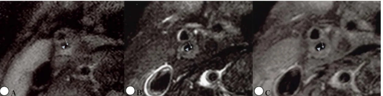
8)은 고해상도 자기공명영상을 이용하여 섬유성 cap의 모양을 관찰하였다. 경화반의 cap이 두껍고 손상없는 경우에는, 저신호강도의 선(band) 이 동맥내경 주위에 일정하고 연속적으로 보이며 T1, T2, 양자밀도(proton density, PD) 강조영상에서 고신호강도의 선이 평편하게 나타나며, cap의 손상이 없이 얇은 경우에는 유체속도강조(time of flight: TOF)에서는 동맥내경 주위에 저신 호강도의 선이 없으며 T1, T2, 양자밀도 강조 영상에서 평편하게 나타났으며, 파열된 경화반 (ruptured plaque)은 TOF 영상에서는 내경 주 위에 저신호강도가 없으며 T1, T2, 양자밀도 강조영상에서 뷸규칙한 동맥 내경가장자리 (irregular lumen boundary)를 보인다(Fig. 2).
고해상도 자기공명영상에서 파열된 섬유성 cap 소견을 보인 경우에는 최근의 일과성 허혈성 뇌졸중이나 뇌졸중과 매우 높은 연관성이 있다 고 판단하였다.
8)지방이 풍부한 괴사성핵(lipid rich necrotic core) 은 T1-강조영상에서 고신 Table 1. MR signal intensity of main components of atherosclerotic plaque
Plaque components T2WI T1WI Intermediate weighted TOF Recent hemorrhage
Lipid-rich necrotic core
Fibrous tissue Calcification
Variable
Variable
Variable Low
High to moderate
High
Moderate Low
Variable
High
High Low
High
Moderate
Moderate to low Low
T2WI, T1-weighted image; T1WI, T1-weighted image; TOF: time of flight
호강도, TOF에서는 중증도의 신호강도, PD에 서는 고신호강도, T2강조영상에서는 다양한 신 호강도를 보인다.
9)3) 경화반내 출혈(intraplaque hemorrhage) 경화반내 출혈은 진행된 경동맥 경화반에서 드물지 않게 나타나고 경화반의 진행에 주요한 원인이 된다. Chu 등
10)은 출혈의 시기에 따라 서 3가지 시기로 분류하였다. 초기 아급성(1주 내), 후기 아급성(1∼6주), 만성(6주 이상) 경화 반 출혈로 분류하였고 초기 아급성 경화반 혈 종은 T1-강조영상에서 고신호, T2-강조영상에 서 저신호 혹은 등신호강도를 보이며 후기 아 급성출혈은 T1-강조영상에서 고신호강도, T2-
강조영상에서도 고신호강도를 보이며, 만성 경 화반내 혈종은 T1-과 T2-강조영상에서 모두 저신호강도를 보인다(Fig. 3). TOF 원천영 상(source image)에서 초기 및 후기 아급성 혈종은 고 신호강도, 만성 경화반내 출혈(old intraplaque hemorrhage)은 저신호강도를 보인다.
10)경화반내 신생혈관증식(plaque neovasculari- zation)
경화반내 신생혈관증식은 경화반의 형성과 진행과정에서 발생이 되는데 파열된 경화반이 나 경화반내 출혈이 있는 경우에 현저하게 증 가되는 것을 볼 수가 있다. 또한 섬유성 cap에 심한 대식세포의 침윤이 있는 경우나 경화반의 Fig. 2. Ruptured fibrous cap of vulnerable carotid plaque. Discontinuous fibrous cap (arrow) surrounding the lumen is noted on T1-(A), T2-(B), and Proton density (C) images. Note thin hyperintense fibrous cap(arrowhead) overlying lipid core.
Fig. 3. Intraplaque hemorrhage of left proximal internal carotid artery. Intraplaque recent hemorrhage (blanked arrow) indentified by hyperintensity in T1-(A) and proton density (B) is noted. T2 weighted image (C) reveals iso-signal intensity (arrowhead) of plaque hemorrhage.
A B C
A B C
어께 부위에 신생혈관의 증식이 많다. 반면에 섬유 및 석회화가 많은 경화반은 상대적으로 신생혈관의 증식이 현저하게 적다. 경화반내 신생혈관증식은 고위험도 경화반과 연관이 있 다고 판단이 된다.
11)자기공명영상에서 조영제 를 주입후 조영증강이 되는 경화반의 섬유세포 성 부위는 미세혈관의 증식과 염증이 있다는 것을 시사할 수 있다(Fig. 4).
궤양성 경화반(ulceration of plaque)
Eliasziw 등
12)에 따르면 궤양성 경화반을 가 진 경동맥협착 환자에서 2년간 약물치료만을 시행했을 시 경동맥협착이 75%인 경우에서 동 측의 뇌졸중 발생율은 26.3% 이나 경동맥협착 이 95%로 증가 시에 동측의 뇌졸중 발생은 73.2
%로 현격하게 증가되었다고 한다. 그러나 궤 양이 없는 환자에서는 협착의 정도와 관계없이 뇌졸중이 21.3%에 불과하였다. 결론적으로 경 동맥 협착이 심한 궤양성 경화반을 가진 환자 에서 뇌졸중의 가능성은 매우 높아진다고 한다.
혈관조영술에서 쉽게 진단이 가능한 경화반 의 궤양은 경동맥의 협착 부위중 근위부에 가 장 흔하게 나타나고 이곳은 혈역학적 힘에 의 해 shear stress가 가장 높은 부위에 해당된다.
또한 경화반의 파열도 동맥의 협착 부위중 근 위부의 어께부위에 가장 흔하게 나타나기 때문 에 shear stress가 경화반의 안정성과 연관이 있는 걸로 알려져 있다.
13)궤양성 경화반은 자 Fig. 4. Contrast enhanced carotid plaque in left
internal carotid artery. T1-weighted image (A) shows iso-signal intensity of carotid plaque (blanked arrow). Strong enhancement of carotid plaque (blanked arrow) is seen on postcontrast T1-weighted image (B). Note focal ruptured fibrous cap (arrowhead)
Fig. 5. Ulceration of carotid plaque in left internal carotid artery. T1-weighted image reveals large ulceration with ruptured fibrous cap (arrow).
Arrowhead is left external carotid artery.
A
B
기공명영상에서 경화반내의 무신호강도(signal void)로 보여 비교적 쉽게 관찰된다(Fig. 5).
경화반내 골형성 혹은 이형성 석회화(dystrophic calcification)
Hunt 등
14)에 따르면 석회화를 동반한 경화 반은 석회화를 동반하지 않은 경동맥 경화반보 다 허혈성뇌졸증 혹은 일과성 허혈성뇌졸증의 가능성이 적다고 보고하였다. 또한 경화반내 골형성은 저 위험도 경화반에서 더욱 흔히 관 찰된다. 석회화 혹은 골형성을 가진 경화반은 초음파에서 고에코로 관찰되나 저에코 경화반 은 지방성분이나 출혈성분에 의해 보인다. 골 형성 혹은 석회화 경화반은 모든 자기공명 영 상에서 저신호강도를 보인다.
지질강하제 치료(lipid-lowering therapy)후 경 화반의 변화
지질강화제 사용 후의 변화를 평가하는 방 법으로 자기공명영상이 이용되고 있다. Corti 등
15)은 simvastatin을 1년간 투여 후 경동맥의 벽두께가 약 8% 감소된 것을 관찰하였으며, 18개월에서 24개월 후 12%의 두께감소가 있었 다고 보고 하였다. 지질강하제 사용 후 고해상 도 자기공명영상을 이용하여 경화반의 두께, 용적, 그리고 성분 변화를 관찰할 수 있다.
결 론
경동맥 죽상경화증의 치료 및 예후의 판정 에 있어서 단순히 동맥 내경의 협착만으로 결 정하기 보다는 최근에 다양한 고해상도 자기공 명영상기법을 이용함으로 인하여 보다 정확하
게 치료방침을 결정할 수가 있게 되었다. 고해 상도 경동맥 자기공명영상을 통하여 경화반의 형태, 구성성분 등을 분석함으로써 초음파나 CT에서는 파악할 수 없는 경화반내 출혈, 섬 유성 cap의 두께를 보다 정확하게 판정하여 경동맥 협착환자의 정확한 치료 방침을 결정할 수가 있다. 또한 지질강하제를 이용한 치료 후 자기공명영상으로 경동맥경화반의 변화를 분석 함으로써 그 치료 효과나 다른 변화 등을 알 수가 있다.
참 고 문 헌
1. Warlow C, Dennis MS, Gijn JV, Hankey GJ, Sandercock PAG, Bamford JM, et al. Stroke:
a practical guide to managemen. 2nd ed.
Oxford: Blackwell Publishing, 2001.
2. Ambrose JA, Tannenbaum MA, Alexopoulos D, Hjemdabl Monsen CE, Leavy J, Weiss M.
Angiographic progression of coronary artery disease and the development of the myocardial infarction. J Am Coll Cardiol 1988 Jul;12(1):
56-62.
3. Glagov S, Weisenberg E, Zarins CK, Stanku- navicius R, Kolettis GJ. Compensatory enlarge- ment of humen artherosclerotic coronary arteries. N Engl J Med 1987 May 28;316(22):
1371-5
4. Nordestgaard BG, Gronholdt ML, Sillesen H.
Echolucent rupture-prone plaques. Curr Opin lipidol 2003 Oct; 14(15):505-12
5. Naghavi M, Libby P, Falk E, Casscells SW, Litovsky S, Rumberger J, et al. From vulnerable palque to vulnerable patient: a call for new definitions and risk assessment strategies:
Part I. Circulation 2003 Oct 7;108(14):1644-72.
6. Ruehm SG, Corot C, Vogt P, Kolb S, Debatin
JF. Magnetic resonance imaging of atherosclerotic plaque with ultrasmall superparamagnetic particles of iron oxide in hyperlipidemic rabbits. Circulation 2001 Jun 23;103(3):415-22.
7. Trivedi RA, U-King-Im JM, Graves MJ, Gross JJ, Horslcy J, Goddard MJ, et al. In vivo detection of macrophages in human carotid atheroma: temporal dependence of ultrasmall superparamagnetic particles of iron oxide- enhanced MRI. Stroke 2004 Jun;35(7):1631-5.
8. Yuan C, Zhang SX, Polissar NL, Echelard D, Ortiz G, Davis JW, et al. Identification of fibrous cap rupture with magnetic resonance imaging is highly associated with recent trasient ischemic attack or stroke. Circulation 2002 Jun 15;105(2):181-5.
9. Yuan C, Mitsumori LM, Ferguson MS, Polissar NL, Echelard D, Ortiz G, et al. In vivo accuracy of multispectral magnetic resonance imaging for identifying lipid-rich necrotic cores and intraplaque hemorrhage in advanced human carotid plaques. Circulation 2001 Oct 23;104 (17):2051-6.
10. Chu B, Kampschulte A, Ferguson MS, Kerwin WS, Yarnykh VL O'Barien KD, et al.
Hemorrhage in the athersclerotic carotid plaque: a high-resolusion MRI study. Stroke 2004 May;35(5):1079-84.
11. Moreno PR, Purushothaman KR, Fuster V, Echeverri D, Truszczynska H, Sharma SK, et al. Plaque neovascularization Is increased in ruptured atherosclerotic lesions of human aorta: implications for plaque vulnerability.
Circulation 2004 Oct 5;110(14):2032-8.
12. Eliasziw M, Streifler JY, Fox AJ, Hachiski VC, Ferguson GG, Barnett HJ, Significance of plaque ulceration in symptomatic patients with high-grade carotid stenosis. North American Symptomatic Carotid Endarterectomy Trial.
Stroke 1994 Feb;25(2):304-8.
13. Lovett JK, Rothwell PM. Site of carotid plaque ulceration in relation to direction of blood flow: an angiographic and pathologic study.
Cerebrovasc Dis 2003;16(4):369-75.
14. Hunt JL, Fairman R, Mitchell ME, Carpenter JP, Golden M Khadapyan T, et al. Bone formation in carotid plaques: a clinicopathological study. Stroke 2002 May 33(5):1214-9.
15. Corti R, Fuster V, Fayad ZA, Worthley SG, Helft G, Smith D et al. Lipid lowering by simvastatin induces regression of human atherosclerotic lesions: two years’ follow -up by high-resolution noninvasive magnetic resonance imaging. Circulation 2002 Dec 3;
106(23):2884-7.