*Corresponding author: Min Kyoung Paik Phone: +82-63-238-3253; Fax: +82-63-238-3837;
E-mail: [email protected]
Korean J Environ Agric. 2015;34(4):350-354. English Online ISSN: 2233-4173
Published online 2015 November 13. http://dx.doi.org/10.5338/KJEA.2015.34.4.40 Print ISSN: 1225-3537
DNA Damage Effect of Botanical Insecticides Using Chinese Hamster Lung Cells
Areumnuri Kim
1, Mihye Jeong
2, Kyung-Hun Park
1, Kyongmi Chon
1, Namjun Cho
1and Min Kyoung Paik
1*1
Chemical Safety Division, National Institute of Agricultural Sciences, Wanju 55365, Korea
2
Research Policy Bureau, Rural Development Administration, Jeonju 54875, Korea
Received: 10 September 2015 / Revised: 26 October 2015 / Accepted: 29 October 2015 Copyright ⓒ 2015 The Korean Society of Environmental Agriculture
This is an Open-Access article distributed under the terms of the Creative Commons Attribution Non-Commercial License (http://creativecommons.org/licenses/by-nc/3.0) which permits unrestricted non-commercial use, distribution, and reproduction in any medium, provided the original work is properly cited.
350
Abstract
BACKGROUND: Botanical insecticides, especially Azadirachta Indica extract (AIE) and Sophorae radix extract (SRE) are widely used in Agriculture field. In our previous studies on genotoxicity test of AIE and SRE samples, a suspicious clastogenic properties was shown.
Herein, we investigated the DNA damage effect of these botanical insecticide samples through the in vitro comet assay.
METHODS AND RESULTS: Chinese hamster lung (CHL) fibroblast cell line was used, and methyl methanesulphonate was as positive control. Respective two samples of AIE and SRE were evaluated using Single Cell Gel Electrophoresis (Comet) assay and measured as the Olive tail moment (OTM). Results from this study indicated that all tested AIE and SRE samples did not show DNA damage in comet assay using CHL cells, compared with control.
CONCLUSION: AIE and SRE samples used in this study were not cause genetic toxicity and are suitable for use as organic materials.
Key words: Botanical insecticide, Comet assay, DNA damage
Introduction
Unlike synthetic chemical pesticides, which leave harmful residues in the aquatic environment, botanical insecticides are believed to be more environmentally friendlier because they are easily biodegraded and leave no residues in the environment and their use is growing in agriculture field (Bhat et al ., 2012). As of August 2013, there are approximately 1,140 environmental-friendly organic materials that has been registered in South Korea.
Especially, botanical insecticides such as Azadirachta Indica extract (AIE) and Sophorae Radix extract (SRE) account for 20% of the registered environmental- friendly organic materials.
Azadirachtin (Aza), as active ingredient of AIE, belongs to the organic compounds group known as tetranortriterpenoid and has known to act as an ecdysone blocker and an insect anti-feedant (Muangphra and Gooneratne, 2011). Matrine, one of the major active ingredient extracted from the traditional medicinal herb Sophora flavescens, has been known to be a very effective for botanical insecticide.
Despite their frequent use, studies on their
Short Communication Open Access
DNA Damage Effect of Botanical Insecticides 351
toxicities and side effects are still sparse. Aza and matrine have generally been regarded as a relatively nontoxic substance for agricultural use and, for instance, there are only few reports on its genotoxic potential in the literature (Vinod et al ., 2011; Cho et al. , 2013; Yoon et al. , 2014).
We previously investigated the genotoxic effects of AIE and SRE through chromosomal aberration (CA) (Yoon et al ., 2014) and in vitro micronucleus (MN) assay (Cho et al ., 2013) using Chinese hamster lung (CHL) cells. Base on those results, all SRE samples had no genotoxic effect in both test, but in the chromosomal aberration test, one of SRE samples had potential clastogen properties, showing a suspicious positive result at 250 ug/ml in the presence of S-9 mix. However, genotoxicity test that is designed to detect changes that occur in a particular index is not possible to detect any genetic toxicity.
Among many genotoxicity tests, comet assay, or single cell gel electrophoresis assay (SCGE) has been reported to evaluate DNA damage in single cells under alkaline conditions (Singh et al ., 1988).
Recently, the popularity of the comet assay has increased because of its relatively simple and rapid procedures and high sensitivity (ability to detect carcinogens as positive). The reason why we focused on the comet assay in this study is that the comet assay was reported to be equally effective at detecting carcinogens that are gene mutagens or clastogens, and only declines slightly in sensitivity with compounds positive for MN in vitro . For instance, some compounds positive for MN in vitro that were negative in the comet assay (Kirkland and Speit, 2008)
In views of the above, we investigated the DNA damage effect of botanical insecticides having a suspicious clastogen property in our previous CA test through the comet assay in order to confirm the genotoxic evaluation of those samples using CHL cells. Respective two samples of SRE and ARE were tested for their possible genotoxic potential according to the alternative in vitro comet assay recently suggested internationally.
Materials and Methods
Cell culture and Materials
Cell culture. Chinese hamster lung (CHL) fibroblast cell line was obtained from the American Type Culture Collection (ATCC, Manassas, USA). Cells were
maintained in Eagle’s minimum essential medium (EMEM, Glbco, Carlsbad, CA) supplemented with 1%
penicillin-streptomycin and 10% heat-inactivated fetal bovine serum at 37℃ in a 5% CO
2atmosphere. The doubling time was about 13 h and cells were subcultured every 2-3 days.
Materials. Respective two samples of AIE and SRE were purchased from commercial products that are circulating in Korea. All AIE samples were from India, and their active ingredient azadirachtin were respectively 0.03% (AIE sample A) and 0.35% (AIE sample B). All SRE samples were form China, and their active ingredient matrine were respectively 0.3%
(SRE sample A) and 0.26% (SRE samples B).
Reagents. Methyl methanesulphonate (MMS, CAS 66-27-3), sodium hydroxide (NaOH, CAS 1310-73-2), sodium chloride (NaCl, CAS 7647-14-5), ethylenediaminetetraacetic acid disodium salt dehydrate (EDTA-Na
2, CAS 6381-92-6), triton X-100 for molecular biology (CAS 9002-93-1), trizma base (CAS 77-86-1), ethidium bromide (EtBr, CAS 1239- 45-8) and dimethyl sulfoxide (DMSO, CAS 67-68-5), normal melting agarose (NMA, CAS 9012-36-6) and low melting agarose (LMA, CAS 39346-81-1) were purchased from Sigma Aldrich (St Louis, MO, USA).
NMA and LMA were diluted to 1% and 0.5% in Phosphate buffer saline (PBS, Glbco, Carlsbad, CA).
MMS and test substances were dissolved in DMSO.
Experimental methods
Treatment. Prior to treatment, cells seeding in 6 well plates at 1×10
5cell/mL and incubated for 24 h at 37℃. Three hours after the treatment of SRE, cells were collected and then gently resuspended with PBS.
The cells were treated with 5 μM MMS, as a positive control.
Slide preparation. First layer on each clean slide was precoated with 1% NMA (200 μL). Second layer containing mixture of cell suspension (500 μL) and 1% LMA (500 μL) were spread onto each first layer and covered using coverslip. Slides were dried at 4℃
for 10 min. Third layer on slide was coated with 0.5%
LMA (200 μL). After hardening third layer, coverslips was removed and then slides were dried 4℃ for 10 min.
Lysis, unwinding and electrophoresis. All steps
were carried out in dark room. Slides were put in a
stain jar that contained a lysis solution (pH 10.0) for 1
h at 4℃. The lysis solution consisted of 2.5 M NaCl,
Fig. 1. Representative images of DNA damage of negative control, positive control, and the tested samples from comet assay.
NC; negative control(DMSO), PC; positive control(5 μM MMS), AIE;
Azadirachta Indica
extract, SRE;Sophorae radix
extract.500 mM Na
2EDTA, 1 M Trizma base% and 10%
DMSO. Prior to unwinding of DNA, the slides were washed three times with distilled water and kept in stain jar with unwinding buffer (pH>13) that consists of 10 N NaOH and 200 mM Na
2EDTA for 30 min at 4℃. Electrophoresis was carried out at 25 V and 300 mA for 20 min, using the alkaline buffer solution used for unwinding of DNA. After electrophoresis, slides were washed three times for 5 min with 0.4 M Tris buffer (pH 7.5), soaked in 70% ethanol and 100%
ethanol for each 5 min, and then dried at room temperature for overnight.
Image analysis. EtBr was used for staining. The cell images were examined at 200 X magnification using a fluorescence microscope (Nikon TE2000-U, Japan). Captured images of 50 cell per slide were analyzed using image analysis software (Andor Komet 7.0, UK) to obtain olive tail moment. Olive tail moment, the parameter of DNA damage, was referred as distance between the center position of the head and the center of % DNA in tail.
Statistical analysis
The data was tested by one way analysis of variance (ANOVA) followed by Duncan’s test using SPSS (Statistical Package for the Social Sciences, version 18.0). A probability of less than 0.05 was considered as statistically significant.
Results and Discussion
Azadirachta Indica extract (AIE)
Comet assay has been known as a simple method for measuring DNA damage in eukaryotic cells. The principle of the assay is based upon the ability of denatured, cleaved DNA fragments to migrate out of the nucleoid under the influence of an electric field, whereas undamaged DNA migrates slower and remains within the confines of the nucleoid when a current is applied (OECD, 2014).
Singh et al. (1988) developed the alkaline version of the comet Assay in which they used the length of DNA migration (tail length) to quantify the extent of damage. However, with time, most of them were not of frequent or wide use. After then, Olive et al. (1990) reported the concept of the tail moment to describe DNA migration. The tail moment came to be known as the Olive tail moment (OTM). This parameter is considered to be particularly useful in describing heterogeneity within a cell population, as OTM can pick up variations in DNA distribution within the tail. Therefore, we measured the OTM to describe DNA damage of CHL cells after treatment of AIE and SRE samples (Fig. 1.).
Table 1 shows the effects of the AIE sample A and
B on the DNA damage of CHL cells. OTM value of
DMSO-treated negative control was 11.29±1.96, and
DNA Damage Effect of Botanical Insecticides 353
Table 1. Effects of the
Azadirachta Indica
extract sample A and B on the DNA damage in Chinese hamster lung cellsTreatment Conc. (μg/ml) OTMa) Negative control
(3% DMSO)
1.29 ± 1.96
Positive control (5μ M MMS)
31.23 ± 15.66*
AIEb) sample A 0.125 2.24 ± 1.90 0.25 1.83 ± 1.83 0.5 2.52 ± 2.52
AIE sample B 0.125 2.56 ± 2.19
0.25 2.83 ± 2.54 0.5 4.00 ± 3.60 DNA damage was measured as the OTM (olive tail moment); tail lengthⅹ% DNA in the tail. a) Value of olive tail moment ± SD of 50 cells per slide of three experiments. *indicates significant at p<0.05. b) AIE;
Azadirachta Indica
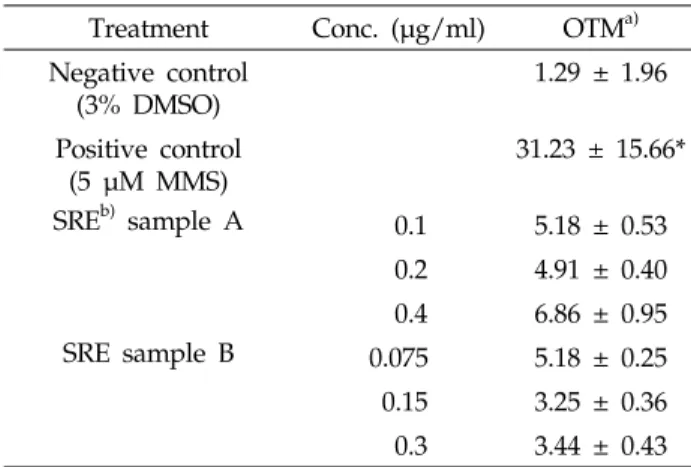
extractTable 2. Effects of the
Sophorae radix
extract sample A and B on the DNA damage in Chinese hamster lung cellsTreatment Conc. (μg/ml) OTMa) Negative control
(3% DMSO)
1.29 ± 1.96
Positive control (5 μM MMS)
31.23 ± 15.66*
SREb) sample A 0.1 5.18 ± 0.53 0.2 4.91 ± 0.40 0.4 6.86 ± 0.95
SRE sample B 0.075 5.18 ± 0.25
0.15 3.25 ± 0.36 0.3 3.44 ± 0.43 DNA damage was measured as the OTM(olive tail moment); tail lengthⅹ% DNA in the tail. a) Value of olive tail moment ± SD of 50 cells per slide of three experiments. *indicates significant at p<0.05. b) SRE;