I. 서론
하나의 세포에서 multicellular organism으로 의 발생(development)과정은 진화의 경이로운 산 물이다. 따라서 인간의 발생과정도 초기에 단순한 세포에서 organized structures로 변화되는 과정 을 필연적으로 가지고 있다. 이러한 발생과정은
크게 5개의 과정으로 설명할 수 있는데 이는 세 포의 분열(cell division), pattern formation, morphogenesis, cell differenciation, 마지막으로 cell growth 과정이다. 이 중에서도 morphogenesis 시기에는 광범위한 세포의 이동이 나타나고 특히 두개안면부의 발생은 좀 더 독특한 과정 을 겪게 된다. 즉 뇌와 척추를 형성하는 neurulation 과정
상순의 발생
고승오
1,2
, 임양희1
, 김기병1
, 신효근1,2
전북대학교 치의학전문대학원 구강악안면외과학교실1, 전북대학교 구강생체과학연구소2
ABSTRACT
Development of the Upper Lip
Seung-O Ko1,2, Yang-Hee Im1, Ki-Byeung Kim1, Hyo-Keun Shin1,2
Department of Oral & Maxillofacial Surgery, School of Dentistry, Chonbuk National University1, Institute of Oral Bioscience, School of Dentistry, Chonbuk National University2
The vertebrate upper lip forms from initially freely projecting maxillary, medial nasal, and lateral nasal prominences at the rostral and lateral boundaries of the primitive oral cavity. These facial prominences arise during early embryogenesis from ventrally migrating neural crest cells in combination with the head ectoderm and mesoderm and undergo directed growth and expansion around the nasal pits to actively fuse with each other.
Initial fusion is between lateral and medial nasal processes and is followed by fusion between maxillary and medial nasal processes. Fusion between these prominences involves active epithelial filopodial and adhering interactions as well as programmed cell death.
Slight defects in growth and patterning of the facial mesenchyme or epithelial fusion result in cleft lip with or without cleft palate, the most common and disfiguring craniofacial birth defect. This review will summarize the current understanding of the basic morphogenetic processes and molecular mechanisms underlying upper lip development.
Key Word : cleft lip, craniofacial development, apoptosis, EMT
중에 neural plate와 ectoderm 사이의 neural plate border에서 유래된 신경능선세포(neural crest cells)가 ventral 쪽으로 이동하여 골조직을 포함한 안면부의 대부분을 형성하고 있다. 척추동 물의 윗입술 역시 신경능선세포에서 유래된 제1인 두궁(1st branchial arch)의 상악돌기(maxillary process) 및 전두비돌기(frontonasal process)에 의해서 형성되고 있다.
본 review에서는 상순의 형태학적인 발생과정과 상순의 발생에 관여하고 있는 molecular mechanism 의 최신 경향에 대하여 간단히 서술하고자 한다.
1. Morphogenesis of the upper lip
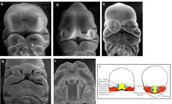
인간의 임신 24일경(쥐의 임신 9일경, 이하 괄 호는 쥐의 embryonic day)이 되면 신경능선세포의 이동이 거의 마무리가 되고 원시구강(stomodeum) 이 상방에서는 발생중인 전뇌(forebrain)와 하방에 서는 제1인두궁의 하악돌기를 경계로 형성된다
1). 임신 26일경(E9.5)에 원시안면(facial primordia) 은 5개의 돌기로 성장하는데 원시구강의 상방에 는 1개의 전두비돌기(frontonasal process)와 1쌍 의 제1인두궁 상악돌기, 원시구강 하방에는 1쌍의 제1인두궁 하악돌기(mandibular process)이다(그 림 1A)
2).
임신 32일경에는(E10.0) 표면외배엽(surface ectoderm)이 전두비돌기의 ventrolateral 방향으 로 성장하여 코의 기원판(nasal placode)을 형성 하고 전두비돌기는 코의 기원판을 중심으로 말발 굽모양으로 성장하여 nasal pits(코오목)을 형성 하면서 내측비돌기와 외측비돌기를 형성한다(그림 1B)
2,3). 임신 35일경(E10.5)이 되면 제1인두궁 상 악돌기의 외배엽성중배엽의 성장과 내측비돌기의 ventrolateral 성장으로 nasal pits을 둥근 형태 에서 얇고 가는 형태로 바꾼다. 그리고 이시기에
외측비돌기는 상악돌기와 내측비돌기사이에 끼어 올라가면서 상순의 경계가 외측은 상악돌기, 내측 은 내측비돌기로 이루어진다(그림 1B). 이때부터 상악돌기는 외측비돌기 하방에 위치하게 되고 내 측비돌기와 외측비돌기의 융합(fusion)이 시작된 다. 임신 38일경(E11.0)에는 제1인두궁의 상악돌 기와 내측비돌기가 계속 성장하면서 외측비돌기 를 더욱 상방으로 밀고 상악돌기와 내측비돌기가 직접 접촉하게 된다. 이때 외측비돌기와 내측비돌 기, 그리고 상악돌기와 내측비돌기의 경계부위가 활발한 융합과정을 거쳐서 입술모양을 갖추게 된 다(그림 1C, 그림 2)
4).
일단 상순의 morphogenesis가 완성이 되면 상 순은 내측비돌기와 상악돌기로부터 유래된 조직 으로 구성되고 비익(alae)은 외측비돌기에 의해서 만들어진다. 그리고 외측비돌기가 최종적인 상순 을 형성하는데 기여하지는 않지만 nostril을 포함 하는 구순열은 내측비돌기가 상악돌기와의 융합 실패 뿐만 아니라 외측비돌기와의 융합실패가 동 반되어야 나타난다고 알려져 있다.
상순의 발생 도중에는 이미 언급한 바와 같이 상악 돌기나 내, 외측비돌기의 활발한 상피융합(epithelial fusion)이 이루어지고 있지만 좌․ 우측 내측비돌 기의 중앙접합부(median groove)는 fusion에 의 한 것이 아닌 것으로 보고되고 있다
2,4,5,6,7,9). 즉 임신 7주경(E11.5-12.0)까지 융합이 일어나는 중 에도 상악돌기는 계속 성장하여 nasal pits와 내 측비돌기를 내전방으로 밀어내면서 상순 정중앙 의 median groove는 점차 얇아져서 최종적으로 는 상순의 정중부를 형성하게 된다
2). 결론적으로 상순의 정중부의 발생은 좌우측 내측비돌기와 상 악돌기의 성장에 의하여 이루어진다(그림 1D).
임신 48일경(E12.5)에, 내측비돌기와 상악돌기
의 융합에 의해 상순의 발생이 완성되면서 상순
중앙부(philtrum)를 형성하는 내측비돌기의 distal
part로부터 intermaxillary segment가 형성되어 oral cavity로 성장하면서 구개의 전방부(․ʻʻprimary palateʼʼ)를 만들게 된다(그림 1D,E)
2,3,10,11,12). 이 전방구개(anterior palate 또는 primary palate) 가 나중에 상악돌기로부터 유래되는 secondary palate와 융합하게 된다. 이차구개의 융합과 일차 구개와 이차구개의 융합은 상순의 발생시 일어나 는 상악돌기와 비돌기의 융합보다 늦게 일어나게 되고 따라서 상순의 융합실패는 종종 구개융합에 영향을 미치어 실제 임상에서 구순열과 구개열이 동반되는 경우를 흔히 볼 수 있다.
정상적으로 상순이 융합(상악돌기와 내측비돌 기, 외측비돌기의 접합부)되는 시기가 되면 현저 한 세포들의 변환(transformation)이 일어난다.
Sun 등에 의하면 닭의 태아에서는 이들 돌기를
덮고 있는 태아표피(periderm)에서 apoptosis가 일어나 떨어져 나간다고 보고하였고
13)Hinrichsen 은 인체의 태아의 SEM 분석에 의하면 다수의 rounded cells가 돌기들의 접합부에 관찰되고 이 들은 융합과정에서 죽은 세포들이 표면으로 밀려 나온 것으로 추측하였다(Fig.3B,C)
2).
또 다른 연구에서는 안면돌기가 서로 가까워 지면서 상피의 filopodia(cytolasmic projections) 가 융합부위에만 고도로 관찰되어 안면돌기들 사 이에서 연결되어 가교를 형성하고 있다는 보고를
하였다
2,4,6,7,9,14). 이들 filopodia가 세포들 사이에
통과하여 anchor역할을 하고 larger cellular extensions 와 adhering junctions들이 침착되어 reinforced 된다는 것이다
7,13).
그림 1. Morphogenesis of the upper lip of mouse. A:E9.5-10. B:E10-10.5, C:E11, D:E12 .5, E:E12-13.5. F:Contribution of each of the prominences to the human face. fnp, frontonasal prominence; man, mandibular process(prominence) of 1st branchial arch; max, maxillary process of 1st branchial arch; mnp, medial nasal process; lnp, lateral nasal process; pp, primary palate.
Lat. nasal prominence Med. nasal prominence Maxillary prominence Mandibular prominence
Eye Nasolacrimal
groove Philtrum
F
그림 2. SEM views of 32~35days cynomolgus monkey(Macaca fascicularis) embryo showing major epithelial bridging.
A: The medial nasal prominence is bridging to the lateral and maxillary nasal prominences. B: A close-up of the epithelial bridging (arrow), which is a result of dynamic fusion of the major prominences.
그림 3. Morphogenesis of the human upper lip. A:
SEM facial view of 4weeks human embryonic head.
B: SEM micrograph of the right nasal pit of a late 5weeks human embryo. C: Enlarged detail of the lower nasal pit shown in B. The boundary between the maxillary and lateral nasal processes is clearly marked by the rounded cells at the surface.
Rounded cells also appear at the contact site between the medial and lateral nasal processes.
그림 4. Apoptosis plays an important role in breakdown of the epithelial seam during lip fusion. A: Frontal section of an embryonic day (E) 11.0 mouse embryo through the telencephalon and the fusing medial and lateral nasal processes. Red signal marks specific anti-active Caspase-3 antibody staining. B: High-magnification view of the fusing epithelial seam between the medial and lateral nasal processes shown in A.
Many of the fusing epithelial cells express active Caspase-3, while very few nasal
mesenchyme cells and epithelial cells in other regions express active Caspase-3,
indicating specific programmed cell death of the fusing epithelial cells. lnp, lateral
nasal process; mnp, medial nasal process.
2. Molecular mechanism of the lip fusion- Programmed cell death, EMT or Both?
상순의 발생시 일어나는 상악돌기와 내측비돌 기, 내측비돌기와 외측비돌기 접합부의 융합 (fusion)의 molecular mechanism은 지금까지 수 많은 연구가 된 이차구개의 MEE(medial edge epithelial) cell의 융합기전과는 달리 그다지 많 은 연구가 보고되지 않고 있는 실정이다. 다만 지 금까지 입술의 융합과 이차구개의 융합기전은 비 슷하다고 알려져 있다
9,13,14).
상순의 융합시에는 돌기들 사이의 접합부에 nasal fin이라고 불리우는 intervening epithelial seam이 형성되고 이들이 점차적으로 파괴되면서 mesenchyme으로 replace 된다고 알려져 있다
5,11,13,14)
. 이들은 TEM을 이용하거나 apoptotic
cells를 관찰할 수 있는 TUNEL assay 등을 통하 여 epithelial seam cells의 운명을 관찰하였는데 Gaare and Langman은 용합부의 상피층에서 다 량의 degenerating cells가 관찰되었고 이들을 ʻʻcell-death zonesʼʼ라고 생각하였다. 그러나 degenerating cells 외에 대부분의 인접상피세포 들은 건강하였고 이들 세포들은 간엽으로 변환되 기 보다는 인접한 상피 linings로 섞였을 것이라 고 추측하였다
14). Sun 등은 유사한 실험결과로 상순 융합에서 epithelial seam cells는 간엽으로 변환(EMT) 된다고 결론지은 바 있다
13).
지금까지 상순이나 이차구개의 융합기전에 관 한 연구는 대부분이 in vitro 상에서의 연구결과 에 의한 것으로 보인다. 그러나 최근에 Cre/loxP transgenic mice를 이용한 in vivo 연구가 이루 어지고 있고 Vaziri Sani 등은 이차구개의 Cre/loxP system을 이용하여 연구한 결과 이차 구개의 융합시에 조기 apoptosis marker인 Caspase-3 가 MEE cells 에 활성화된다고 보고 하였다
15). 또한 상순의 융합에서도 비슷한 실험결
과가 보고되었는데 Jiang 등은 이차구개 융합과 유사하게 입술의 융합에서도 programmed cell death가 중요한 역할을 한다고 주장한 바 있다 (그림 4)
16).
아직도 입술이나 이차구개의 융합기전은 apoptosis에 의한 것인지 EMT에 의한 것인지 또는 둘 다인지 아니면 인접상피세포로의 incorporation 에 의한 것인지 등 여전히 그 molecular mechanism이 명확하게 밝혀지지 않았고 현재까 지도 논란의 대상이 되고 있어서 앞으로도 더 많 은 연구가 필요할 것으로 생각된다.
참고문헌