WWW.KJOG.ORG 817
SCHWANNOMA OF THE CLITORIS: A CASE REPORT
Yu-Suk Yu, MD, Do-Young Kwon, MD, Ji-Min Park, MD, Seok-Cheol Choi, MD, Sang-Young Ryu, MD
Department of Obstetrics and Gynecology, Korea Cancer Center Hospital, Korea Institute of Radiological and Medical Sciences, Seoul, Korea
Schwannomas arise from schwann cells in the peripheral nerve sheath, without involving the nerve fibers. Schwannomas are benign, slow-growing tumors that infrequently recur and rarely undergo malignant change. Also schwannomas of the female genitalia are considered extremely rare. We report a case of schwannoma of the clitoris. A 76-year-old woman without neurofi bromatosis presented with a 2.0 × 1.7 cm, painless mass of the clitoris. Simple surgical excision of the mass was undertaken, and fi nal pathology revealed schwannoma of the clitoris. No evidence of local recurrence was noted after 1 year of follow-up.
Keywords:
Schwannoma; Clitoris
CASE REPORT
Received: 2011. 6.10. Revised: 2011. 9.23. Accepted: 2011.10.14.
Corresponding author: Seok-Cheol Choi, MD
Department of Obstetrics and Gynecology, Korea Cancer Center Hospital, Korea Institute of Radiological and Medical Sciences, 75 Nowon-gil, Nowon-gu, Seoul 139-706, Korea
Tel: +82-2-970-2157 Fax: +82-2-970-2157 E-mail: [email protected]
Th is is an Open Access article distributed under the terms of the Creative Commons Attribution Non-Commercial License (http://creativecommons.org/licenses/
by-nc/3.0/) which permits unrestricted non-commercial use, distribution, and reproduction in any medium, provided the original work is properly cited.
Copyright © 2011. Korean Society of Obstetrics and Gynecology Korean J Obstet Gynecol 2011;54(12):817-819
http://dx.doi.org/10.5468/KJOG.2011.54.12.817 pISSN 2233-5188 · eISSN 2233-5196
Schwannoma, neurinoma or neurilemmoma arise from schwann cells in the peripheral nerve sheath, without involving the nerve fi bers.
Its most frequent sites are the extremities, posterior mediastinum, retroperitoneum, head and neck. On the other hand, schwannoma is a benign tumor which rarely affects the female genitalia [1,2].
Especially, despite the rich nerve supply of the clitoris, schwanno- mas of the clitoris are extremely rare [3-5]. We report a case of schwannoma of the clitoris.
Case Report
A 76-year-old postmenopausal Korean woman, who had four chil- dren, was referred to our hospital in 9 February 2010 for evalua- tion of vaginal spotting.
Physical examination revealed a 2.0 ×1.7 cm sized, movable, non- tender mass in the clitoris. But, she had not noticed the clitoral mass before; it was not painful.
Except for this lesion, her external genitalia was normal. She un- derwent hysterectomy 15 years before due to uterine prolapse.
Her vagina was clear. There was no defi nite bleeding focus. Also, no abnormalities were detected on neurologic examination. She had no urinary or gastrointestinal problems. And there was no evidence of virilism and neurofi bromatosis. About 7 years ago, she was diagnosed with myocardial infarction, and she has been tak- ing heart medicines.
There was no family history of virilism or neurofi bromatosis. And there was no family history of other hereditary disease. But, her brother died of lung cancer.
On 9 February 2010, simple surgical excision of the clitoral mass was performed under local anesthesia.
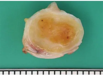
The specimen was a well demarcated, encapsulated and yellowish myxoid nodule measuring 2.0 ×1.7 ×1.5 cm. There was no hemor- rhage or necrosis (Fig. 1).
Microscopically, features of schwannoma were observed. The tu- mor was composed of many spindle cells with nuclear palisading arranged in whorls (Fig. 2).
There was no postoperative complication. And no evidence of local
recurrence was noted after 1 year of follow-up.
WWW.KJOG.ORG 818
KJOG Vol. 54, No. 12, 2011
Discussion
Schwannoma, also known as neurinoma, neurilemmoma, neu- rolemmoma [6] and perineural fibroblastoma [7] is a peripheral nerve sheath tumor characterised by proliferation of schwann cells [7-10].
Schwannomas rarely affect the female genitalia [1,2]. Especially, despite the rich nerve supply of the clitoris, schwannomas of the clitoris are extremely rare [3-5].
Schwannomas are benign, slow-growing tumors that infrequently recur and rarely undergo malignant change [11].
Histologically, the distinctive feature of schwannoma is the pat- tern of alternating Antoni A and B areas [7-10]. Antoni A areas are
composed of compacted spindle cells arranged in palisades or in an organoid arrangement (verocay bodies). Antoni B areas consist of tumor cells suspended in a loosely textured matrix with a more rounded cell morphology. Our case also showed that the tumor was composed of many spindle cells with nuclear palisading ar- ranged in whorls.
Also, an immunohistochemical examination can be used in the diagnosis. In addition to the S-100 protein, positivity tumor cells express basal lamina components (such as laminin and type IV collagen), vimentins, and sometimes KP1 (CD68) and glial fi brillary acidic proteins [5].
The most common types of benign peripheral nerve sheath tumors are schwannoma and neurofi broma [7,8]. But microscopically, the presence of encapsulation, two types of Antoni areas, and diffusely strong immunostaining for S-100 protein distinguish schwanno- mas from neurofi bromas [12]. And clinically, most of the clitoral neurofi bromas occur in a background of neurofi bromatosis, mostly neurofibromatosis type 1 [13], whereas none of the reported cases of clitoral schwannoma had neurofibromatosis. Neither in this case. In conclusion, despite its rarity, a schwannoma should be included in the differential diagnosis of a clitoral mass [14].
And simple surgical excision and follow-up considered as most adequate treatment [3].
References
1. Obeidat BR, Amarin ZO, Jallad MF. Vaginal schwannoma: a case report. J Reprod Med 2007;52:341-2.
2. Yamashita Y, Yamada T, Ueki K, Ueki M, Sugimoto O. A case of vulvar schwannoma. J Obstet Gynaecol Res 1996;22:31-4.
3. Cheng WC. Nerve tumour of the clitoris. Report of a case and commentary. J Obstet Gynaecol Br Commonw 1966;73:1016-7.
4. Huang HJ, Yamabe T, Tagawa H. A solitary neurilemmoma of the clitoris. Gynecol Oncol 1983;15:103-10.
5. Llaneza P, Fresno F, Ferrer J. Schwannoma of the clitoris. Acta Obstet Gynecol Scand 2002;81:471-2.
6. Kao GF, Laskin WB, Olsen TG. Solitary cutaneous plexiform neurilemmoma (schwannoma): a clinicopathologic, immu- nohistochemical, and ultrastructural study of 11 cases. Mod Pathol 1989;2:20-6.
7. Enzinger FM, Weiss SW. Benign tumors of peripheral nerves.
In: Enzinger FM, editor. Soft tissue tumors. 3rd ed. St Louis:
Mosby; 1995. p.821-63.
8. Scheithauer BW, Woodruff JM, Erlandson RA. Schwannoma.
Fig. 1. Gross feature of specimen. Figure shows a 2.0 × 1.7 × 1.5 cm sized mass. Cut surface is well demarcated, encapsulated and yellowish myxoid.
Fig. 2. Pathologic findings of specimen. Light microscopic appearance of tumor shows proliferation of many spindle cells with whorling pattern (H&E, ×200).
WWW.KJOG.ORG 819
Yu-Suk Yu, et al. Schwannoma of the clitoris: A case report
In: Scheithauer BW, Woodruff JM, Erlandson RA, editors. Tu- mors of the eripheral nervous system. Atlas of tumor patholo- gy. 3rd series, Fascicle 24. Washington: Armed Forces Institute of Pathology; 1997. p.105-56,177-218.
9. Fletcher CD, Davies SE. Benign plexiform (multinodular) schwannoma: a rare tumour unassociated with neurofi broma- tosis. Histopathology 1986;10:971-80.
10. Fletcher CD. Peripheral nerve sheath tumors: a clinicopatho- logic update. In: Rosen PP, Fechner RE, editors. Pathology an- nual. Part 1. Norwalk: Appleton and Lange; 1990. p.51-74.
11. Woodruff JM, Selig AM, Crowley K, Allen PW. Schwannoma
(neurilemoma) with malignant transformation. A rare, distinctive peripheral nerve tumor. Am J Surg Pathol 1994;18:882-95.
12. Weiss SW, Goldblum JR. Benign tumors of peripheral nerves.
In: Weiss SW, Goldblum JR, editors. Enzinger and Weiss’s soft tissue tumors. 4ed. St. Louis: Mosby, 2001. p.1111-1208.
13. Yüksel H, Odabaşi AR, Kafkas S, Onur E, Turgut M. Clitoro- megaly in type 2 neurofi bromatosis: a case report and review of the literature. Eur J Gynaecol Oncol 2003;24:447-51.
14. Chuang WY, Yeh CJ, Jung SM, Hsueh S. Plexiform schwannoma of the clitoris. APMIS 2007;115:889-90.
음핵에서 발생한 신경집종 1예
한국원자력의학원 산부인과
유유숙, 권도영, 박지민, 최석철, 유상영
신경집종은 말초신경초에 있는 슈반세포에서 발생하는 양성종양으로서 재발과 악성변화를 거의 하지 않고, 천천히 자라며, 여성의 생식 기관에서는 드물게 발생한다. 우리는 2.0 × 1.7 cm 음핵의 종괴를 단순 수술적 절제하였고, 종괴는 병리학적으로 신경집종 소견을 보였다.
1년 추적 관찰 후 국소 재발의 증거는 보이지 않았다. 이에 저자 등은 음핵에 드물게 발생하는 신경집종에 대해 보고하는 바이다.
중심단어: 신경집종, 음핵