WWW.KJOG.ORG 47
PRIMARY YOLK SAC TUMOR OF OMENTUM: A CASE REPORT
Do-Young Kwon, MD, Yu-Suk Yu, MD, Su-Hyun Baek, MD, Seok-Cheol Choi, MD, Sang-Young Ryu, MD
Department of Obstetrics and Gynecology, Korea Cancer Center Hospital, Korea Institute of Radiological and Medical Sciences, Seoul, Korea
Yolk sac tumor (YST) is one of the rare malignant germ cell tumor and usually occurs in gonad. Extragondal sites of YSTs are reported in mediastinum, vagina, brain, and retroperitoneum but are extremely rarely in omentum. The clinicopathologic feature of primary omental YST is not well known and there are only 5 cases reported currently. Recently we experienced a primary YST of omentum in 27-year-old woman who was performed exploratory laparotomy due to abdominal distension and pain. She has remained free of diseases for 2 years with normal menstruation after the fertility-saving surgery and postoperative adjuvant chemotherapy with bleomycin, etoposide, cisplatin regimen. The subject of primary YST of omentum is reviewed, and the possible histogenesis of the tumor is discussed.
Keywords:
Yolk sac tumor; Germ cell tumor; Omentum
CASE REPORT
Received: 2011.10.25. Revised: 2011.11. 7. Accepted: 2011.11. 8.
Corresponding author: Sang-Young Ryu, MD
Department of Obstetrics and Gynecology, Korea Cancer Center Hospital, Korea Institute of Radiological and Medical Sciences, 75 Nowon-gil, Nowon-gu, Seoul 139-706, Korea
Tel: +82-2-970-2114 Fax: +82-2-970-2449 E-mail: [email protected]
Th is is an Open Access article distributed under the terms of the Creative Commons Attribution Non-Commercial License (http://creativecommons.org/licenses/
by-nc/3.0/) which permits unrestricted non-commercial use, distribution, and reproduction in any medium, provided the original work is properly cited.
Copyright © 2012. Korean Society of Obstetrics and Gynecology Korean J Obstet Gynecol 2012;55(1):47-50
http://dx.doi.org/10.5468/KJOG.2012.55.1.47 pISSN 2233-5188 · eISSN 2233-5196
Among the malignant germ cell tumor, yolk sac tumor (YST) is known to be the third most common malignant germ cell tumor, following the dysgerminoma and immature teratoma [1-3]. YST simulate the yolk sac histologically and functionally in production of alphafetoprotein (AFP) [4]. Although most of YST occurs in the ovaries, 10 to 15% of YST may arise in extragonadal sites, such as the mediastinum, the pineal region, and sacrococcygeal region and the female reproductive tract [5]. Among them primary YST of omentum is extremely rare and only 5 cases are reported currently, and the pathogenesis and the clinical characteristics are unknown [1,2,4-6].
We report a case of primary YST of the omentum in 27-year-old women and discuss the clinical and histologic features in conjunc- tion with a review of the literature.
Case Report
A 27-year-old woman, single of nullipara, was referred to depart- ment of gynecology due to aggravating abdomen distension and pain that develop incidentally a month ago. On history taking, her mentral cycle was regular with 28 day cycle without any menor- rhagia or dysmenorrhea. On physical examination, fetal head sized pelvic mass was palpated with a tenderness but without rebound tenderness. Laboratory test showed negative urine-human chori- onic gonado trophin and no leukocytosis. Computerized tomogra-
phy (CT) scan showed a large lobulating contoured pelvic mass in- fi ltrating the omentum with ascites and a low attenuating nodule at right pericardiophrenic area (Fig. 1).
On the exploratory laparotomy, there was a 13 × 12 cm multi- loculated solid, yellowish-grey mass in the greater omentum with multiple small nodules measuring 1-1.5 cm in greatest dimension.
Another 7 × 5 cm white yellowish and friable multiloculated mass was found in cul-de-sac with small amount of ascites. The uterus, both fallopian tube and right ovary are grossly normal, but there was a 1.5 × 0.5 cm small nodule at left ovary. The frozen biopsy showed a malignancy favoring YST. In abdominal and pelvic cavity, disseminating bean sized small nodules were also found.
Debulking surgery with the resection of left ovarian surface
WWW.KJOG.ORG 48
KJOG Vol. 55, No. 1, 2012
masses and the cul-de-sac mass, pelvic lymphadenectomy, omen- tectomy and resection and reanastomosis of the small bowel were performed saving the uterus and both ovary. Emergency laboratory test showed an elevated alpha fetoprotein (AFP) 6,065 U/mL.
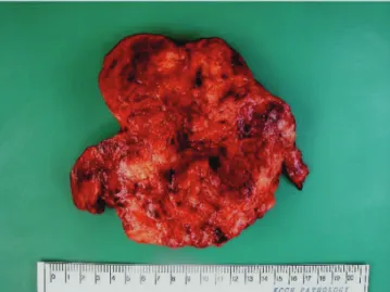
On pathological examination, the omental mass measured 13.5 × 13 × 7 cm and 603 g weight. The cut surface is white, soft, hemorrhagic, and partly shows necrosis (Fig. 2).
Histological evaluation showed the YST exhibiting the Schiller- Duval body (Fig. 3A) and hyaline globules (Fig. 3B). Special immu- nohistochemical staining of the tumor showed positive for AFP (Fig.
3C), cytokeratin and negative for β-HCG.
On the 10th day after surgery, adjuvant bleomycin, etoposide, cisplatin (BEP) chemotherapy consisting of bleomycin (20 mg iv on days 2, 9, and 16), etoposide (100 mg/m² iv for 5 consecutive days), and cisplatin (15 mg/m² iv for 5 consecutive days) was be- gun. A total of four cycles of chemotherapy were repeated every
3 weeks. The patient showed complete response after 6 cycles of chemotherapy and showed no evidence of disease on physical examination, serum AFP level, and abdomino-pelvic CT scan for 2 years.
Discussion
The extragonadal YST is rare malignant germ cell tumor arising outside ovary or testes. The most common site of extragonadal YST are reported in sacrococcygeal region followed by anterior medi- astinum, vagina, vulva, cervix, and uterine corpus [4].
However, primary YST of omentum is extremely rare and only 5
Fig. 1. Computerised tonography showed a large lobulating abdominal mass infi ltrating the omentum with small amount of ascites.
Fig. 2. Gross feature of specimen showed a multinodularomental mass measuring 13.5×13×7 cm. The cut surface is white, soft, hemorrhagic, and partly necrotic.
Fig. 3. Histological evaluation of the specimen shows typical Schiller Duval bodies (arrows) in microcystic area (A, H&E, ×200) and hyaline globules (ar- rows) (B, H&E, ×400). Special immunohistochemical staining of the tumor showed positive for AFP (C, immunohistochemical staining, ×400).
A B C
WWW.KJOG.ORG 49
Do-Young Kwon, et al. Primary yolk sac tumor of omentum: A case report
cases are reported currently [1,2,4-6].
Omental YST shows histologic characteristics similar to that of gonadal YST such as microcystic, endodermal sinus, solid, alveo- larglandular, polivesicular vitelline, myxomatous, papillary, mac- rocystic, hepatoid, and glandular or primitive endoderm patterns [5]. Schiff positive intracytoplasmic and extracytoplasmic globules are typical features of YST and these globules are usually positive immunohistochemically for AFP, A1AT, transferrin, and basement membrane components such as fibronectin, type IV collagen, vimentin, and laminin [4,5]. In our case, typical histologic charac- teristics of YST such as Schiller–Duval bodies and positive immu- nohistochemical staining for AFP and cytokeratin.
The histogenesis of extragonadal YST remains controversial. One hypothesis is that these tumors arise from aberrant differentia- tion of somatic cells, and the other one is that the germ cells have been arrested in their embryonic migration. The arrested embryon- ic migration hypothesis explains that when the primitive gonadal ridge expanded to the region of the external genitals, some of germ cell arrested anywhere along the migration course and can be the possible site of germ cell tumor in the future [4]. Metastasis from an occult focus in the ovary may contribute to primary YST of omentum, but in our case, thorough pathologic examination has excluded this possibility.
In this case, omental mass was huge main mass and left ovarian mass was located at ovarian surface without invasion, grossly. Al- though in pathologic examination small nodule at left ovary shows yolk sac tumor, ovarian capsule infiltration was not confirmed.
Mass of ovary was one of metastatic seeding nodules as like those disseminated in abdominal and pelvic cavity.
Generally, gonadal YST is a disease with a peak incidence at 20 years [5], however, the ages of in w primary YST of omentum omen previous reported were is 37, 44, 45, and 46 years implicat- ing a rather older women’s disease [2,4,6]. However, primary YST of omentum in our report showed a general young age of gonadal YSTs.
Because omental YST also affects women of child bearing age, the treatment of ometnal YST may not different from that of gondal YST [7]. Any gross metastases should be resected if possible, and
“fertility preserving” procedure can be considered even when the tumor may remain after surgery [3,8]. Preservation of uterus and ovaries are strongly recommended because the high response rate of chemotherapy with BEP [3,9,10]. In previous report, the average age of the 4 women with a primary YST of omentum was 43 years and there was no need to ‘fertility-saving surgery’. Actu- ally total hysterectomy was performed in all the 4 women with
primary YST of omentum [2,4,6]. However, in this report, we per- formed fertility saving surgery consisting of removal of the pelvic tumor, pelvic lymphadenectomy, omentectomy and resection of abdominal metastases with preservation of the uterus and both ovaries. Although the child bearing outcome is not confi rmed yet, this is the fi rst report that fertility saving surgery was performed in primary YST of omentum.
Primary YST of omentum responds well to combination chemo- therapy [2,4,6]. A regimen of BEP has been a major advance in the therapy of advanced and localized germ cell tumors [3]. Our patient also showed complete response with 6 cycyles of BEP combination chemotherapy.
Although long term outcome of omental YST is not well known, all the 5 patients with primary YST of omentum reported showed a favorable outcome [2,4,6]. In this report, the patient showed no evidence of disease on physical examination and serum a-fetopro- tein level and imaging studies for 2 years without any menstrual abnormality.
In conclusion, we report a 27-year-old woman with a primary YST of omentum successfully with fertility saving surgery and postop- erative adjuvant chemotherapy with a brief review of literature.
References
1. Rossi R, Stacchiotti D, Bernardini MG, Calvieri G, Lo Voi R.
Primary yolk sac tumor of the endometrium: a case report and review of the literature. Am J Obstet Gynecol 2011;204:e3-4.
2. Park NH, Ryu SY, Park IA, Kang SB, Lee HP. Primary en- dodermal sinus tumor of the omentum. Gynecol Oncol 1999;72:427-30.
3. Pectasides D, Pectasides E, Kassanos D. Germ cell tumors of the ovary. Cancer Treat Rev 2008;34:427-41.
4. Kim SW, Park JH, Lim MC, Park JY, Yoo CW, Park SY. Primary yolk sac tumor of the omentum: a case report and review of the literature. Arch Gynecol Obstet 2009;279:189-92.
5. Pasternack T, Shaco-Levy R, Wiznitzer A, Piura B. Extraovarian pelvic yolk sac tumor: case report and review of published work. J Obstet Gynaecol Res 2008;34:739-44.
6. Zhang B, Gao S, Chen Y, Wu Y. Primary yolk sac tumor arising in the pancreas with hepatic metastasis: a case report. Korean J Radiol 2010;11:472-5.
7. Piura B, Dgani R, Zalel Y, Nemet D, Yanai-Inbar I, Cohen Y, et
al. Malignant germ cell tumors of the ovary: a study of 20
cases. J Surg Oncol 1995;59:155-61.
WWW.KJOG.ORG 50
KJOG Vol. 55, No. 1, 2012
8. Berek JS, Hacker NF. Berek and Hacker’s gynecologic oncology.
Philadelphia (PA): Lippincott Williams & Wilkins; 2010.
9. Weinberg LE, Lurain JR, Singh DK, Schink JC. Survival and re- productive outcomes in women treated for malignant ovarian
germ cell tumors. Gynecol Oncol 2011;121:285-9.
10. Seli E, Tangir J. Fertility preservation options for female patients with malignancies. Curr Opin Obstet Gynecol 2005;17:299-308.
복막에 발생한 난황낭종양의 1예
한국원자력의학원 산부인과
권도영, 유유숙, 백수현, 최석철, 유상영
난황낭종양은 생식 세포에 속하는 종양으로서 비교적 흔한 암종은 아니지만 악성도가 높다. 종양의 대부분은 생식 기관(난소, 고환)에서 기원하며, 난황낭종양의 약 10-15% 정도가 생식기관 이외의 위치에서 발생한다. 복막에서 난황낭종양이 발생한 경우는 매우 드물며, 현 재까지 5명의 환자가 보고된 바 있다. 27살의 비교적 젊은, 본 경우의 환자는 복부팽만을 주소로 내원하였고 우리는 시험적 개복술과 수 술 후 보존적 항암치료를 시행하였다. 수술은 향후 임신 가능성을 고려하려 자궁과 난소를 보존하는 방향으로 이루어졌다. 항암치료 종료 2년 동안 추적 관찰하였으며, 현재까지 검사 소견상 재발이나 전이의 어떤 소견도 보이지 않고 있다.
중심단어: 난황낭종양, 생식세포종양, 복막