136
Copyrights © 2014 The Korean Society of RadiologyINTRODUCTION
Paratesticular tumors are infrequent tumor of mesenchymal origin that can affect the testicular tunics, epididymis, and sper- matic cord. The most common neoplasm arising in paratesticu- lar area is adenomatoid tumor (1). Schwannoma of the scrotum and testis is extremely rare. We report the case of an intrascrotal and extratesticular tumor that was histopathologically shown to be schwannoma.
CASE REPORT
A 59-year-old man presented with a painless, slowly growing, palpable, scrotal tumor of ten years of duration. On physical ex- amination, 2 cm sized round and movable mass was palpated in the lower extratesticular region within the left scrotum. It was located just below scrotal skin but was not attached to the skin or any other regional structure. In particular, the mass was sepa- rated from the left testis, and both testes appeared to be normal.
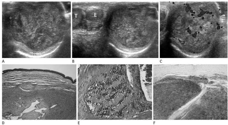
Lymphadenopathy was not detected. Ultrasonography depicted a 2 × 2 cm sized smooth, round, well-circumscribed, heteroge-
neous, hyperechoic mass within the left scrotum (Fig. 1A). The tumor was separate from the left testis and epididymis (Fig. 1B).
Doppler examination showed that the mass was well-vascular- ized (Fig. 1C). We initially considered this extratesticular mass a benign tumor such as adenomatoid tumor or leiomyoma.
Therefore, curative surgical excision of tumor was performed.
At the resection, the tumor was observed to be separated from the testis, based on ultrasound findings. Grossly, the resected mass appeared yellowish, round, and soft with focal hemor- rhage. Microscopically, tumor showed to be a well demarcated solid tumor with multiple dilated hyalinized vessels (Fig. 1D).
The tumor was comprised of spindle cells with elongated nuclei and fibrillary cytoplasm. Tumor cells showed hypercellular areas with nuclear palisading and alternating with hypocellular areas (Fig. 1E). Immunohistochemically, tumor cells were diffusely positive for S-100 protein (Fig. 1F). Histological and immuno- histochemical findings were consistent with schwannoma.
DISCUSSION
Schwannoma is a benign encapsulated neoplasm that usually
Case Report
pISSN 1738-2637 / eISSN 2288-2928 J Korean Soc Radiol 2014;71(3):136-138 http://dx.doi.org/10.3348/jksr.2014.71.3.136
Received June 2, 2014; Accepted July 7, 2014 Corresponding author: Seung Joon Choi, MD Department of Radiology, Gachon University Gil Medical Center, 21 Namdong-daero 774beon-gil, Namdong-gu, Incheon 405-760, Korea.
Tel. 82-32-460-3060 Fax. 82-32-460-3065 E-mail: [email protected]
This is an Open Access article distributed under the terms of the Creative Commons Attribution Non-Commercial License (http://creativecommons.org/licenses/by-nc/3.0) which permits unrestricted non-commercial use, distri- bution, and reproduction in any medium, provided the original work is properly cited.
Schwannoma is a benign tumor arising from Schwann cells of the peripheral nerve sheath. It usually develops in the head, neck, mediastinum or retroperitoneum, but seldomly in the scrotum. Here, the authors report the imaging findings of a patient with a ten year history of a slowly growing scrotal tumor that was histopathologi- cally proven to be schwannoma.
Index terms Schwannoma
Intrascrotal and Extratesticular Tumor Ultrasonography
Intrascrotal and Extratesticular Schwannoma: A Case Report
1 음낭내 고환외 신경초종: 증례 보고1Min Jung Kim, MD
1, Seung Joon Choi, MD
1, Young Sup Shim, MD
1, Hyung Sik Kim, MD
1, Jeong Ho Kim, MD
1, Hye-Young Choi, MD
1, Hyunchul Kim, MD
2Departments of 1Radiology, 2Pathology, Gachon University Gil Medical Center, Incheon, Korea
Min Jung Kim, et al
137
jksronline.org J Korean Soc Radiol 2014;71(3):136-138
ic, slowly growing scrotal mass with duration of several months to a year (3-6). Only one patient complained of local pain in the scrotum for 20 days before presentation (7). Chan et al. (3) re- ported ultrasonographic findings of a well-circumscribed ovoid heterogeneous mass separate from the testis, which measured 7 cm in greatest diameter. Bergeron et al. (4) reported a solid vo- luminous, heterogeneous, lobulated mass, with greatest diame- ter of 5.5 cm; and Shahid et al. (5) also described a well-circum- scribed heterogeneous mass. On ultrasound, all tumors were depicted as a well-circumscribed heterogeneous mass, well-sep- arated from testis and epididymis. There has been no case re- porting of a cystic portion or calcification.
The most common extratesticular tumor is adenomatoid tu- mor, which occurs at a peak age of between 20 and 50 years.
These patients usually present with an asymptomatic scrotal tu- mor. Adenomatoid tumors are smooth, round, well-defined, and varying in size. On ultrasound, they are typically homoge- neous and hyperechogenic in nature (1). Leiomyoma is the sec- arises from cranial, spinal, and peripheral nerves. They mostly af-
fect the head and neck region along a nerve sheath (2). However, although schwannoma is the most common peripheral nerve sheath tumor, it is extremely rare in the paratesticular area. To date, intrascrotal schwannomas have been reported in only seven cases in the medical literature written in English (3-9).
Histologically, schwannoma is an encapsulated tumor with bi- phasic architectural pattern composed of Antoni A and Antoni B areas. Antoni A areas correspond to compacted spindle cells that are often arranged in palisades or adopt an organoid ar- rangement (Verocay bodies), whereas Antoni B areas are char- acterized by loose-textured tissue in a myxomatous matrix that may appear microcystic. Immunohistochemistry studies show uniform positive staining for S-100 protein (2).
The diagnosis of schwannoma is challenging, and the clinical and radiographic findings of scrotal schwannoma are non-spe- cific, which means the tumor can be easily misdiagnosed as an- other solid tumor. Patients usually present with an asymptomat-
Fig. 1. A 59-year-old man presented with a slowly growing scrotal tumor of duration 10 years.
A. Ultrasonograph showed a 2 × 2 cm sized smooth, round, well-circumscribed, heterogeneous hyperechoic mass within the left scrotum.
B. The mass was separated from the left testis (T) and epididymis (E) at longitudinal scan.
C. Color Doppler revealed a well vascularized mass.
D. The tumor showed a well demarcated solid tumor with multiple dilated hyalinized vessels (H&E, × 40).
E. The tumor was comprised of spindle cells with elongated nuclei and fibrillary cytoplasm. Tumor cells showed hypercellular areas with nuclear palisading alternating with hypocellular areas (H&E, × 200).
F. Tumor cells were diffusely positive for S-100 protein by immunohistochemical staining (× 100).
E B
D A
F C
Intrascrotal and Extratesticular Schwannoma
138
J Korean Soc Radiol 2014;71(3):136-138 jksronline.org3. Chan PT, Tripathi S, Low SE, Robinson LQ. Case report--an- cient schwannoma of the scrotum. BMC Urol 2007;7:1 4. Bergeron M, Bolduc S, Labonté S, Moore K. Intrascrotal
extratesticular schwannoma: a first pediatric case. Can Urol Assoc J 2014;8:E279-E281
5. Shahid M, Ahmad SS, Vasenwala SM, Mubeen A, Zaheer S, Siddiqui MA. Schwannoma of the scrotum: case report and review of the literature. Korean J Urol 2014;55:219-221 6. Arciola AJ, Golden S, Zapinsky J, Fracchia JA. Primary in-
trascrotal nontesticular schwannoma. Urology 1985;26:
304-306
7. Jiang R, Chen JH, Chen M, Li QM. Male genital schwanno- ma, review of 5 cases. Asian J Androl 2003;5:251-254 8. Ikari R, Okamoto K, Yoshida T, Johnin K, Okabe H, Okada Y.
A rare case of multiple schwannomas presenting with scrotal mass: a probable case of schwannomatosis. Int J Urol 2010;17:734-736
9. Bhanvadia V, Santwani P. Intrascrotal extratesticular schwannoma. J Cytol 2010;27:37-39
10. Hertzberg BS, Kliewer MA, Hertzberg MA, Distell BM. Epi- didymal leiomyoma: sonographic features. J Ultrasound Med 1996;15:797-799
ond most common tumor of the epididymis. This tumor most commonly manifests as a slow-growing, nontender scrotal mass, which occurs at a peak age during the fifth decade of life. Leio- myomas usually present as well-demarcated tumor surrounded by a gray-white fibrous capsule and would range from 1 to 4 cm in size. This tumor has a variable sonographic appearance whether it is predominantly solid or cystic, and it may contain calcifications. Leiomyomas can be associated with a hydrocele in half of the cases (1, 10).
However, it is difficult to accurately diagnose paratesticular tu- mors, preoperatively. Despite its rarity in the scrotum, schwanno- ma should be included in the differential diagnosis of paratestic- ular tumor with other more common benign tumors.
REFERENCES
1. Akbar SA, Sayyed TA, Jafri SZ, Hasteh F, Neill JS. Multimo- dality imaging of paratesticular neoplasms and their rare mimics. Radiographics 2003;23:1461-1476
2. Skovronsky DM, Oberholtzer JC. Pathologic classification of peripheral nerve tumors. Neurosurg Clin N Am 2004;15:
157-166
음낭내 고환외 신경초종: 증례 보고1
김민정
1· 최승준
1· 심영섭
1· 김형식
1· 김정호
1· 최혜영
1· 김현철
2신경초종은 말초신경초의 슈반 세포(Schwann cell)에서 기원하는 양성 종괴이다. 이 종괴는 일반적으로 두경부, 종격동이 나 후복강에 생기며 음낭에 생기는 경우는 매우 드물다. 저자들은 10년 동안 천천히 자라는 고환 종괴를 주소로 내원한 59세 남자 환자에서 발견된 음낭내 고환외 신경초종을 경험하였기에 이의 초음파 영상을 보고하고자 한다.
가천대학교 의학전문대학원 길병원 1영상의학과, 2병리과