Treatment of May-Thurner Syndrome with Catheter-Guided Local Thrombolysis and Stent Insertion
Jong-Youn Kim, MD1, Donghoon Choi, MD1, Young Guk Ko, MD1 Sungha Park, MD1, Yangsoo Jang, MD1 and Do Yun Lee, MD2
1Cardiology Division, Yonsei Cardiovascular Center and Cardiovascular Research Institute,
2Department of Diagnostic Radiology, Yonsei University College of Medicine, Seoul, Korea
ABSTRACT
Background:May-Thurner syndrome is an uncommon disease entity in which the left common iliac vein is com- pressed by the right common iliac artery, with the subsequent development of deep vein thrombosis and chronic venous insufficiency. Herein, our experience on the treatment of extensive iliofemoral deep venous thrombosis due to May-Thurner syndrome, using endovascular techniques, is reported. Methods:Twenty-one symptomatic patients, 8 men and 13 women, with a mean age of 51 years, were referred for treatment. Eighteen of these patients were treated with catheter-guided thrombolysis, but three, with short segment involvement, did not require thro- mbolysis. After completion of the thrombolytic therapy, the residual venous narrowing was treated by balloon angioplasty and/or self-expandable stent placement. Patients were then followed-up by clinic visits and veno- graphy. Results:The mean total dose of urokinase and duration of infusion were 4.28±1.89 million units and 72±35 hours, respectively. Eighteen of the 21 patient received stent deployments. The mean diameter of the stents was 12.9±2.0 mm. Initial technical success, with immediate symptom resolution, was achieved in 20 of the 21 patients (95%). Among the patients who received stent implantation, two had recurrent thrombotic occlusion during the follow-up period. (mean 10.8 months); all three patients who did not receive stent implantation had recurrent thromboses There were no major bleeding complications, with the exception of one patient who developed a retroperitoneal hematoma. Conclusions:Catheter-guided thrombolysis and angioplasty with stent implanta- tion are safe and effective for the treatment of May-Thurner syndrome. (Korean Circulation J 2004;34 (7):655-659) KEY WORDS:Venous thrombosis;Thrombolytic therapy;Stents.
Introduction
In 1956, May and Thurner first described a spur-like formation of the left common iliac vein in 22% of autop- sies.1) Chronic pulsatile compression of the left common iliac vein, between the right common iliac artery and the lumbar vertebral body, may induce excessive local intimal proliferation, resulting in impaired venous return and ma-
ssive venous thrombosis.2) Usually, these patients present with left leg edema, pain or deep vein thrombosis. Bec- ause of the chronic physical compression by the right common iliac artery and the large size of the thrombus that develops at the iliac vein, anticoagulation alone is usually ineffective in resolving the deep vein thrombosis and the subsequent prevention of pulmonary embolisms.
Previous reports have shown anticoagulation alone, and a thrombectomy combined with prospective anticoagu- lation, to have rethrombosis rates of up to 73% in patients with a venous spur.3) Therefore, corrective treatment is indicated to prevent the development of a pulmonary embolism and the long-term complications of post-throm- botic syndrome, including chronic leg edema, pain, hyper- pigmentation and skin ulcers. Recently, catheter-directed Received:February 11, 2004
Accepted:May 18, 2004
Correspondence:Donghoon Choi, MD, Cardiology Division, Yonsei Cardiovascular Center and Research Institue, Yonsei Uni- versity College of Medicine, 134 Shinchon-dong, Seodaemun- gu, Seoul 120-752, Korea
Tel:82-2-361-7071, Fax:82-2-393-2041 E-mail:cdhlyj@yumc.yonsei.ac.kr
thrombolysis and endovascular angioplasty with stents have been used to treat DVT in these patients. Herein, our experience and treatment efficacy of endovascular venous angioplasty in patients with deep vein thrombo- sis due to May-Thurner syndrome is reported.
Methods
A retrospective analysis was performed on 21 patients diagnosed with May-Thurner syndrome, and treated with catheter guided local thrombolysis and endovascular ve- nous angioplasty, between August 1999 and December 2002. Sixteen patients presented with acute iliofemoral venous thrombosis and 5 with chronic venous outflow obstruction. The characteristic May-Turner syndrome lesion was diagnosed with a venogram. These lesions are defined by stenosis of the proximal left common iliac vein, including the portion underlying the right common iliac artery, as well as the presence of significant venous collateral vessels (Figure 1). After an ipsilateral puncture of the popliteal vein by a modified Seldinger’s method (Early in this series, other access sites were utilized, in- cluding the right internal jugular (n=2), right common
femoral vein (n=1), left common femoral vein (n=2), a 6.5 F hemostatic sheath was inserted. Through the hem- ostatic sheath, a 5F multi-sideport infusion catheter (60 cm, COOK, Bloomington, USA) was inserted and passed through the thrombotic occlusion of the left common iliac vein. In the case of total occlusion, the infusion cath- eter was placed just distal to the occlusion, through which urokinase infusion was performed. When the lesion was partially reanalyzed at follow-up, the infusion catheter was passed across the lesion and the urokinase infusion continued. In addition to urokinase infusion, heparin in- fusion was performed through the hemostatic sheath to maintain the aPTT between 60 to 90 second. A venogram was performed daily to assess the status of the throm- botic occlusion. The endpoints of thrombolytic therapy were near clot dissolution, lytic stagnation (no interval change for 24 hours) or development of a major compli- cation (in one case, retroperitoneal bleeding developed).
Residual stenosis combined with an organic change of the vein was treated by angioplasty and stent implant- ation (Figure 2). The stent size was determined by mea- suring the diameter of patent distal common iliac vein, with the stenting performed through an 8 F hemostatic
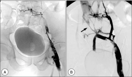
Figure 1. Digital subtraction angiography (DSA) on admission showed a total occlusion of the left iliac and femoral veins (A). Follow-up DSA after urokinase infusion for 3 days showed remaining thrombi and an obstructive lesion at the left common iliac vein (arrow), with extensive cross-pelvic and hypogastric collateralization (B).
A B
sheath exchanged for the 6.5 F sheath. Through the sheath, a 0.035-inch hydrophilic guidewire was passed across the lesion, and angioplasty with stent implantation performed with self expanding Wallstent® (Boston Sci- entific, Waterstone, Massachusetts, USA) with average profile of 12.9±2 mm and length of 65.4±18.2 mm.
10000 unit of heparin was administered at the start of the procedure, and systemic heparinization performed after the procedure to maintain the aPTT between 60 to 90 seconds for 5 days. Coumadization was simultaneously started with heparinization to maintain an INR of 2-3, and performed for 6 months. A venogram was perfor- med 1 week after the procedure, immediately before discharge and 6 months after the procedure, to assess the patency rate.
Results
Out of the 21 patients, there were 8 males and 13 female, with an average age of 51 years. Three of the patients demonstrated complications due to pulmonary thromboembolism on a lung perfusion scan and a further 3 did not undergo catheter guided thrombolysis due to the short length of their lesion. The average infusion rate
and total dose of urokinase were 92000±30000 IU/hr and 4,280,000±1890000 IU, respectively, with a total infusion time of 72±35 hours. For the treatment of resi- dual stenosis, balloon angioplasty alone was performed on 3 patients, and stent implantation on 18. Wallstent® (Boston Scientific, Waterstone, Massachusetts, USA) was used for stent implantation in all the patients, with an average profile of 12.9±2 mm and length of 65.4±
18.2 mm. After the procedure, all patients had good pa- tency of the common iliac vein. The follow-up venogram, performed 7-10 days after the procedure, showed abse- nce of residual thrombi in all 18 patients implanted with stents, with resolution of the leg edema. One of the 3 patients who underwent balloon angioplasty alone showed severe residual stenosis, but his colon cancer was in the terminal stage, so no further management, only IVC filter insertion was performed. There was a single case of major bleeding as a complication that resulted in a retroperi- toneal hematoma, which resolved spontaneously in 13 days. After the procedure, all 21 patients showed no evidence of newly developed pulmonary embolism in the follow-up lung perfusion scans performed within 7 days of the procedure. The follow-up rate was high, with follow-up venogram performed on 17 of the 18 patients Figure 2. After deployment of 2 Wallstents (12×46 mm and 10×83 mm)(A). Completion digital subtraction angio- graphy showed a satisfactory angiographic result, with abolition of collaterals and the rapid in-line flow, superiorly, into the inferior vena cava (B).
A B
who underwent stent implantation, with 2 patients showing recurrent thrombi formation. All 3 patients that underwent balloon angioplasty alone showed recurrent thrombotic occlusion, with 2 of these developing signs of postthrom- botic syndrome, resulting in debilitating pain, leg edema and recurrent ulcer formation. One patient implanted with stent showed leg edema, with postthrombotic syndrome, despite the absence of recurrent thrombi.
Discussion
May and Thurner syndrome is a spur-like formation of the left common iliac vein due to chronic pulsatile compression of the left common iliac vein, between the right common iliac artery and the lumbar vertebral body, resulting in excessive local intimal proliferation, impaired venous return and venous thrombosis.1)2) This may acc- ount for the higher frequency of left rather than right side common iliac vein thromboses.6) Chronic vibratory puls- ation of the common iliac artery on the venous wall may result in frictional damage of the intimal wall, which may result in subsequent intimal proliferation and venous thrombosis.5) This disease is reported to be more frequent in women, which was also the case in our study.7) Beca- use of the chronic nature of the disease process, patients typically present with symptoms and signs of postthrom- botic syndrome, such as pigmentation, varicose vein, ch- ronic leg pain, phlebitis and recurrent skin ulcers.2) Also, acute thrombosis may develop in patients at high risk of developing deep vein thrombosis, such as in patients undergoing orthopedic surgery.2) In our study, sixteen patients presented with acute deep vein thrombosis, 5 had a history of cerebrovascular disease or trauma resulting in prolonged immobilization and 4 were in an early post- operative state following surgical treatment for maligna- ncies.
The two major treatment goals with deep vein throm- bosis are the prevention of pulmonary embolism and postthrombotic syndrome development. The pathogenesis of postthrombotic syndrome is known to be due to chr- onic venous obstruction and venous valve insufficiency
due to the formation of thrombi. Previous, Eklof and Kistner showed that patients with deep vein thrombosis treated by thrombectomy showed no evidence of post thrombotic syndrome, whereas 18% of those treated with anticoagulation showed signs of post thrombotic synd- rome after 4 years of follow-up.8) The results of that study show that it is imperative to remove the thrombi as early as possible. Thrombolytics are an effective treat- ment for the resolution of venous thrombi, with systemic thrombolytic therapy reported as being superior to anti- coagulation in preventing postthrombotic syndrome.9) However, the use of systemic thrombolytic therapy is limited by the possibility of severe bleeding complica- tions, which make it contraindicatory for patients at a high risk of deep vein thrombosis formation, such as in preg- nancy, surgery and cerebrovascular disease.10)11) Localized thrombolytic therapy is commonly being performed to decrease the risk of major bleeding. Theoretically, local infusion of thrombolytic agents at the site of thrombotic occlusion maximizes the therapeutic effect while mini- mizing the risk of major bleeding. The successful treat- ment of deep vein thrombosis with local thrombolytic therapy has already been shown.12)13) Urokinase is used in our catheter laboratory because of its lower cost and lower rate of bleeding complications compared to t-PA.
All the patients in the study group showed successful recanalization, with only one case of major bleeding re- sulting in a retroperitoneal hematoma, which resolved uneventfully.
The popliteal vein is the usual site of access used by us, with the internal jugular vein (n=2) and right com- mon femoral vein (n=1) being the other sites used. Use of these sites entails passing catheters in a retrograde fashion, so infusion of the thrombolytic drug may be less effective. Direct puncture of the left common femoral vein is inadvisable as thrombolysis or intervention will some-times involve the access site. In 6 cases of this study, the left common femoral vein had a thrombus and required stent insertion. Therefore, the common femoral vein is used as the access site only when the involved site is short and limited within the femoral vein.
The rationale for angioplasty with stent implantation in residual stenosis is that the implant of a stent is effe- ctive therapy for a venous obstruction14) and superior to balloon dilatation alone.15) The possible problem of valve destruction by the stents, and the subsequent develop- ment of venous insufficiency, does not apply to femoral veins due to the absence of valves in the femoral veins.
Complete resolution of the residual stenosis may not be possible with balloon angioplasty, which may result in higher risk of rethrombosis. In fact, all 3 patients who underwent balloon angioplasty alone showed restenosis and rethrombosis, which reemphasizes the importance of complete resolution of the residual stenosis. This was demonstrated by the fact that only 2 of the 17 patients showed thrombus formation at the site of stent implant- ation at follow-up. Because of the inherent thrombogeni- city of metallic stents before endothelization and the high risk of recurrent venous thrombosis, all the patients rece- ived standard prophylactic treatment with coumadine for 6 months. It is our belief the duration of treatment is adequate unless there are associated conditions requiring prolonged or indefinite anticoagulation.
There was a single case of major bleeding complication that resulted in a retroperitoneal hematoma, which reso- lved spontaneously in 13 days. After the procedure, all 21 patients showed no evidence of newly developed pul- monary embolism in the follow-up lung perfusion scans performed within 7 days of the procedure. This study has demonstrated the safety of local catheter guided thro- mbolysis and stenting, with minimal risk of major bleed- ing complications and iatrogenic embolism during the procedure.
This study has shown excellent immediate results and patency rates, with minimal complications, using locali- zed catheter-guided thrombolysis plus angioplasty with stent implantation. Further studies will be needed to demonstrate the long term patency rate in a larger popul-
ation of patients.
REFERENCES
1) May R, Thurner J. Ein Gef ssporn in der vena iliaca com- munis sinistra als wahrscheinliche ursache der überwiegende linksseitigen beckenvenenthrombose. Z Kreisl-Forsch 1956;
45:912-22.
2) Baron HC, Sharms J, Wayne M. Iliac vein compression syn- drome: a new method of treatment. Am Surg 2000;66:653-5.
3) Burroughs KE. New considerations in the diagnosis and th- erapy of deep vein thrombosis. South Med J 1999;92:517-20.
4) Binkert CA, Schoch C, Stuckmann G, Largiader J, Wigger P, Schoepke W, et al. Treatment of pelvid venous spur(May- Thurner syndrome) with self-ezpanding metallic endoprost- heses. Cardiovasc Intervent Radiol 1998;21:22-6.
5) Gerarld JO, Charles PS, Craig B, Stephen TK, Manmood KR, Daniel YS, et al. Endovascular management of iliac vein compression(May-Thurner) syndrome. JVIR 2000;11:
823-36.
6) Heijmen RH, Bollen TL, Duyndam DA, Overtoom TT, Van den Berg JC, Moll FL. Endovascular venous stenting in May- Thurner syndrome. J Cardiovasc Surg 2001;42:83-7.
7) Steinberg JB, Jacocks MA. May-Thurner syndrome. a prev- iously unreported variant. Ann Vasc Surg 1993;7:577-81.
8) Eklof B, Kistner RL. Is there a role for thrombectomy in iliofemoral venous thrombolysis? Semin Vase Surg 1996;9:
34-45.
9) O’Donnell TF Jr, Browse NL, Burnand KG, Thomas ML.
The socioeconomic effects of iliofemoral thrombosis. J Surg Res 1987;22:483-8.
10) Strandness ED, Manzo RA, Markel A. Is there a role for thrombolytic treatment of deep vein thrombosis? Cardiol Board Rev 1993;10:25-7.
11) Sherry S. Thrombolytic therapy for DVT. Semin Interv Radiol 1985;2:331-7.
12) Jung IH, Choi DH, Kim HJ, Kyung HD, Koo BK, Cho SY, et al. A case of eosinophilia associated with massive deep vein thrombosis treated with local urokinase infusion. Korean Circ J 2001;31:256-61.
13) Kim HJ, Chung IH, Cho DK, Cho JR, Choi DH, Koo BK, et al. May-Thurner syndrome: two cases treated with cath- eter-directed thrombolysis and stent placement. Korean Circ J 2001:31:1324-29.
14) Nazarian GK, Bjarnason H, Dietz CA, Bernadas CA, Hunter DW. Iliofemoral venous stenosis: effectiveness of treatment with metallic endovascular stents. Radiology 1996;200:193-9.
15) Whittemore AD, Donaldson MC, Polak JF, Mannick JA.
Limitations of balloon angioplasty for vein graft stenosis. J Vasc Surg 1991;14:340-5.