Korean J Radiol 12(3), May/Jun 2011
kjronline.org 400
RE: Role of Duplex Doppler US for Thyroid Nodules: Looking for the
“Sword” Sign
Alexis Lacout, MD
1, Pierre Yves Marcy, MD
2, Juliette Thariat, MD
31Department of Radiology, Centre d’imagerie Médicale, Centre Médico Chirurgical (CMC), France; 2Head & Neck and Interventional Radiology, Department Antoine Lacassagne Cancer Research Institute, France;
3Department of Radiation Oncology, Antoine Lacassagne Cancer Research Institute, France
Duplex Doppler US may be useful for the detection of thyroid malignancies that show either anarchic winding or penetrating “sword like” neoangiogenic vessels. It may be helpful in selecting nodules that should undergo fine needle aspiration biopsy.
Index terms: Doppler US; Ultrasonography; Anaplastic carcinoma; Thyroid
Dear Editor:
We read with great interest the article by Algin et al. (1)
“Role of duplex power Doppler ultrasound in differentiation between malignant and benign thyroid nodules”, in the December 2010 issue of the Korean Journal of Radiology.
The authors concluded that the vascularity pattern was not useful to distinguish malignant from benign thyroid nodules.
In this article, it is worthy to note that the cytology examinations were dichotomized between the benign and malignant samples. May we suggest that the use of the cytologic Bethesda system 2008 classification (2) would probably have shown some correlations between one or more of each of the Bethesda classification categories and the pattern of thyroid nodule vascularity. As the management of thyroid nodules is currently based upon
Received February 10, 2011; accepted after revision March 3, 2011.
Corresponding author: Alexis Lacout, MD, Department of Radiology, Centre d’imagerie Médicale, Centre Médico Chirurgical, 83 Avenue Charles de Gaulle, 15000 Aurillac, France.
• Tel: (33) 4 71 48 00 50 • Fax: (33) 4 71 48 53 48
• E-mail: [email protected]
the Bethesda classification, highlighting such a correlation would probably have been useful and more up-to-date for nodule selection.
Furthermore, as the decision to perform fine-needle aspiration biopsy (FNAB) or surgery for thyroid nodules is made based on the presence of at least one malignant criteria on B-mode ultrasonography (US) (e.g.,
microcalcifications, irregular margins, hypoechogenicity, etc.), a Doppler US pattern analysis would also have been useful for the nodules that showed only benign features.
We do emphasize that some Doppler US features in this subgroup may have correlation either with malignancy or a category of the 2008 Bethesda classification.
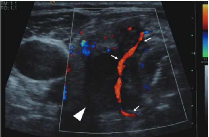
Another point to consider is that the authors’ analysis was restricted to the distribution of vascularization within the thyroid nodule. We stress that quantitative analysis of the nodule vascularization could be helpful to differentiate benign from malignant nodules (3). Furthermore, two other true qualitative Doppler US criteria that reflect angiogenesis may strongly suggest malignancy. The first is anarchic intralesional vascularization with winding vessels, and the second is the presence of a peripheral large afferent (penetrating) vessel (4) (Fig. 1). As a matter of fact, penetrating “sword-like” vessels seen on Doppler US may be present in poor differentiated papillary or anaplasic thyroid nodules at an early stage, and this can lead to a diagnosis
Letter to the Editor
DOI: 10.3348/kjr.2011.12.3.400 pISSN 1229-6929 · eISSN 2005-8330
Fig. 1. Patient who presented with poorly-differentiated papillary thyroid carcinoma.
Doppler US shows large vessel (arrows) crossing from side-to-side in suspicious left hypoechoic thyroid nodule (arrowhead), as sword would do (“sword sign”). Fine-needle aspiration biopsy confirmed high grade malignancy of this aggressive nodule.
Korean J Radiol 12(3), May/Jun 2011
kjronline.org 401
pre-FNA requirements: a synopsis of the National Cancer Institute Thyroid Fine-Needle Aspiration State of the Science Conference. Diagn Cytopathol 2008;36:390-399
3. Lyshchik A, Moses R, Barnes SL, Higashi T, Asato R, Miga MI, et al. Quantitative analysis of tumor vascularity in benign and malignant solid thyroid nodules. J Ultrasound Med 2007;26:837-846
4. Spiezia S, Colao A, Assanti AP, Cerbone G, Picone GM, Merola B, et al. Usefulness of color echo Doppler with power Doppler in the diagnosis of hypoechoic thyroid nodules: work in progress.
Radiol Med 1996;91:616-621
of early-stage malignancy and the subsequent optimizing of
these patients’ therapeutic management.
REFERENCES
1. Algin O, Algin E, Gokalp G, Ocakoglu G, Erdogan C,
Saraydaroglu O, et al. Role of duplex power Doppler ultrasound in differentiation between malignant and benign thyroid nodules. Korean J Radiol 2010;11:594-602
2. Cibas ES, Alexander EK, Benson CB, de Agustin PP, Doherty GM, Faquin WC, et al. Indications for thyroid FNA and