It is well known that chronic hyperglycemia is a major contributor to microvascular complications. Diabetic nephropathy is one of the most serious microvascular complications and leading cause of mortality and morbidity in diabetic patients.
There are several mechanisms of diabetic nephropathy including the activation of protein kinase C (PKC), increased advanced glycosylation end products, the
Aldose Reductase Inhibitor Ameliorates Renal Vascular Endothelial Growth Factor Expression
in Streptozotocin-Induced Diabetic Rats
Joong Kyung Sung,
1*Jang Hyun Koh,
2*Mi Young Lee,
1Bo Hwan Kim,
1Soo Min Nam,
3Jae Hyun Kim,
1Jin Hee Yoo,
1So Hee Kim,
1Sun Won Hong,
4Eun Young Lee,
5Ran Choi,
1and Choon Hee Chung
11Department of Internal Medicine, Yonsei University Wonju College of Medicine, Wonju; 2Center for Health Promotion, Samsung Medical Center, Sungkyunkwan University, Seoul; 3Department of Internal Medicine, Sun General Hospital, Daejeon;
4Department of Pathology, Yonsei University College of Medicine, Seoul; 5Department of Internal Medicine, Soonchunhyang University College of Medicine, Cheonan, Korea.
Purpose:The vascular endothelial growth factor (VEGF) expression of podocyte is one of the well-known major factors in development of diabetic nephropathy. In this study, we investigated the effects of aldose reductase inhibitor, fidarestat on diabetic nephropathy, and renal VEGF expression in a type 1 diabetic rat model.
Materials and Methods: Twenty four Sprague-Dawley male rats which were performed intraperitoneal injection of streptozotocin and normal six rats were divided into four groups including a normal control group, untreated diabetic control group, aldose reductase (AR) inhibitor (fidarestat, 16 mg ˙ kg-1˙ day-1) treated diabetic group, and angiotensin receptor blocker (losartan, 20 mg ˙ kg-1˙ day-1) treated diabetic group. We checked body weights and blood glucose levels monthly and measured urine albumin-creatinine ratio (ACR) at 8 and 32 weeks.
We extracted the kidney to examine the renal morphology and VEGF expressions.
Results:The ACR decreased in fidarestat and losartan treated diabetic rat groups than in untreated diabetic group (24.79 ± 11.12, 16.11 ± 9.95, and 84.85 ± 91.19, p
< 0.05). The renal VEGF messenger RNA (mRNA) and protein expression were significantly decreased in the fidarestat and losartan treated diabetic rat groups than in the diabetic control group.Conclusion:We suggested that aldose reductase inhibitor may have preventive effect on diabetic nephropathy by reducing renal VEGF overexpression.
Key Words: Aldose reductase inhibitor, vascular endothelial growth factor, albumin creatinine ratio, diabetic nephropathy
Received: June 4, 2009 Revised: August 8, 2009 Accepted: August 10, 2009
Corresponding author: Dr. Choon Hee Chung, Department of Internal Medicine,
Yonsei University Wonju College of Medicine, 162 Ilsan-dong, Wonju 220-701, Korea.
Tel: 82-33-741-0506, Fax: 82-33-731-5884 E-mail: [email protected]
* These authors contributed equally to this work.
∙The authors have no financial conflicts of interest.
© Copyright:
Yonsei University College of Medicine 2010 This is an Open Access article distributed under the terms of the Creative Commons Attribution Non- Commercial License (http://creativecommons.org/
licenses/by-nc/3.0) which permits unrestricted non- commercial use, distribution, and reproduction in any medium, provided the original work is properly cited.
INTRODUCTION
upregulation of cytokines and growth factors, and activa- tion of rennin angiotensin system.1-4These mechanisms are known to participate in the increase of renal vascular endo- thelial growth factor (VEGF) production.5-9
It has been reported that VEGF is a mitogenic factor for vascular endothelial cells and involved in vasopermea- bility.10Recent data showed that VEGF gene expression in- creased in diabetic kidneys and could be reduced by VEGF antibodies or angiotensin receptor blockers (ARB).6,11-14So, it has been considered that VEGF may be an important factor in the progression of diabetic nephropathy and regula- tion of VEGF expression is a potential therapeutic target for treatment of diabetic nephropathy.
Intracellular hyperglycemia activates the polyol pathway, which is one of the pathogenic pathways involved in dia- betic microvascular complications.15,16Aldose reductase (AR) is the key enzyme that catalyzes nicotinamide adenine dinucleotide phospate (NADPH) in the polyol pathway, which eventually leads to the reduction of glucose to sorbi- tol.15It was reported that increased AR activity derived from hyperglycemia could induce retinal VEGF over- expression and administration of fidarestat, a highly specific AR inhibitor (ARI), could ameliorate retinal VEGF expression in a diabetic rat model.17
In this study, we investigated the effects of ARI on diabetic nephropathy and renal VEGF expression in a type 1 diabetic rat model and compared the effects with angio- tensin receptor blocker which could reduce renal VEGF expression.
Animals
Eight-week-old male Sprague-Dawley rats weighing 200- 250 g were purchased. At nine weeks of age, diabetes was induced by a single injection of streptozotocin (70 mg/kg body weight) into the intraperitoneum. One week later, rats with glucose levels higher than 200 mg/dL were consi- dered diabetes mellitus. The experimental rats were divided into four groups, including a normal control group (CON, n = 6), a diabetic control group (DM, n = 10), a fidarestat treated diabetic group (DM + ARI, 16 mg ˙ kg-1˙ day-1, n = 10), and an angiotensin II receptor blocker treated diabetic group (DM + ARB, losartan, 20 mg ˙ kg-1˙ day-1, n = 10) as a positive control. Fidarestat was administered via a stan- dard diet, while losartan was administered by orally through a gavage tube from 9 to 32 weeks of age.
We monitored body weights monthly and blood glucose levels (Surestep®, Lifescan Inc., Milpitas, CA, USA) from the tail vein weekly. Twenty four hours urine was collected for assessment of albumin (Roche Molecular Biochemicals,
Indianapolis, IN, USA) and creatinine levels at 8 and 32 weeks of age.
Kidney extraction
At 33 weeks of age, rats were anesthetized with ketamine hydrochloride (70 mg/kg) by intraperitoneal injection and both kidneys were extracted. One kidney was preserved using a quick freeze method with liquid nitrogen while the contralateral kidney was fixed in 4% paraformaldehyde for 48 hours, and then embedded in paraffin for histological examination and immunohistochemical staining of VEGF.
Measurement of glomerular volumes and mesangial matrix index
Paraffin embedded tissues were cut into 5 µm thick sections and stained with periodic acid-Schiff (PAS) stain. We examined these sections with an optical microscope that was equipped with a charge coupled device camera (Pulnix, Sunnyvale, CA, USA) in order to obtain pictures of glo- meruli, which were subsequently sent to a computer moni- tor. We measured 40 glomerular areas per rat using an image analysis system (GmbH, SIS, Minster, Germany).
In addition, we calculated glomerular volume by the Weibel and Gomez formula:18Glomerular volume (Gv) = Area1.5×1.38/1.01 (1.38: shape coefficient, 1.01: size distribution coefficient).
Glomerular matrix index was assessed in 40 glomeruli per each rat after locating the slices on the center of the camera and scored as grade 0 to 4: grade 0, normal glome- ruli; grade 1, mesangial expansion area up to 25%; grade 2, 25% to 50%; grade 3, 50% to 75%; grade 4, 75% to 100%; estimated and put into 0 to 4 scale.19A kidney slide was assessed 10 times.
Calculation of optical density of VEGF
After paraffin embedded tissues were cut into 5 µm-thick slices, they were placed on slides and the paraffin was removed in order to carry out immunohistochemical stain- ing. Shortly after paraffin removal, the slides were trans- ferred into a 10 mm/L citrated buffer solution (pH 6) and washed with distilled water. The slides were then immersed in a 0.05% H2O2-methanol solution for 15 minutes, and were then placed in an anti-VEGF antibody solution (Santa Cruz Biotechnology, Inc., California, CA, USA), which was diluted 1 : 1000 at room temperature. The slides were then processed with a biotinylated secondary antibody using a Rat ABC staining system (Santa Cruz Biotech- nology, Inc., California, CA, USA) and avidin-biotinpero- xidase complex (ABC reagents). Finally, the slides were incubated with peroxidase substrates, which included 0.05%
3, 3’-diaminobenzidine tetrahydrochloride (DAB).
We investigated the stained tissues using the same opti-
MATERIALS AND METHODS
cal microscope setup described above (Pulnix, Sunnyvale, CA, USA). We measured the optical density of stained VEGF using an image analysis system (GmbH, SIS, Minster, Germany).
RNA extraction and preparation of cDNA
Total RNA was isolated from the frozen kidney tissues using TRIzol reagent (Invitrogen Life Technologies Inc., Gaithersburg, MD, USA) as described in the product ma- nual; the amount of RNA obtained was determined by spectrophotometry. A total of 1 µg of RNA was mixed with 500 µg/mL oligo d(T)15primer, 200 U/mL of moloney murine leukemia virus (MMLV) reverse transcriptase (RT), 10 mM dNTPs, 40 U/µL RNase inhibitor, and MMLV RT 5× buffer (Promega, San Luis Obispo, CA, USA) to make a final reaction volume of 25 µL. The mix- ture was incubated at 42˚C for 30 min and the reaction was terminated by raising the temperature to 95˚C for 5 min.
Reverse transcriptase-polymerase chain reaction (RT- PCR) reactions were carried out via automatic thermo- cycling (MJ Mini Thermal Cycler, BIO-RAD, Hercules, CA, USA).
Real time RT-PCR for VEGF
Prepared complementary DNA (cDNA) and primer of rat glyceraldehyde-3-phosphate dehydrogenase (GAPDH) or VEGF were mixed with 10× QuantiTect Primer Assay (Qiagen, Valencia, CA, USA) and a Syber Green RT-PCR kit (Qiagen, Valencia, CA, USA). Samples were subjected to real-time RT-PCR using a Roter-Gene RG-3000 cycler (Corbett Research, Mortlake, NSW, Australia). The Syber Green RT-PCR conditions were as follows: 15 sec at 94˚C, 30 sec at 58˚C, and 30 sec at 72˚C, with the total number of cycles ranging from 40 to 45. Data was analyzed with the software provided by Roter-Gene. Syber Green Ct values were determined using GAPDH normalization. To avoid contamination, all assays were performed according to universal thermal cycling parameters and all experiments were performed in triplicate.
Statistical analysis
All results are presented as means ± SD. Data was analyzed
using SPSS version 11. Statistical significance was evaluated using analysis of variance (ANOVA) with Tukey’s post- test (multiple comparisons). p values of less than 0.05 were considered statistically significant.
Clinical characteristics in experimental animals At eight weeks of age, there was no difference in body weight between the control and diabetic groups (all rats had body weights of 200-250 g). However, body weights were significantly lower in the diabetic control, as well as all of the medication treated diabetic groups, when compared with the normal control group at 32 weeks of age. Blood glucose levels increased approximately 4 to 5 fold in dia- betic rats compared with control rats. There were no statis- tically significant differences between untreated diabetic and ARI or ARB treated diabetic rats with respect to blood glucose concentration (Table 1).
Changes of 24 urinary albumin levels and ACR At 8 weeks of age, 24 hour urinary albumin excretion and albumin creatinine ratios (ACR) were not different among the experimental rat groups. At 32 weeks of age, a progres- sive increase of 24 hour urinary albumin and ACR were observed in all rat groups compared with those of the 8 weeks. In the fidarestat and losartan treated groups, 24 hour urinary albumin excretion and ACR were signifi- cantly lower than that of the diabetic rat group at 32 weeks (ACR: DM + ARI; 9.96 ± 10.68, DM + ARB; 6.61 ± 9.05, DM; 84.85 ± 91.19 mg/gCr). The decrease of ACR was approximately 64% of the ARI treated DM group and 81%
of the ARB treated DM group compared with the diabetic control group (Table 2).
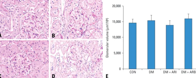
Glomerular volumes and glomerular matrix index Although the calculated glomerular volumes did not show significant differences among the experimental groups, glomerualr mesangial expansion was observed in the DM group compared with other groups in PAS staining of glomeruli (Fig. 1). Also, glomerular matrix index (GMI)
RESULTS
Table 1. Body Weights (g) and Blood Glucose Levels (mg/dL) at 32 Weeks
CON (n = 6) DM (n = 5) DM + ARI (n = 10) DM + ARB (n = 9) Body weights 700.28 ± 67.45 480.00 ± 75.07* 503.00 ± 59.18* 445.20 ± 63.83*
Blood glucose 95.6 ± 8.31 436.4 ± 59.67* 462.3 ± 49.98* 411.9 ± 77.95*
CON, control; DM, diabetes; ARI, aldose reductase inhibitor; ARB, angiotensin II receptor blocker.
The body weights of diabetic rats were significantly reduced compared to the control rats. Blood sugar was also markedly increased in diabetic rats compared to normal control rats, and ARI and ARB administration didn't have an effect on the reduction of blood sugar.
Data are mean ± SD.
*p < 0.05 compared with CON.
scores significantly decreased in the CON group (1.05 ± 0.05) and all medication treatment groups (DM + ARI; 1.088
± 0.153, DM + ARB; 1.075 ± 0.175, p < 0.001 vs. DM group) compared with the DM group (2.1 ± 1.122) (Fig. 2).
Renal VEGF expression
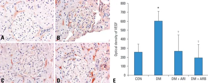
In the DM group, immunohistochemical stain for VEGF in the kidney showed darker brown pigmentation than other groups (Fig. 3). Compared with the CON group, the optical density of immunohistochemical staining for renal VEGF in the DM group significantly increased and restored in ARI and ARB treated rat groups (Fig. 3).
Renal VEGF mRNA expression
For quantification of renal VEGF mRNA expression, we examined real time RT-PCR. The VEGF/GAPDH ratio was 8.32 fold in the DM group, 1.52 fold in the DM + ARI group, and 1.35 fold in the DM + ARB group compared with the CON group. The ARI treatment decreased VEGF mRNA expression by 82% compared with the DM group.
Significant differences in VEGF mRNA expression bet- ween the ARI and ARB treated diabetic groups were not observed (Fig. 4).
Table 2. Changes of 24 Hour Urinary Albumin (mg/day) and ACR (mg/mmol)
Albumin ACR
8th 32nd 8th 32nd
CON 197.43 ± 218.20 579.72 ± 229.31 3.78 ± 7.48 11.14 ± 10.5
DM 170.43 ± 264.17 1782.94 ± 1495.75* 4.64 ± 4.34 84.85 ± 91.19*
DM+ARI 174.68 ± 181.71 626.27 ± 128.33� 9.96 ± 10.68 24.79 ± 11.12� DM+ARB 285.05 ± 375.59 330.19 ± 143.02� 6.61 ± 9.05 16.11 ± 9.95� CON, control; DM, diabetes; ARI, aldose reductase inhibitor; ARB, angiotensin II receptor blocker; ACR, albumin creatinine ratio.
Urine albumin excretion and ACR significantly increased in 32-week-old diabetic control rats compared to the eight-week-old rats. ARI and ARB treated diabetic rats showed that urine albumin excretion and ACR were significantly reduced compared to the diabetic control rats.
Data are presented as means ± SD.
*p < 0.05 compared with CON.
�p < 0.05 compared with DM.
Fig. 1. Representative examples of PAS staining of glomeruli are shown for (A) CON, (B) DM, (C) DM + ARI, and (D) DM + ARB. (E) is calculated glomerular volumes at 32nd week. The calculated glomerular volumes did not differ among the experimental groups. Scale bar, 100 µm. CON, control; DM, diabetes; ARI, aldose reductase inhibitor; ARB, angiotensin II receptor blocker.
Fig. 2. Comparison of glomerular matrix indices (GMI). Although calculated glomerular volumes did not differ among the experimental groups, the glomerular mesangium significantly expanded in untreated diabetic rats compared to the control rats, and it was ameliorated by ARI and ARB treatments. CON, control; DM, diabetes; ARI, aldose reductase inhibitor; ARB, angiotensin II receptor blocker. *p < 0.05 compared with CON, �p < 0.05 compared with DM.
A B
C D E
In this study, we found that aldose reductase inhibitor, fida- restat, could reduce the albumin excretion by ameliorating the VEGF expression in streptozotocin-induced diabetic rats as like angiotensin receptor blocker, losartan.
Diabetic nephropathy is the most serious complication of diabetes, and is the most common cause of end-stage renal disease. During the early phase of diabetic nephropathy, glomerular hyperplasia and thickening of glomerular base-
ment membranes with mesangial protein accumulation may appear.3These processes could lead to glomerular hyper- filtration and microalbuminuria initially, and eventually progress to renal failure and end stage renal disease.20 Many factors contributed to these processes such as advanced glycation end products, PKC activation, transforming growth factor-β, and other growth factors.2,11,21In addition, it has been demonstrated that AR activity increased in diabetic nephropathy.22 There is a positive relationship between the severity of glomerular structural changes and the immunoreactivity of AR.23 Donnelly, et al. reported that tolrestat could prevent glomerular hyperfiltration and extracelluar matrix accumulation in streptozotocin-induced diabetic rats.24 Furthermore, Isogai, et al.25 demonstrated that epalrestat could rescue glomerular basement mem- brane shrinkage and reduce urinary albumin excretion in a diabetic rat model.
In this study, the glomerular volume was not different among all experimental groups, but mesangial expansion was observed in the diabetic control group compared with the normal control and medication treated rat groups. This data could be explained by glomerular sclerosis because glomerular sclerosis might be derived from long term hyper- glycemic conditions. However, we could suggest that fida- restat could affect the renal structure by decreasing mesan- gial expansion.
AR activity may induce diabetic nephropathy by several mechanisms such as increased lipid peroxidation, depletion of major nonenzymatic antioxidants, and downregulation of superoxide dismutase activity.21,26-28The polyol pathway which is activated by hyperglycemic conditions may in- crease the production of superoxide - and nicotinamide adenine dinucleotide - induced reactive oxygen species (ROS), and could enhance VEGF mRNA expression.29
Fig. 4. Quantification of renal VEGF mRNA by real time RT-PCR. The VEGF / GAPDH ratio in the diabetic control group increased compared to the normal control rats. Treatment with ARI decreased VEGF mRNA expression. In addition, the ARB treated diabetic group decreased VEGF mRNA expression.
However, there were no significant differences in VEGF mRNA expression between the ARI and ARB treated diabetic groups. *p < 0.05 compared with CON, �p < 0.05 compared with DM. VEGF, vascular endothelial growth factor;
GAPDH, glyceraldehyde-3-phosphate dehydrogenase; mRNA, messenger RNA; RT-PCR, reverse transcriptase-polymerase chain reaction; CON, control;
DM, diabetes; ARI, aldose reductase inhibitor; ARB, angiotensin II receptor blocker.
Fig. 3. Immunohistochemical staining for VEGF from (A) CON, (B) DM, (C) DM + ARI, (D) DM + ARB. Scale bar, 100 µm. (E) is optical density of the glomerular VEGF. The optical density of immunohistochemical staining for VEGF in the diabetic control group increased significantly compared to the normal control group. Furthermore, the ARI and ARB treated diabetic groups had significantly lower expression of VEGF than the diabetic control group. *p < 0.05 compared with CON, �p < 0.05 compared with DM.
VEGF, vascular endothelial growth factor; CON, control; DM, diabetes; ARI, aldose reductase inhibitor; ARB, angiotensin II receptor blocker.
A B
C D E
DISCUSSION
Various data has shown that VEGF expression is in- creased in diabetic rat models and administration of anti- VEGF antibodies to diabetic rats could suppress urinary albumin excretion by reducing hyperpermeability, glome- rular basement membrane thickness, and mesangial expan- sion.12,13,30 There were many factors that could increase VEGF levels. It is known that various cytokines and growth factors including transforming growth factor-β, reactive oxygen species, and activation of rennin-angiotensin system could increase renal VEGF expression.1-4Previously, we demonstrated that diabetic nephropathy could be improved by treatment of theangiotensin receptor blocker by decrea- sing renal VEGF expression.14For these reasons, VEGF is thought to be an important factor in the development of dia- betic nephropathy.
In the present study, we showed that 24 hour urinary albumin creatinine ratio (ACR) and renal VEGF expression were significantly increased in diabetic rats and fidarestat treatment could improve the diabetic nephropathy concor- dance with decreasing renal VEGF expression similar to losartan treatment. Our data could suggest that fidarestat could improve diabetic nephropathy by reducing renal VEGF expression.
The first discovered aldose reductase inhibitor, tolrestat, was withdrawn because of the side effects of hepatic nec- rosis.31 In our experiment, the fidarestat feeding diabetic rats all survived. In spite of our data, a safety profile should be provided in future studies.
In this study, losartan was used as a positive control. It has been reported that VEGF expression could be inhibited by treatment with ARB.14,32Our data showed that the ability of VEGF reduction did not distinguish between fidarestat and losartan treated rat groups, indicating that fidarestat could affect the prevention of diabetic nephropathy.
In conclusion, our findings suggested that fidarestat treatment could improve the diabetic nephropathy by reduc- ing renal VEGF expression in type 1 diabetic rat model.
Further clinical studies should take place for the establish- ment of fidarestat treatment in diabetic nephropathy.
Fidarestat compound was kindly supported by Sanwa Kaga- ku Kenkyusho Co., Ltd, Japan.
1. Derubertis FR, Craven PA. Activation of protein kinase C in glomerular cells in diabetes. Mechanisms and potential links to the pathogenesis of diabetic glomerulopathy. Diabetes 1994;43:1-8.
2. Yamagishi S, Inagaki Y, Okamoto Y, Amano S, Koga K, Take- euchi M, et al. Advanced glycation end product-induced apop- tosis and overexpression of vascular endothelial growth factor and monocyte chemoattractant protein-1 in human-cultured mesangial cells. J Biol Chem 2002;227:20309-15.
3. Natarajan R, Bai W, Lanting L, Gonzales N, Nadler J. Effects of high glucose on vascular endothelial growth factor expression in vascular smooth muscle cells. Am J Physiol 1997;273:H2224-31.
4. Keogh RJ, Dunlop ME, Larkins RG. Effect of inhibition of aldose reductase on glucose flux, diacylglycerol formation, pro- tein kinase C, and phospholipase A2 activation. Metabolism 1997;46:41-7.
5. Kim NH, Jung HH, Cha DR, Choi DS. Expression of vascular endothelial growth factor in response to high glucose in rat me- sangial cells. J Endocrinol 2000;165:617-24.
6. Tsuchida K, Makita Z, Yamagishi S, Atsumi T, Miyoshi H, Obara S, et al. Suppression of transforming growth factor beta and vascular endothelial growth factor in diabetic nephropathy in rats by a novel advanced glycation end product inhibitor, OPB- 9195. Diabetologia 1999;42:579-88.
7. Lu M, Kuroki M, Amano S, Tolentino M, Keough K, Kim I, et al. Advanced glycation end products increase retinal vascular endothelial growth factor expression. J Clin Invest 1998;101:
1219-24.
8. Wang L, Kwak JH, Kim SI, He Y, Choi ME. Transforming growth factor-beta1 stimulates vascular endothelial growth factor 164 via mitogen-activated protein kinase kinase 3-p38alpha and p38delta mitogen-activated protein kinase-dependent pathway in murine mesangial cells. J Biol Chem 2004;279:33213-9.
9. Kuroki M, Voest EE, Amano S, Beerepoot LV, Takashima S, Tolentino M, et al. Reactive oxygen intermediates increase vas- cular endothelial growth factor expression in vitro and in vivo. J Clin Invest 1996;98:1667-75.
10. Ferrara N. Role of vascular endothelial growth factor in the regulation of angiogenesis. Kidney Int 1999;56:794-814.
11. Cha DR, Kim IS, Kang YS, Han SY, Han KH, Shin C, et al. Uri- nary concentration of transforming growth factor-beta-inducible gene-h3(beta ig-h3) in patients with Type 2 diabetes mellitus.
Diabet Med 2005;22:14-20.
12. Cooper ME, Vranes D, Youssef S, Stacker SA, Cox AJ, Rizkalla B, et al. Increased renal expression of vascular endothelial growth factor (VEGF) and its receptor VEGFR-2 in experimental dia- betes. Diabetes 1999;48:2229-39.
13. de Vriese AS, Tilton RG, Elger M, Stephan CC, Kriz W, Lameire NH. Antibodies against vascular endothelial growth factor improve early renal dysfunction in experimental diabetes. J Am Soc Nephrol 2001;12:993-1000.
14. Lee EY, Shim MS, Kim MJ, Hong SY, Shin YG, Chung CH.
Angiotensin II receptor blocker attenuates overexpression of vascular endothelial growth factor in diabetic podocytes. Exp Mol Med 2004;36:65-70.
15. Fonseca VA. Clinical diabetes: translating research into practice.
Philadelphia: Saunders Elsevier; 2006.
16. Tilton RG, Kawamura T, Chang KC, Ido Y, Bjercke RJ, Stephan CC, et al. Vascular dysfunction induced by elevated glucose levels in rats is mediated by vascular endothelial growth factor. J Clin Invest 1997;99:2192-202.
17. Obrosova IG, Minchenko AG, Vasupuram R, White L, Abatan OI, Kumagai AK, et al. Aldose reductase inhibitor fidarestat prevents retinal oxidative stress and vascular endothelial growth
REFERENCES
ACKNOWLEDGEMENTS
factor overexpression in streptozotocin-diabetic rats. Diabetes 2003;52:864-71.
18. Lane PH, Steffes MW, Mauer SM. Estimation of glomerular volume: a comparison of four methods. Kidney Int 1992;41:1085-9.
19. Saito T, Sumithran E, Glasgow EF, Atkins RC. The enhancement of aminonucleoside nephrosis by the co-administration of prota- mine. Kidney Int 1987;32:691-9.
20. Ziyadeh FN. The extracelluar matrix in diabetic nephropathy. Am J Kidney Dis 1993;22:736-44.
21. Craven PA, DeRubertis FR. Protein kinase C is activated in glo- meruli from streptozotocin diabetic rats. Possible mediation by glucose. J Clin Invest 1989;83:1667-75.
22. Ghahary A, Luo JM, Gong YW, Chakrabarti S, Sima AA, Mur- phy LJ. Increased renal aldose reductase activity, immunoreac- tivity, and mRNA in streptozocin-induced diabetic rats. Diabetes 1989;38:1067-71.
23. Raptis AE, Viberti G. Pathogenesis of diabetic nephropathy. Exp Clin Endocrinol Diabetes 2001;109 Suppl 2:S424-37.
24. Donnelly SM, Zhou XP, Huang JT, Whiteside CI. Prevention of early glomerulopathy with tolrestat in the streptozotocin-induced diabetic rat. Biochem Cell Biol 1996;74:355-62.
25. Isogai S, Inokuchi T, Ohe K. Effect of an aldose reductase inhibi- tor on glomerular basement membrane anionic sites in strepto-
zotocin-induced diabetic rats. Diabetes Res Clin Pract 1995;30:
111-6.
26. Obrosova IG. How dose glucose generate oxidative stress in peri- pheral nerve? Int Rev of Neurobiol 2002;50:3-35.
27. Lee AY, Chung SS. Contributions of polyol pathway to oxidative stress in diabetic cataract. FASEB J 1999;13:23-30.
28. Obrosova IG, Van Huysen C, Fathallah L, Cao XC, Greene DA, Stevens MJ. An aldose reductase inhibitor reverse early diabetes- induced changes in peripheral nerve function, metabolism, and antioxidative defense. FASEB J 2002;16:123-5.
29. Kuroki M, Voest EE, Amano S, Beerepoot LV, Takashima S, Tolentino M, et al. Reactive oxygen intermediates increase vas- cular endothelial growth factor expression in vitro and vivo. J Clin Invest 1996;98:1667-75.
30. Hoshi S, Shu Y, Yoshida F, Inagaki T, Sonoda J, Watanabe T, et al. Podocyte injury promotes progressive nephropathy in zucker diabetic fatty rats. Lab Invest 2002;82:25-35.
31. Foppiano M, Lombardo G. Worldwide pharmacovigilance sys- tem and tolrestat withdrawal. Lancet 1997;349:399-400.
32. Otani A, Takagi H, Suzuma K, Honda Y. Angiotensin II poten- tiates vascular endothelial growth factor-induced angiogenetic activity in retinal microcapillary endothelial cells. Circ Res 1998;
82:619-28.