• 검색 결과가 없습니다.

Paracrine influence of human perivascular cells on the proliferation of adenocarcinoma alveolar epithelial cells

N/A
N/A
Protected

Academic year: 2022

Share "Paracrine influence of human perivascular cells on the proliferation of adenocarcinoma alveolar epithelial cells"

Copied!
8
0
0

로드 중.... (전체 텍스트 보기)

전체 글

(1)

Author contributions: E.B.K., S.H.N., B.R.A., S.R.Y., J.Y.L. and W.J.K.

contributed to the study design, performed the statistical analyses, contributed to the interpretation of data, and drafted the manuscript;

K.W.H., W.S.P., C.M.L., and E.T.H. contributed to the acquisition of the data and to the critical review of the manuscript; S.J.L. and S.H.H. contributed to the study design, to the interpretation of the data, and to the critical review of the manuscript. All authors read and approved the final manuscript.

This is an Open Access article distributed under the terms of the Creative Commons Attribution Non-Commercial License, which permits unrestricted non-commercial use, distribution, and reproduction in any medium, provided the original work is properly cited.

Copyright © Korean J Physiol Pharmacol, pISSN 1226-4512, eISSN 2093-3827

INTRODUCTION

The tumor microenvironment (TME) is the noncancerous cellular region in which tumors exist [1]. The TME is composed of the extracellular matrix and various cell types, including endothelial cells (ECs), fibroblasts, macrophages, and perivascular cells (PVCs) [1,2]. These cells are involved in regulating tumor growth, invasion, angiogenesis, and metastasis, both through direct cell-to-cell contact and by a paracrine signaling mechanism [3]. Therefore, understanding the communication between the tumor and TME is critical to identifying therapeutic agent(s) for

the prevention of tumor growth and metastasis [1,3].

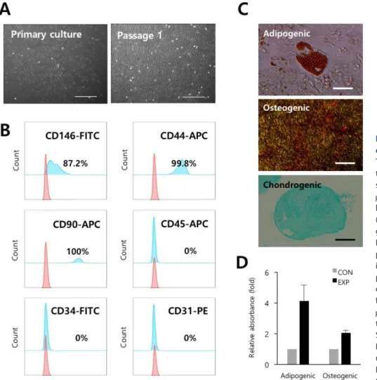
PVCs are enclosed within the basement membrane of blood vessels. Crisan et al. and others [4-6] have reported that PVCs are ancestors of multipotent mesenchymal stem cells (MSCs) in multiple tissues of both fetal and adult origin, and have greater regenerative potential compared to MSCs. The use of PVCs has increasingly gained attention as an alternative therapeutic candidate for the treatment of various diseases [4- 6]. In normal and developmental contexts, PVCs contribute to tissue homeostasis and repair by regulating vascular stability and contractility [3]. PVCs are also key components of the TME,

Original Article

Paracrine influence of human perivascular cells on the proliferation of adenocarcinoma alveolar epithelial cells

Eunbi Kim

1,#

, Sunghun Na

2,#

, Borim An

1

, Se-Ran Yang

3

, Woo Jin Kim

1

, Kwon-Soo Ha

4

, Eun-Taek Han

5

, Won Sun Park

6

, Chang-Min Lee

7

, Ji Yoon Lee

8

, Seung-Joon Lee

1,

*, and Seok-Ho Hong

1,

*

Departments of

1

Internal Medicine,

2

Obstetrics & Gynecology,

3

Thoracic and Cardiovascular Surgery,

4

Molecular and Cellular Biochemistry,

5

Medical Environmental Biology and Tropical Medicine,

6

Physiology, School of Medicine, Kangwon National University, Chuncheon 24341, Korea,

7

Department of Molecular Microbiology and Immunology, Department of Medicine, Alpert Medical School, Brown University, Providence, Rhode Island 02912, US,

8

Department of Biomedical Laboratory Science, College of Health Sciences, Sanji University, Wonju 26339, Korea

ARTICLE INFO

Received September 7, 2016 Revised December 20, 2016 Accepted January 9, 2017

*Correspondence

Seok-Ho Hong

E-mail: [email protected] Seung-Joon Lee

E-mail: [email protected] Key Words

Cancer Paracrine effect Perivascular cells Proliferation

#

These authors contributed equally to this work.

ABSTRACT Understanding the crosstalk mechanisms between perivascular cells

(PVCs) and cancer cells might be beneficial in preventing cancer development and

metastasis. In this study, we investigated the paracrine influence of PVCs derived

from human umbilical cords on the proliferation of lung adenocarcinoma epithelial

cells (A549) and erythroleukemia cells (TF-1a and K562) in vitro using Transwell

co-culture systems. PVCs promoted the proliferation of A549 cells without inducing

morphological changes, but had no effect on the proliferation of TF-1a and K562

cells. To identify the factors secreted from PVCs, conditioned media harvested from

PVC cultures were analyzed by antibody arrays. We identified a set of cytokines,

including persephin (PSPN), a neurotrophic factor, and a key regulator of oral

squamous cell carcinoma progression. Supplementation with PSPN significantly

increased the proliferation of A549 cells. These results suggested that PVCs produced

a differential effect on the proliferation of cancer cells in a cell-type dependent

manner. Further, secretome analyses of PVCs and the elucidation of the molecular

mechanisms could facilitate the discovery of therapeutic target(s) for lung cancer.

(2)

and are therefore able to induce the onset of angiogenesis and apoptosis through crosstalk with ECs in pathological conditions such as cancer and hyperglycemia [1].

For example, in tumors, angiopoietin-2 (ANG2) is primarily secreted from activated ECs to destabilize blood vessels by inducing PVC detachment and EC sprouting via the antagonizing factor ANG1 [7-9]. In ECs exposed to high glucose, miR-503 transcriptionally activated and modulated p75NTR, affecting the migration and proliferation of PVCs via the reduction of ephrin-B (EFNB2) and vascular endothelial growth factor A (VEGFA) expression [10]. Therefore, the modulation of PVC-EC interactions could provide a therapeutic strategy to inhibit cancer growth and/or manage diabetes complications.

Although the underlying molecular mechanisms of the angiogenic contribution of PVCs are relatively well defined, the effects and molecular mechanisms of PVCs on the proliferation of cancer cells are still unclear. Paracrine signaling pathways between PVCs and cancer cells have been suggested as an important mechanism in tumor growth and metastasis [1].

Therefore, the identification of key secretory factors that regulate the proliferation of cancer could provide valuable insight into the prevention and development of tumors. In the present study, we isolated PVCs from human umbilical cords (HUCs) and investigated their paracrine influences on the proliferation of human lung adenocarcinoma (A549) and erythroleukemia cell (TF-1a and K562) lines in vitro using Transwell

co- culture systems. PVCs promoted the proliferation of lung adenocarcinoma cells, but not erythroleukemia cells, which was mediated by the release of soluble factors from the PVCs.

METHODS

Cell isolation and cultures

HUC tissues were obtained from full term births after Caesarian section with informed consent using the guidelines approved by IRB (IRB approval number: KNUH-2012-11-003- 008) at the Kangwon National University Hospital. PVCs were isolated and cultured as previously described [11]. A549, K-562, and TF-1a cell lines were purchased from American Type Culture Collection (ATCC, Manassas, VA, USA). A549 cells were grown in Dulbecco's Modified Eagle Medium (DMEM; Hyclone, Logan, UT, USA) supplemented with 10% fetal bovine serum (FBS; Hyclone) and 1% penicillin/streptomycin (Sigma-Aldrich, St. Louis, MO, USA). K562 cells were grown in Iscove's Modified Dulbecco's Media (IMDM; Hyclone) supplemented with 10%

FBS and 1% penicillin/streptomycin. TF-1a cells were cultured in RPMI (Hyclone) supplemented with 10% FBS and 1% penicillin/

streptomycin. The cells were cultured at 37

o

C and 5% CO

2

and subcultured at 80~90% confluency.

Flow cytometry

The phenotypes of PVCs were analyzed by flow cytometry as previously described [12]. Briefly, single cell suspensions (passage 2) were incubated with CD31-phycoerythrin, CD34-fluorescein isothiocyanate (FITC), CD45-allophycoerythrin (APC), CD44- APC, CD90-APC, and CD146-FITC for 60 minutes at 4

o

C in the dark. The cells were rinsed with 1% FBS-phosphate- buffered saline (PBS) and analyzed on a BD FACSCanto

TM

II flow cytometer (BD Biosciences, San Jose, CA, USA). Dead cells were excluded by staining with 7AAD viability staining solution (BD Biosciences). Acquired data were analyzed using FlowJo software (Tree Star, Inc., Ashland, OR, USA). All antibodies were purchased from BD Biosciences.

Osteogenic and adipogenic differentiation

The multilineage differentiation potentials of the PVCs were evaluated as previously described [13]. Briefly, PVCs from passage 3 were seeded at 4×10

4

cells/well in expansion medium.

When the cells reached 80% confluency, they were treated with osteogenic or adipogenic induction medium (StemPro

, ThermoFisher Scientific, San Jose, CA, USA) for 21 days at which time the medium was changed every 3 days. PVC cultures were stained with Alizarin Red S (CM-0058; Lifeline Cell Technology, Carlsbad, CA, USA) or Oil Red O (CM-0055; Lifeline Cell Technology) and the dye contents were quantified using a spectrophotometer.

Transwell

co-cultures

Transwell

plates (Corning, Corning, NY, USA) with 0.4 mm pore polycarbonate membrane inserts were used for the co-culture experiments. A549 cells (7×10

4

) were co-cultured for 48 hrs with PVCs at a ratio of 1:1 and 1:3 (A549:PVCs). TF- 1a and K562 cells (2×10

5

and 3×10

5

, respectively) were also co- cultured for 48 hrs with PVCs at a ratio of 1:1 and 1:3 (TF-1a or K562:PVCs, respectively). PVCs were seeded into the lower chamber, and cancer cells were placed in the upper chamber. Cell number and size were measured using the MOXI Z automated cell counter kit (ORFLO Technologies, Carlsbad, CA, USA).

Antibody arrays

Antibody arrays (Human L507 array kit, AAH-BLG-1; Ray-

Biotech, Norcross, GA, USA) were used to profile the secretions

from PVCs. When PVCs reached 90% confluency, the cells were

cultured in defined serum-free medium for 24 hrs to condition

the growth medium. The resulting conditioned medium (CM)

was harvested, centrifuged, and filtered using 0.2 mm filters

to remove any cell debris. The CM was concentrated using an

Amicon Ultra-0.5 centrifugal filter unit with a cutoff of 3 kDa

(3)

(EMD Millipore, Hayward, CA, USA), and the concentrated CM was stored at –80

o

C. Non-CM was used as a control. The concentrated medium was used for the antibody array analyses according to the manufacturer’s instructions. Secretion factors that were 1.5-fold or greater relative to the control were scored as significant.

Persephin (PSPN) treatment and inhibition of PSPN

To determine the effect of PSPN on the proliferation of A549 cells, A549 cells were seeded at a density of 2×10

5

cells into 6-well culture plates and cultured in the presence of PSPN (10, 20, and 40 ng/ml) for 48 hrs. In experiment blocking PSPN signals, anti- PSPN antibody (MyBioSource, San Diego, CA, USA) at a final concentration of 500 ng per mL was added in A549 cultures.

Cell number and morphology were evaluated using the MOXI Z automated cell counter (ORFLOW Technologies).

Real-time quantitative PCR

Total RNA from cancer cell lines was extracted with the

RNeasy Mini Kit (Qiagen Inc., Valencia, CA) and converted to first strand cDNA with TOPscript

TM

RT DryMIX kit (Enzynomix). The cDNA of GFRa4 mRNA was amplified and quantified using AccuPower

PCR PreMix reagents (Bioneer Corp). Primer sequence was as follow: GFRa4, for wa rd 5’-AGGAGGCATCT TGGT TGTA A-3’ a nd reverse 5’-GGGTCTCCAGAGAGGTCTCA-3’, GAPDH, forward 5’-TGCACCACCAACTGCTTAGC-3’ and reverse 5’-GGCATGGACTGTGGTCATGAG-3’. The expression level of GFRa4 was normalized to GAPDH and data analysis was performed by the comparative CT method.

Statistical analyses

Data are presented as the mean±SD. Statistically significant differences were determined by the Student’s t-test. Significance levels were set at p<0.05.

Fig. 1. Isolation and characterization of PVCs derived from HUCs. (A) The morphology of the PVCs in cul- ture. Fibroblast-like colonies were subcultured and expanded. Bars=100 mm. (B) Representative flow cytometry histograms for phenotypes of PVCs (passage 2) are shown. The pink histo- gram indicates the isotype control. (C) Evaluation of multilineage differentiation potentials of the PVCs. Representative images of Oil Red O staining for adi- pocytes, Alizarin Red S staining for osteocytes, and Alcian Blue staining for chondrocytes are shown. Bars=100 mm, or 500 mm. (D) Quantification of the dye (Oil Red O and Alizarin Red S) content using spectrophotometry.

PVCs, perivascular cells; HUCs, human umbilical cords; CON, control cultures;

EXP, experimental cultures with differen-

tiation conditions.

(4)

RESULTS

Phenotypic and functional identification of human PVCs derived from human UCs

PVCs were isolated and cultured from vessels of umbilical cords by a non-enzymatic isolation method [11]. Isolated PVCs displayed spindle-shaped, fibroblast-like morphology as seen in MSCs (Fig. 1A). PVCs were phenotypically identified as strongly positive for the expression of CD44 (99.8%, MSC marker), CD90 (100%, MSC marker), and CD146 (87.2%, PVC marker) and as negative for the CD34 (hematopoietic progenitor marker), CD31 (endothelial cell marker), and CD45 (mature hematopoietic cell marker) (Fig. 1B). In addition, we confirmed that the PVCs had multilineage differentiation potentials, including for adipocytes, osteocytes, and chondrocytes (Fig. 1C and 1D). These results indicated that the isolated and expanded cells consisted of PVCs.

PVCs significantly increased the proliferation of human lung adenocarcinoma cells

To determine the effects of PVCs on the proliferation of cancer cells, PVCs were indirectly co-cultured with human lung

adenocarcinoma cells (A549) and erythroleukemia cells (TF-1a and K562) using Transwell

co-culture systems. We assumed that PVCs can differentially regulate the growth of adherent (A549) and non-adherent (TF-1a and K562) cancer cells. Cancer cell lines were co-cultured with PVCs at a ratio of 1:1 and 1:3 for 48 hrs. The proliferation of all cancer cells was not affected by co-culture at a ratio of 1:1. The proliferation of TF-1a and K562 cells was also not affected by co-culture at a ratio of 1:3. However, the number of A549 cells increased approximately two-fold after indirect co-culturing with PVCs at a ratio of 1:3 (Fig. 2A). Cell morphology and diameter of all cancer cell lines were not altered by co-culturing with PVCs at both ratios (Fig. 2B and 2C). These findings suggested that PVCs had differential effects on the proliferation of cancer cells in a cell-type dependent manner, and they promoted the proliferation of human lung adenocarcinoma cells via paracrine actions.

Paracrine action of PSPN on the proliferation of A549 cells

To identify the secreted factors, the PVC-CM and non-CM were analyzed using 507 antibody array chips for secreted soluble factors. Secreted factors that had 1.5-fold or more expression

Fig. 2. PVCs significantly increase the proliferation of human lung adenocarcinoma cells. (A) Indirect co-culturing of PVCs with human

erythroleukemia cell lines (TF-1a and K562) and lung adenocarcinoma cell line (A549). The error bars indicate the standard deviation. **p<0.01. (B)

The morphology of the cancer cells co-cultured with PVCs for 48 hrs. Bars=50 mm. (C) The diameter of the cancer cells was measured after co-culturing

with PVCs for 48 hrs. The error bars indicate the standard deviation. CON, control group; PVCs, perivascular cells.

(5)

relative to the non-CM controls were considered as significantly abundant. We found that 14 cytokines and growth factors were significantly abundant in the PVC-CM compared with the non- CM controls (Fig. 3A). Some cellular functions of these factors are shown in Table 1. Some cytokines and growth factors, such as IGFBP-7, THBS1, PSPN, INHBA, WIF-1, FGF20, and HB-EGF play important roles in regulating tumor growth and metastasis [14-20]. Of these identified factors, we further investigated the effects of PSPN on the proliferation of A549 cells because its effects on tumorigenesis are less understood. A549 cells were treated with different concentrations of PSPN (10, 20, and 40

ng/ml) for 48 hrs. Supplementation with 20 ng/ml or 40 ng/ml of PSPN significantly promoted the proliferation of A549 cells without inducing morphological changes (Fig. 3B and 3C). These results indicated that the growth of lung adenocarcinoma cells was influenced by PVCs and by PSPN via paracrine signaling. To determine if the paracrine effect of PSPN on A549 proliferation is mediated via its receptor signaling pathway, we compared expression level of GFRa4 in A549, TF-1a and K562 cell cultures.

We found that GFRa4 transcripts was significantly more abundant in A549 cells than TF-1a and K562 cells (Fig. 4A). We also confirmed the suppressive effect of a neutralizing anti-PSPN

Fig. 3. PSPN contributes to the pro- liferation of human lung adenocar- cinoma cells. (A) Secretome analyses of PVC-CM using antibody arrays. A list of selected cytokines that had 1.5 fold or more expression relative to the control sample (non-CM) is shown. (B) A549 cells were plated at a density of 2×10

5

cells and treated with a variety of concentrations of PSPN (10, 20, and 40 ng/ml) for 48 hrs. The error bars indicate standard deviation. *p<0.05, **p<0.01.

(C) Representative images of A549 cells cultured in the presence and absence of PSPN for 48 hrs. Bars=50 mm. CON, control group; PSPN, Persephin; PCV-CM, perivascular cell-conditioned medium;

non-CM, non-conditioned control me- dium.

Table 1. Cellular functions for the selected secreted cytokines

Symbol Name Cellular functions Reference

IGFBP-7 Insulin-like growth factor

binding protein-7 Cell adhesion, tumorigenesis, and angiogenesis [14]

THBS1 Thrombospondin-1 Platelet aggregation, angiogenesis, and tumorigenesis [15]

MIP-2 Macrophage inflammation protein 2 Chemotactic activity for inflammatory and immune effector cells [30]

PSPN Persephin Cell proliferation and tumorigenesis [24]

[16]

Tarc Thymus and Activation-Regulated

Chemokine (CCL17) Chemotaxis in T cells [31]

INHBA Activin A Reproduction, inflammation, and tumorigenesis [17]

GPC5 Glypican 5 Control cell division and growth regulation [32]

WIF-1 Wnt Inhibiting Factor-1 Tumor growth and induction of apoptosis [18]

Progranulin Pro-epitheliun and prostate cancer (PC)

cell-derived growth factor Cell growth, wound healing, and anti-inflammation [33]

FGF20 Fibroblast growth factor 20 Cell growth, morphogenesis, and tumor growth [19]

DCN Decorin Angiogenesis and cell cycle regulation [34]

[35]

IL-21 Interleukin-21 Regulation of immune cells and cell proliferation [36]

IL-7 Interleukin-7 Development and homeostasis of immune cells [37]

HB-EGF Heparin-binding EGF-like growth factor Regulation of mitotic activity and cancer metastasis [20]

(6)

antibody on the proliferation of A549 cells (Fig. 4B), suggesting that PSPN promotes proliferation of human adenocarcinoma cells by its receptor-mediated pathway.

DISCUSSION

In the present study, we performed secretome analyses of PVC-CM and identified a set of secreted cytokines known to be important in regulating the cell cycle, tumorigenesis, angiogenesis, and inflammation. The most abundant cytokine in PVC-CM compared with non-CM was IGFBP-7, which is linked to the suppression of growth and metastasis of tumors.

For example, the activation of the IGFBP-7 gene suppresses the invasion of head and neck squamous cell carcinoma and acute myeloid leukemia (AML) cells [21,22]. These studies suggest that IGFBP-7 could be a prognostic factor for the probability of invasion as well as a therapeutic target for the improvement of AML patient survival. THBS1 and PSPN are also highly detected in the PVC-CM. THBS1, a glycoprotein, is implicated in the regulation of tumor growth and metastasis [15,23]. PSPN was first identified as a glial cell line-derived neurotrophic factor (GDNF) that had neurotrophic effects on some neuronal cell types. For example, PSPN enhanced the morphological differentiation and phenotypic induction of dopaminergic neurons and could be considered as a cell therapy for Parkinson’s disease [24-27].

However, very little is known about the role of PSPN in tumor progression. To the best of our knowledge, only one study has explored the effects of PSPN on the proliferation of cancer.

Baba et al. reported that PSPN was a key factor in proliferation of human oral cancer through activation of RET-mitogen- activated protein kinase [16]. Our study showed that, while supplementation of PSPN promoted the proliferation of human lung adenocarcinoma cells, it did not affect the proliferation of erythroleukemia cells, because PVC co-culturing did not change the growth of TF-1a and K562 cells. These results suggested a differential effect of PVCs on the proliferation of cancer cells in a

cell-type dependent manner. Thus, an investigation of PSPN in a broader range of tumor cell types should be done.

Although multipotent MSCs for use in clinical trials have been shown to be safer and more effective than pluripotent stem cells, their roles in cancer development remain controversial.

This could be attributed to the different MSC sources, variations in donors, and the injection timing and dosage of MSCs [28].

Because PVCs have been proposed as common ancestors of MSCs, they could provide an alternative source due to their greater regeneration potential compared with other MSCs [4- 6]. Thus, the role of PVCs in cancer development should be addressed prior to clinical applications, especially for patients with cancer who are undergoing or have undergone treatment.

Sinha et al. reported that co-culturing with PVCs increased the proliferation of ovarian cancer cells in vitro [29]. Similarly, our results also showed that the proliferation of lung adenocarcinoma cells was increased by co-culturing with PVCs, and this effect was mediated by secreted factors from the PVCs. Thus, identification of secreted factors and their functional evaluation related with tumor growth could be critical to the discovery of novel anticancer therapy.

In conclusion, PVCs significantly contributed to lung cancer growth, and this could have been by paracrine signaling.

Further study is required using in vivo animal models and direct injections of PVC-CM or selected paracrine factors to define the role of PVCs in lung cancer growth and metastasis.

ACKNOWLEDGEMENTS

This study was supported by the Basic Science Research Program through the National Research Foundation of Korea (NRF) funded by the Ministry of Education, Science and Technology (2014-R1A1A2055021), the Ministry of Science, ICT

& Future Planning (2015R1A4A1038666) and Kangwon National University.

Fig. 4. Neutralization of PSPN in- hibits proliferation of human lung adenocarcinoma cells. (A) qPCR analysis of relative GFRa4 expression in A549, TF- 1a and K562 cells. (B) PSPN neutralizing antibody (500 ng/mL) suppressed the increased proliferation of A549 cells by PSPN (40 ng/mL) treatment. The error bars indicate the standard deviation.

**p<0.01, a, p<0.01. (control vs PSPN), b,

p<0.01. (PSPN vs PSPN+Ab).

(7)

CONFLICTS OF INTEREST

The authors declare no conflicts of interest.

REFERENCES

1. Ribeiro AL, Okamoto OK. Combined effects of pericytes in the tumor microenvironment. Stem Cells Int. 2015. doi: 10.1155/2015/

868475.

2. Hall AP. Review of the pericyte during angiogenesis and its role in cancer and diabetic retinopathy. Toxicol Pathol. 2006;34:763-775.

3. Hay E. Unusual presentation of pheochromocytoma. Am J Emerg Med. 1991;9:399-402.

4. Crisan M, Yap S, Casteilla L, Chen CW, Corselli M, Park TS, Andriolo G, Sun B, Zheng B, Zhang L, Norotte C, Teng PN, Traas J, Schugar R, Deasy BM, Badylak S, Buhring HJ, Giacobino JP, Lazzari L, Huard J, Péault B. A perivascular origin for mesenchymal stem cells in multiple human organs. Cell Stem Cell. 2008;3:301-313.

5. Baksh D, Yao R, Tuan RS. Comparison of proliferative and multilineage differentiation potential of human mesenchymal stem cells derived from umbilical cord and bone marrow. Stem Cells.

2007;25:1384-1392.

6. Zebardast N, Lickorish D, Davies JE. Human umbilical cord peri- vascular cells (HUCPVC): A mesenchymal cell source for dermal wound healing. Organogenesis. 2010;6:197-203.

7. Augustin HG, Koh GY, Thurston G, Alitalo K. Control of vascular morphogenesis and homeostasis through the angiopoietin-Tie system. Nat Rev Mol Cell Biol. 2009;10:165-177.

8. Scharpfenecker M, Fiedler U, Reiss Y, Augustin HG. The Tie-2 ligand angiopoietin-2 destabilizes quiescent endothelium through an internal autocrine loop mechanism. J Cell Sci. 2005;118:771-780.

9. Thomas M, Augustin HG. The role of the Angiopoietins in vascular morphogenesis. Angiogenesis. 2009;12:125-137.

10. Caporali A, Meloni M, Nailor A, Mitić T, Shantikumar S, Riu F, Sala-Newby GB, Rose L, Besnier M, Katare R, Voellenkle C, Verkade P, Martelli F, Madeddu P, Emanueli C. p75(NTR)-dependent activation of NF-κB regulates microRNA-503 transcription and pericyte-endothelial crosstalk in diabetes after limb ischaemia. Nat Commun. 2015;6:8024.

11. An B, Na S, Lee S, Kim WJ, Yang SR, Woo HM, Kook S, Hong Y, Song H, Hong SH. Non-enzymatic isolation followed by supple- mentation of basic fibroblast growth factor improves proliferation, clonogenic capacity and SSEA-4 expression of perivascular cells from human umbilical cord. Cell Tissue Res. 2015;359:767-777.

12. Hong SH, Maghen L, Kenigsberg S, Teichert AM, Rammeloo AW, Shlush E, Szaraz P, Pereira S, Lulat A, Xiao R, Yie SM, Gauthier- Fisher A, Librach CL. Ontogeny of human umbilical cord perivascular cells: molecular and fate potential changes during gestation. Stem Cells Dev. 2013;22:2425-2439.

13. An B, Heo HR, Lee S, Park JA, Kim KS, Yang J, Hong SH. Supple- mentation of growth differentiation factor-5 increases proliferation and size of chondrogenic pellets of human umbilical cord-derived perivascular stem cells. Tissue Eng Regen Med. 2015;12:181-187.

14. Kashyap MK. Role of insulin-like growth factor-binding proteins in the pathophysiology and tumorigenesis of gastroesophageal cancers.

Tumour Biol. 2015;36:8247-8257.

15. Sid B, Sartelet H, Bellon G, El Btaouri H, Rath G, Delorme N, Ha ye B, Martiny L. Thrombospondin 1: a multifunctional protein impli- cated in the regulation of tumor growth. Crit Rev Oncol Hematol.

2004;49:245-258.

16. Baba T, Sakamoto Y, Kasamatsu A, Minakawa Y, Yokota S, Higo M, Yokoe H, Ogawara K, Shiiba M, Tanzawa H, Uzawa K. Persephin: A potential key component in human oral cancer progression through the RET receptor tyrosine kinase-mitogen-activated protein kinase signaling pathway. Mol Carcinog. 2015;54:608-617.

17. Wijayarathna R, de Kretser DM. Activins in reproductive biology and beyond. Hum Reprod Update. 2016;22.

18. Huang Y, Du Q, Wu W, She F, Chen Y. Rescued expression of WIF-1 in gallbladder cancer inhibits tumor growth and induces tumor cell apoptosis with altered expression of proteins. Mol Med Rep. 2016;

14:2573-2581.

19. Koga C, Adati N, Nakata K, Mikoshiba K, Furuhata Y, Sato S, Tei H, Sakaki Y, Kurokawa T, Shiokawa K, Yokoyama KK.

Characterization of a novel member of the FGF family, XFGF-20, in Xenopus laevis. Biochem Biophys Res Commun. 1999;261:756-765.

20. Raab G, Klagsbrun M. Heparin-binding EGF-like growth factor.

Biochim Biophys Acta. 1997;1333:F179-199.

21. Chen LH, Liu DW, Chang JL, Chen PR, Hsu LP, Lin HY, Chou YF, Lee CF, Yang MC, Wen YH, Hsu WL, Weng CF. Methylation status of insulin-like growth factor-binding protein 7 concurs with the malignance of oral tongue cancer. J Exp Clin Cancer Res. 2015;34:20.

22. Verhagen HJ, de Leeuw DC, Roemer MG, Denkers F, Pouwels W, Rutten A, Celie PH, Ossenkoppele GJ, Schuurhuis GJ, Smit L.

IGFBP7 induces apoptosis of acute myeloid leukemia cells and synergizes with chemotherapy in suppression of leukemia cell survival. Cell Death Dis. 2014;5:e1300.

23. Teraoku H, Morine Y, Ikemoto T, Saito Y, Yamada S, Yoshikawa M, Takasu C, Higashijima J, Imura S, Shimada M. Role of thrombo- spondin-1 expression in colorectal liver metastasis and its molecular mechanism. J Hepatobiliary Pancreat Sci. 2016;23:565-573.

24. Milbrandt J, de Sauvage FJ, Fahrner TJ, Baloh RH, Leitner ML, Tansey MG, Lampe PA, Heuckeroth RO, Kotzbauer PT, Simburger KS, Golden JP, Davies JA, Vejsada R, Kato AC, Hynes M, Sherman D, Nishimura M, Wang LC, Vandlen R, Moffat B, Klein RD, Poulsen K, Gray C, Garces A, Johnson EM Jr. Persephin, a novel neurotrophic factor related to GDNF and neurturin. Neuron. 1998;20:245-253.

25. Zihlmann KB, Ducray AD, Schaller B, Huber AW, Krebs SH, Andres RH, Seiler RW, Meyer M, Widmer HR. The GDNF family members neurturin, artemin and persephin promote the mor- phological differentiation of cultured ventral mesencephalic dopa- minergic neurons. Brain Res Bull. 2005;68:42-53.

26. Roussa E, Oehlke O, Rahhal B, Heermann S, Heidrich S, Wiehle M, Krieglstein K. Transforming growth factor beta cooperates with persephin for dopaminergic phenotype induction. Stem Cells.

2008;26:1683-1694.

27. Murata T, Tsuboi M, Koide N, Hikita K, Kohno S, Kaneda N.

Neuronal differentiation elicited by glial cell line-derived neurotro- phic factor and ciliary neurotrophic factor in adrenal chromaffin cell line tsAM5D immortalized with temperature-sensitive SV40 T-antigen. J Neurosci Res. 2008;86:1694-1710.

28. Yagi H, Kitagawa Y. The role of mesenchymal stem cells in cancer

development. Front Genet. 2013;4:261.

(8)

29. Sinha D, Chong L, George J, Schlüter H, Mönchgesang S, Mills S, Li J, Parish C, Bowtell D, Kaur P; Australian Ovarian Cancer Study Group. Pericytes promote malignant ovarian cancer progression in mice and predict poor prognosis in serous ovarian cancer patients.

Clin Cancer Res. 2016;22:1813-1824.

30. Driscoll KE. Macrophage inflammatory proteins: biology and role in pulmonary inflammation. Exp Lung Res. 1994;20:473-490.

31. Imai T, Baba M, Nishimura M, Kakizaki M, Takagi S, Yoshie O. The T cell-directed CC chemokine TARC is a highly specific biological ligand for CC chemokine receptor 4. J Biol Chem. 1997;272:15036- 15042.

32. Li F, Shi W, Capurro M, Filmus J. Glypican-5 stimulates rhabdomy- osarcoma cell proliferation by activating Hedgehog signaling. J Cell Biol. 2011;192:691-704.

33. Nicoletto BB, Canani LH. The role of progranulin in diabetes and kidney disease. Diabetol Metab Syndr. 2015;7:117.

34. Järveläinen H, Sainio A, Wight TN. Pivotal role for decorin in

angiogenesis. Matrix Biol. 2015;43:15-26.

35. Buraschi S, Neill T, Goyal A, Poluzzi C, Smythies J, Owens RT, Schaefer L, Torres A, Iozzo RV. Decorin causes autophagy in endothelial cells via Peg3. Proc Natl Acad Sci U S A. 2013;110:E2582-2591.

36. Parrish-Novak J, Dillon SR, Nelson A, Hammond A, Sprecher C, Gross JA, Johnston J, Madden K, Xu W, West J, Schrader S, Burkhead S, Heipel M, Brandt C, Kuijper JL, Kramer J, Conklin D, Presnell SR, Berry J, Shiota F, Bort S, Hambly K, Mudri S, Clegg C, Moore M, Grant FJ, Lofton-Day C, Gilbert T, Rayond F, Ching A, Yao L, Smith D, Webster P, Whitmore T, Maurer M, Kaushansky K, Holly RD, Foster D. Interleukin 21 and its receptor are involved in NK cell expansion and regulation of lymphocyte function. Nature.

2000;408:57-63.

37. Muegge K, Vila MP, Durum SK. Interleukin-7: a cofactor for V(D)J

rearrangement of the T cell receptor beta gene. Science. 1993;261:93-

95.

수치

Fig. 1. Isolation and characterization  of PVCs derived from HUCs. (A)  The morphology of the PVCs in  cul-ture
Fig. 2. PVCs significantly increase the proliferation of human lung adenocarcinoma cells
Table 1. Cellular functions for the selected secreted cytokines
Fig. 4. Neutralization of PSPN in- in-hibits proliferation of human lung  adenocarcinoma cells

참조

관련 문서

In this study, we have fabricated the 660 nm red LED illumination controller and LED array module and evaluated the proliferation and differentiation

In this Thesis, an empirical study on the Influence of Consumer's Preferences and Satisfaction on Purchase Intention of Korean Automobile in Vietnamese Consumers

In this study, we investigated the effects of low-power CO 2 laser on proliferation on human gingival fibroblast cells so that determine laser

Determining optimal surface roughness of TiO₂blasted titanium implant material for attachment, proliferation and differentiation of cells derived from

The major contributions of this study are identifying the influence on the performance of venture business by means of structural equation model and the

 Often found connected to other molecules on the outsides of cells --- cellular recognition, cell signaling, cell

• Generation of different specialized kinds of cells from zygote (fertilized egg) or other precursor cells.. – Generate blood cells, muscle

4 &gt; Effects of Taro on COX-2 expression and iNOS expression(hot water) in human thyroid cancer cells. The cells were pretreated for 48hours with either