This is an Open Access article distributed under the terms of the Creative Commons Attribution Non-Commercial License (http://creativecommons.org/
licenses/by-nc/3.0) which permits unrestricted non-commercial use, distribution, and reproduction in any medium, provided the original work is properly cited.
Lactobacillus plantarum
L67의 항알러지 활성신미영1․송수연1․고종호2․김광현․오세종1,*
1전남대학교 동물자원학부, 2한국폴리텍특성화대학
Suppressive Effect of
Lactobacillus plantarum
L67 on Allergic Disease Risk FactorsMiyoung Shin1, Sooyeon Song1, Jongho Koh, Kwang-Hyun Kim1 and Sejong Oh1,*
1Division of Animal Science, Chonnam National University, Gwangju 61186, Korea
2Department of Bio-Food Processing & Analysis, Bio-Campus, Korea Polytechnic College, Nonsan-si 32943, Korea
Abstract
In this study, we found that the glycoprotein (18 kDa) isolated from Lactobacillus plantarum L67 (L67 protein) inhibits allergy-related inflammatory mediators in BPA treated rat basophilic leukemia 2H3 (RBL-2H3) cells and primary cultured splenocy- tes. First, we measured the levels of ERK, p38, and AP-1 by Western blotting and we evaluated the expression of IL-1β, IL-6, and IL-10 cytokines by real-time quan- titative PCR. Finally, we evaluated the applicability of L. plantarum L67 as a yogurt starter culture by measuring pH, enumeration of bacteria, and sensory scores. The results of this study shows that L67 protein inhibits the phosphorylation of ERK and p38 MAPK, as well as the activation of AP-1 (c-Fos and c-Jun). With inflammation- related factors (Th2-related cytokines), L67 protein inhibits BPA-induced IL-1β, IL-6, and IL-10 activity. Bisphenol A stimulates MAPK signaling, which induces increased GATA-3 expression, and GATA-3 induces the differentiation of Th2 cells and secretion of Th2-related cytokines. Our findings showed that L67 protein may regulate the activation of GATA3 and T-bet through the induction of MAPK signal- ing, and it relieves the Th1/Th2 cytokine profile imbalance. Taken together, these findings demonstrate that the L. plantarum L67 strain is a potential food supplement and starter culture, that could help in the prevention of Th2-related immune disorders.
Keywords
allergy, bisphenol A, Lactobacillus plantarum L67, probiotics Received: Dec 12, 2016
Revised: Dec 16, 2016 Accepted: Dec 22, 2016
* Corresponding author : Sejong Oh,
Division of Anima; Science, Chonnam National University, 62286, Republic of Korea.
Tel: +82-62-530-2116 Fax: +82-62-530-2129 E-mail: [email protected]
서 론
프로바이오틱스는 인간 건강에 유익한 효과가 있는 살아있는 미생물로서 숙주에게 유익한 장내 미생물 들의 균형을 향상시킬 수 있다(Sornplang and Piyadeatsoontorn, 2016). 특히 Lactobacillus
plantarum은 항암작용, 항응고 활성, 항바이러스 작용, 라디칼 소 거활성을 나타내는 항산화 작용을 하는 것으로 알려져 있다 (Andersson et al., 2010; Li et al., 2012; Giardina et al., 2014; Kassayova et al., 2014). 또한 일부 lactobacilli는 면역 시스템을 조절하여 1형 알러지 질환을 억제하는데(Segawa et al., 2008; Hong et al., 2014; Konieczna et al., 2015), 이러한 연 구결과는 특정 프로바이오틱스가 면역 조절을 통해 알러지 반응을 억제시킬 수 있음을 나타낸다. 특정 Lactobacillus 속 유산균들은 미성숙 수지상 세포를 조절하고, T-cell의 기능을 조절하며, 장내 병원성 균의 부착을 억제하는 것과 같은 유익한 효과를 갖는 것으 로 알려져 있는데 주로 이들 유산균이 생산하는 당단백질이 이러한 기능이 있는 것으로 보고되었다(Rhee and Park, 2001; John- son-Henry et al., 2007; Konstantinov et al., 2008; Song et al., 2015). Song 등(2016a, 2016b)은 L. plantarum L67로부터 분리된 당단백질(18 kDa)에서 항염증 및 항알러지 효과가 있음을 밝혔으며, 발효유제품을 포함한 식품에 적용이 가능한 것으로 보고 하였다.
Bisphenol A (BPA)는 에스트로겐성 내분비 교란 물질로서 den- tal, 식품 포장재, 식품 캔 및 수도관의 광택제에 광범위하게 사용 되는데(Vandenberg et al., 2007), 사람과 다른 동물 모두의 면역 시스템에 영향을 미친다. 최근의 연구에 따르면 BPA는 대식세포에 서 cytokine과 immunoglobulin의 생산을 통해 면역 시스템에 영향을 줄 수 있음이 밝혀졌는데, BPA는 IL-6 및 IL-10을 포함한 T hepler (Th) type 2 (Th2) 관련 cytokine의 발현을 조절할 뿐 만 아니라, IL-4 및 antigen-specific IgE의 생성을 증가시킨다 (Yan et al., 2008; Lee and Lim, 2010; O’Brien et al., 2014).
따라서, BPA를 비롯한 일상생활에서 흔히 사용되는 여러 환경 화 학 물질이 인간의 내분비 및 면역계에 영향을 미치며(Chalubin- ski and Kowalski, 2006), 아토피성 피부염, 알러지성 비염 및 기관지 천식과 같은 질병을 유발하는 것이다(Bornehag et al., 2004; Jaakkola et al., 2004).
알러지성 염증은 Th2-related cytokines을 통하여 유도된다.
Th1과 Th2 세포는 T-cell (T-bet) 및 GATA-binding 단백질 3 (GATA-3)에서 발현되는 T-box와 같은 특이적인 전사 인자에 의 해 분화되어진다. T-bet은 Th1 세포의 분화 및 IFN-γ 분비와 연 관되어 있으며(Szabo et al., 2003), GATA-3가 Th2 세포 분화에 중추적인 역할을 하고, Th1 세포 분화를 억제한다(Szabo et al., 2000; Zhu et al., 2006).
Song 등(2016a)은 마우스 비장 세포에서 IL-12 및 IFN-γ의 과발 현을 유도시키는 L. plantarum이 생산하는 당단백질(18 kDa)을 확인하였고, histamine과 β-hexoaminidase을 포함한 알러지
관련 factor들의 생산을 억제하였다(Song et al., 2016a). 본 연구 팀은 L. plantarum L67에서 분리된 당 단백질이 BPA처리된 rat basophilic leukemia 2H3 (RBL-2H3) 세포와 직접 채취한 비장 세포에서 알러지 관련 염증 매개체를 억제한다는 것을 발견했다.
따라서, 본 연구는 Western blotting을 통해 ERK, p38, AP-1의 농도를 측정하였으며, T helper 세포의 분화를 연구하기 위해 GATA-3와 T-bet의 발현을 평가하고자 하였다.
재료 및 방법
1. 유산균 분리
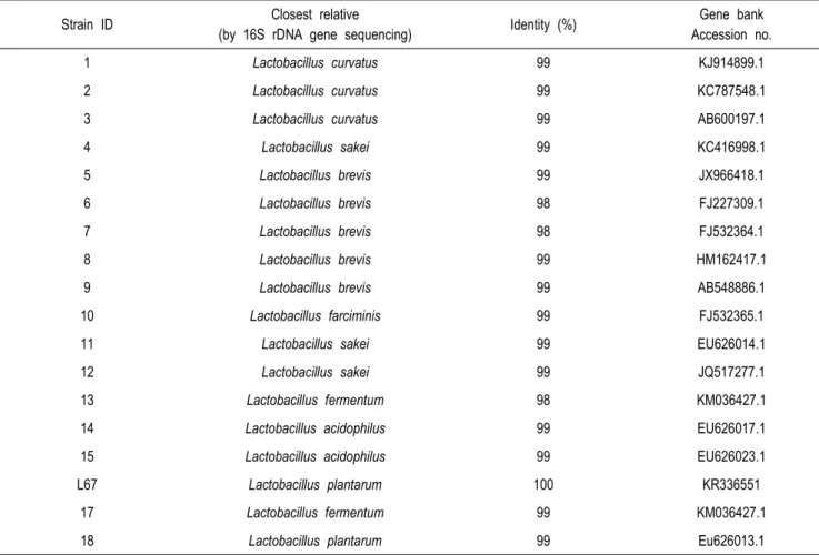
본 연구에서 사용된 Lactobacillus 균주는 유아분변 및 김치로부 터 총 18종의 유산균을 분리하였다(Table 1). Lactobacillus 의 확인은 catalase 활성 유무 및 Gram 염색으로 선별한 다음, 16S rDNA 서열 분석을 통하여 species를 결정하였다. 선발된 Lacto- bacillus 균주는 de Man, Rogosa, and Sharpe (MRS) broth (Difco, Detroit, MI)에서 37℃, 18시간 동안 배양한 다음, 원심 분리하여 세포를 회수한 후 Skim milk (10%)를 첨가하여 —80℃
에 저장하여 보관하였다.
2. 마우스 비장세포로부터 IL-12 and IFN-γ 생산
BALB/c 마우스로부터 얻은 배양된 비장 세포를 사용하여 IL-12와 IFN-γ의 생산을 유도하는 능력을 평가하였다. 10% fetal bovine serum, 100 U/mL penicillin 및 100 μg/mL streptomycin 을 함유하는 RPMI 1640 배지에서 배양한 비장 세포를 2.5×10⁶ cells/mL의 농도로 접종한 다음, 유산균(2.5×10⁶ CFU/mL)을 첨 가하여 7℃, 5 % CO2 조건에서 배양한 후 IL-12와 IFN-γ를 측정 하였다.
3. L. plantarum L67로부터 분리된 당 단백질 제조 당 단백질의 제조를 위해 10 mL의 L. plantarum 37℃에서 18시 간 동안 배양한 후 L67 배양액을 얻었다. 세포를 4℃, 12,000×g 의 조건에서 원심 분리한 후 1% SDS 용액을 첨가하여 10분간 열 을 가한 다음, 상등액을 4℃에서 12,000 ×g에서 15분간 원심 분 리하였다. 이를 동결 건조기를 사용하여 동결 건조시켜 —70℃에 저장하였다(Song et al., 2016a). 단백질 농도는 Bradford 분석 법을 사용하여 측정되었다(Bradford, 1976).
4. 세포배양
RBL-2H3에 세포는 한국 세포주은행으로부터 제공받았다(Seoul, Korea). 세포는 fetal bovine serum (10 %), penicillin (100
Strain ID Closest relative
(by 16S rDNA gene sequencing) Identity (%) Gene bank Accession no.
1 Lactobacillus curvatus 99 KJ914899.1
2 Lactobacillus curvatus 99 KC787548.1
3 Lactobacillus curvatus 99 AB600197.1
4 Lactobacillus sakei 99 KC416998.1
5 Lactobacillus brevis 99 JX966418.1
6 Lactobacillus brevis 98 FJ227309.1
7 Lactobacillus brevis 98 FJ532364.1
8 Lactobacillus brevis 99 HM162417.1
9 Lactobacillus brevis 99 AB548886.1
10 Lactobacillus farciminis 99 FJ532365.1
11 Lactobacillus sakei 99 EU626014.1
12 Lactobacillus sakei 99 JQ517277.1
13 Lactobacillus fermentum 98 KM036427.1
14 Lactobacillus acidophilus 99 EU626017.1
15 Lactobacillus acidophilus 99 EU626023.1
L67 Lactobacillus plantarum 100 KR336551
17 Lactobacillus fermentum 99 KM036427.1
18 Lactobacillus plantarum 99 Eu626013.1
* This table was reprinted with permission of publisher.
Table 1. Genotypic methods applied for identification of lactobacilli
U/mL) 및 streptomycin (100 μg/mL)을 첨가한 Dulbecco’s modified Eagle 배지에서 37℃, 5% CO2에서 배양하였다. 세포 (2.3×10⁶ cells/mL)를 6-well plate로 나누어 배지를 주 2회 교 체해 주었다. 또한 무균 상태에서 BALB/c 마우스의 비장 세포를 Hanks’ balanced salt 용액을 사용하여 회수하였으며, 채취한 비 장은 40 -μm cell strainer (Falcon, Corning Inc., Corning, NY)를 사용하여 파쇄하였다. 균질된 세포의 상층액에 ammo- nium chloride (0.8% wt/vol)을 첨가하여 침전시켜 원심분리 후 세포를 PBS로 세척하고, fetal bovine serum (10 %), penicillin (100 U/mL)와 streptomycin (100 mg/mL)가 첨가된 RPMI 1640 배지에 5% CO2, 37℃에서 재현탁시켰다. 그런 다음 세포 (1×10⁶ cells/mL)를 6-well 또는 96-well flatbottomed plates 로 나누어 30분 동안 50 μM BPA와 L67 단백질(5-100 μg/mL) 을 함께 처리하여 사용하였다.
5. Western Blotting을 위한 Cell Extract 준비
세포를 BPA(50 μM) 또는 L67 단백질(5~100 μg/mL)와 함께 30분간 처리하였다. 배지를 제거한 후 세포를 PBS로 세척하였으 며, Oh와 Lim(2008) 방법에 따라 단백질을 분리하였다. 단백질 농도는 Lowry 방법(Lowry et al., 1951)에 따라 측정하였고, 모든 시료는 —70℃에서 보관하였다.
6. Western Blot 분석
단백질은 Mini-Protean Ⅱ electrophoresis cell (Bio-Rad, Her- cules, CA)를 사용하여 실온에서 2시간 100 V로 10% poly- acrylamide mini-gell 전기영동으로 분석하였다. Nitrocellu- lose membranes (Millipore, Bedford, MA)에 단백질을 옮긴 후, membrane을 4℃에서 Tris-buffered saline Tween 20 (TBS-T) 용액에 rabbit polyclonal antibodies [1:3,000, T-
(A)
(B)
(C)
(D)
Fig. 1. Inhibitory effect of Lactobacillus plantarum L67 protein on ERK (Western blot in A; relative band intensity in B) and p38 mi- togen-activited protein kinase (MAPK; Western blot in C; relative band intensity in D) phosphorylation in bisphenol A (BPA)- reated RBL2H3 cells. This figure was reprinted with permission of pu- blisher.
bet, GATA-3, mitogen-activated protein kinase (MAPK), AP-1, and α-tubulin]를 넣고 18시간 배양하였다. TBS-T로 3번 세척한 후, TBS-T 용액에 horseradish peroxidase-conjuga-
ted goat anti-rabbit IgG (1:10,000; Cell Signaling, Danvers, MA)를 용해시켜 membrane과 함께 실온에서 1시간 30분 동안 배양시켰다. 단백질 밴드는 chemiluminescence (ELPIS Biote- chnology, Taejeon, Korea)를 이용하여 확인하였으며, Scion imaging software (Scion Image Beta 4.02, MD)를 이용하여 밴드의 강도를 측정하였다.
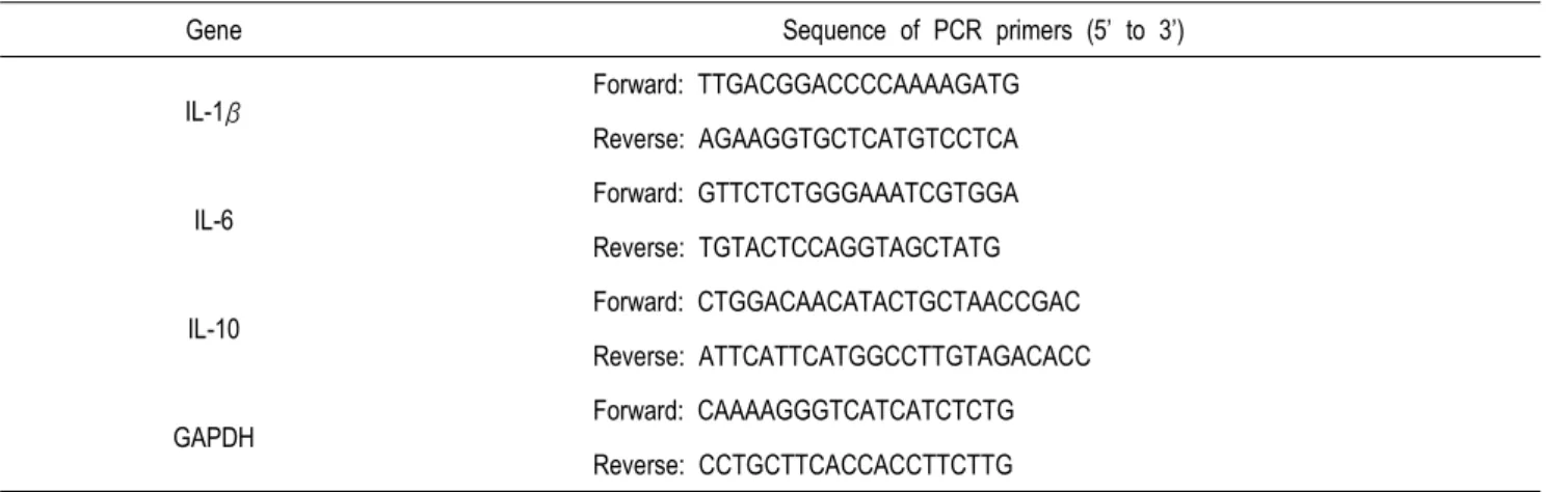
7. RNA 분리 및 Real-Time Quantitative PCR
Total RNA는 RNeasy Mini Kit (Qiagen, Valencia, CA)에서 제공된 방법(www.qiagen.com/goto/microRNAprotocols)에 따라 분석하였다. Maxime RT Premix Oligo (dT) real-time quantitative PCR kit (Intron, Seongnam, Korea)를 사용하여 1 μL의 total RNA (1 μg/20 μL)로 cDNA를 준비하였고, 본 실험에 사용한 프라이머는 Table 2에 나타내었다. 각각의 유전자 의 발현 수준을 2X Prime Q Master Mix (Kapa Biosystems, Boston, MA)를 사용하여 real-time PCR로 측정하고, CFX- 96TM Real-Time system (Bio-Rad)을 사용하여 분석하였다. 증 폭은 IL-1β, IL-6, IL-10, and GAPDH (95℃에서 30초, 56℃에 서 30초, 그리고 72℃에서 60초; Table 2)에 대하여 50 사이클로 진행되었다. IL-1β, IL-6 또는 IL-10에 대한 Ct 값과 BPA 단독으 로 처리되거나 또는 BPA와 L67 단백질이 함께 처리된 세포의 GAPDH (reference)값의 차이로 Sample Δ cycle threshold (S ΔCt) 값을 계산하였다. 처리군과 대조군 사이의 상대적 유전자 발 현 수준은 다음 공식을 사용하여 계산되었다 : 2 - (SΔCt - CΔ Ct) (Livak and Schmittgen, 2001).
결 과
1. 비장 세포에서 IL-12 및 IFN-γ 분비를 유도하는 Lactoba- cillus 균주의 선발
총 18개의 Lactobacillus 균주를 대상으로 비장 세포의 Th1/
Th2 균형을 향상시키는 능력을 측정하였다. 비장 세포에서 Th1 관련 사이토카인으로 알려진 IL-12 및 IFN-γ의 분비를 ELISA로 측정하였으며, Table 3에 나타낸 바와 같이, IL-12 및 IFN-γ 생 산을 유도하는 활성은 Lactobacillus 균주에 따라 차이가 있었다.
본 연구에 사용한 균주 중, 마우스 비장 세포에서 IL-12와 IFN-γ 둘다 분비를 유도한 L. plantarum L67을 최종 선발하여 다음실험 에 사용하였다(Table 3).
2. BPA 처리한 RBL-2H3 세포에서 p38 MAPK와 ERK의 발현에 미치는 L67 단백질의 영향
Table 2. Oligonucleotides used for real-time PCR
Gene Sequence of PCR primers (5’ to 3’)
IL-1β Forward: TTGACGGACCCCAAAAGATG
Reverse: AGAAGGTGCTCATGTCCTCA
IL-6
Forward: GTTCTCTGGGAAATCGTGGA Reverse: TGTACTCCAGGTAGCTATG
IL-10 Forward: CTGGACAACATACTGCTAACCGAC
Reverse: ATTCATTCATGGCCTTGTAGACACC
GAPDH Forward: CAAAAGGGTCATCATCTCTG
Reverse: CCTGCTTCACCACCTTCTTG
* This table was reprinted with permission of publisher.
L67 단백질이 BPA 처리된 RBL-2H3 세포에서 p38의 인산화를 억제할 수 있는지 여부를 알아보기 위해, western blotting에 의 한 p38 인산화를 분석하였다. P38의 인산화는 BPA를 처리한 RBL-2H3 세포에서 대조군에 비해 4.5배 증가하였다. 그러나 이러 한 인산화의 증가는 L67 단백질을 각각 5, 20, 50, 및 100 μg /mL의 농도로 처리했을 때 3.67, 3.62, 3.43, 0.87 배로 감소하는 것으로 나타났다. 또한, ERK 활성은 BPA을 30분 동안 처리함으로 써 증가하였고, 이 증가된 활성은 L67 단백질을 처리한 농도에 비 례하여 감소되었다.
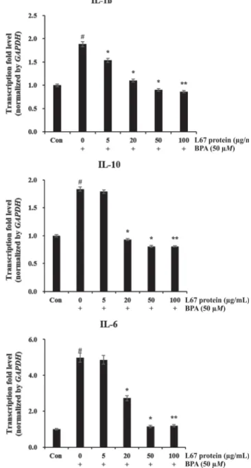
3. BPA 처리된 RBL-2H3 세포에서 IL-6, IL-10, IL-1β 유전자 발현에 미치는 L67 단백질의 억제 효과
L67 단백질 치료가 알러지 반응에 미치는 영향을 연구하기 위해, BPA가 처리된 RBL-2H3 세포에서 IL-6, IL-10 및 IL-1β 유전자 의 발현 양상을 조사하였다. Fig. 2에 나타낸 바와 같이, BPA로 인해 자극된 세포는 대조군에 비해 IL-6, IL-10 및 IL-1β의 발현 이 유의하게 증가된 것으로 나타났다. 그러나 L67 단백질 첨가의 경우, BPA만을 단독으로 처리한 것에 비하여 IL-6, IL-10 및 IL-1 β 유도를 용량 의존적으로 억제시켰다. BPA는 IL-6 유전자 전사 를 4.5배 증가시켰으며, L67 단백질을 각각 5, 20, 50 및 100 μg/mL 첨가했을 때 4.2, 3.6, 2.8, 1.9, 1.8 배의 감소를 보였다.
이와 비슷하게 IL-10, IL-1β 유전자 전사는 BPA 처리에 반응하여 1.8배 증가했지만, L67 단백질을 첨가한 경우 감소하는 것으로 나 타났다.
고 찰
본 연구에서는 L67 단백질이 BPA 처리된 RBL-2H3 세포와 마우 스에서 채취된 비장 세포에서 알러지 관련 매개체의 활성뿐만 아니 라, cytokine의 유도를 억제하는지 여부를 조사하였다. 여러 연구 에 의하면 lactobacilli가 대식세포 활성화를 조절하고, Th1/ Th2 균형을 조절하는 것으로 보고되었는데(Segawa et al., 2008;
Won et al., 2011), 특정 Lactobcillus 균주들은 Th1 유도 활성 을 가지며, ovalbumin-specific IgE 생성을 감소시키고, strain- dependent하게 관련 cytokine 패턴을 변화시키는 것으로 확인되 었다(Lee et al., 2013).
T-cell이 분화하는 동안 Th2의 두드러진 반응을 초래하는 매커니 즘은 아직 완전히 밝혀지지 않았지만, Th2 분화와 관련된 많은 요 인들 중 MAPK는 IL-4, IL-10과 같은 Th2 관련 사이토카인의 발 현의 주요 매개체이다(Yamashita et al., 2005; Watanabe et al., 2005). 따라서 MAPK 신호 전달 경로는 Th2 면역 반응에 의 해 유발된 알러지 질환에서의 표적이 될 수 있다(Abdel-Raheem et al., 2005). 우리는 ERK와 p38의 활성을 측정하여 L67단백질 이 MAPK 신호 전달을 억제하는지, IL-1β, IL-6 및 IL-10의 발현 을 억제하는지 여부를 살펴보았다. L67 단백질이 RBL-2H3 세포 에서 BPA에 의해 유도된 ERK와 p38 MAPK 인산화를 저해한다 는 것으로 나타났다. 또한 다양한 프로바이오틱스가 MAPK 및 AP-1과 같은 염증 마커 또는 Lipopolysaccharides와 같은 다른 염증성 유도체에 의해 유도될 수 있는 것으로 보고되었다(Kumar et al., 2014; Li et al., 2014; Jeong et al., 2015).
분화된 Th1 세포는 IFN-γ와 lymphotoxin을 생산하는데, 이는 세포 내 병원균에 대한 면역 저항에 필수적이라고 알려져 있다 (Glimcher and Murphy, 2000). 이와 대조적으로 Th2세포는 IL-1β, IL-6, IL-10을 방출하고, 이는 아토피, 알러지 반응과 관련
Table 3. Production of IL-12 and IFN-γ from mouse spleno- cytes induced by various strains of lactobacilli
Strain ID IL-12 (pg/mL) IFN-γ (pg/mL)
Control 0±0.12 200±7.8
1 35.0±1.2 1,900±75.2
2 38.4±1.1 1,300±65.2
3 35.4±0.3 2,050±10.3
4 25.6±0.2 2,000±10.2
5 55.8±1.7 1,000±8.7
6 17.3±0.4 800±10.5
7 15.2±0.5 1,310±54.3
8 10.9±0.3 400±5.6
9 11.6±0.2 408±7.8
10 64.3±0.6 1,200±21.3
11 48.0±2.3 550±6.4
12 68.9±8.7 1,400±12.3
13 18.1±0.4 1,950±15.3
14 13.3±0.3 200±4.3
15 36.0±0.3 300±3.2
L67 68.7±4.3 1,800±43.2
17 16.2±0.4 210±5.6
18 14.3±0.3 1,850±20.2
This table was reprinted with permission of publisher.
이 있으며(Tillie-Leblond et al., 1999; Herrick et al., 2000), IL-1β는 Th2 및 B 세포를 조절하기 위해 IgE를 대체하는 클래스 전환 과정에서 중요한 자극제, IL-10은 Th1 세포의 분화를 억제하 는 IL-12 및 IFN-γ (Th1 사이토카인)의 중요한 억제제로서 기능 한다는 것을 알 수 있다(Ritter et al., 2014; Murphy et al., 2000). BPA가 Th2 반응을 유도하기 때문에, RBL-2H3 세포에서 의 IL-1β, IL-6 및 IL-10의 유전자 발현을 조사한 결과, BPA 처리 가 IL-1β, IL-6 및 IL-10의 발현을 촉진한다는 것을 발견하였다.
이것은 BPA가 IL-1β, IL-6 및 IL-10을 통해 Th0에서 Th2 세포 로의 분화를 촉진한다는 것을 나타낸다. 그러나 BPA 처리에 의해 유도된 IL-1β, IL-6 및 IL-10의 증가된 발현은 L67 단백질 처리 에 의해 유의하게 억제되었다.
본 연구는 L67 단백질이 ERK와 p38 MAPK의 인산화를 억제하는
Fig. 2. Inhibitory effect of Lactobacillus plntarum L67 protein on IL-1β, IL-6, and IL-10 expression in bisphenol A (BPA)-treated RBL2H3 cells. This figure was reprinted with permission of pu- blisher.
것뿐만 아니라, 염증 관련 인자(Th2 관련 사이토카인)가 있는 L67 단백질이 BPA에 의한 IL-1β, IL-6, IL-10의 활성을 억제시킬 수 있음을 확인한 것으로 L. plantarum L67 균주가 Th2 관련 면역 질환의 예방에 도움이 될 수 있는 새로운 식품 소재로써의 가치가 있을 것으로 판단되었다.
감사의 글
Journal of Dairy Science (99:9372-9382, 2016; http://dx.
doi.org/10.3168/jds.2016-11809)에 게재된 결과를 사용하도록 허가해준 Elsevier (License Number: 4007431144815; Li- cense Date: Dec 14, 2016)에 감사드리며, 자세한 내용은 원문 을 참조하길 바란다.
참고문헌
1. Abdel-Raheem IT, Hide I, Yanase Y, Shigemoto- Mogami Y, Sakai N, Shirai Y, Saito N, Hamada FM, El-Mahdy NA, Elsisy AD, Sokar SS, and Nakata Y.
(2005) Protein kinase C-alpha mediates TNF release process in RBL-2H3 mast cells. Br. J. Pharmacol. 145, 415-423.
2. Andersson U, Branning C, Ahrne S, Molin G, Alenfall J, Onning G, Nyman M, and Holm C. (2010) Probiotics lower plasma glucose in the high-fat fed C57BL/6J mouse. Benef. Microbes 1, 189-196.
3. Banerjee A, and Gerondakis S. (2007) Coordinating TLR-activated signaling pathways in cells of the im- mune system. Immunol. Cell Biol. 85, 420-424.
4. Bornehag CG, Sundell J, Weschler CJ, Sigsgaard T, Lundgren B, Hasselgren M, and Hagerhed-Engman L.
(2004) The association between asthma and allergic symptoms in children and phthalates in house dust: A nested case control study. Environ. Health Perspect.
112, 1393-1397.
5. Bradford MM. (1976) A rapid and sensitive method for the quantitation of microgram quantities of protein utilizing the principle of protein-dye binding. Anal.
Biochem. 72, 248-254.
6. Chalubinski M, and Kowalski ML. (2006) Endocrine disrupterspotential modulators of the immune system and allergic response. Allergy 61, 1326-1335.
7. Cheng H. (2010) Volatile flavor compounds in yogurt:
A review. Crit. Rev. Food Sci. Nutr. 50, 938-950.
8. Cruz AG, Faria JAF, Walter EHM, Andrade RR, Caval- canti RN, Oliveira CAF, and Granato D. (2010) Pro- cessing optimization of probiotic yogurt containing
glucose oxidase using response surface methodology.
J. Dairy Sci. 93, 5059-5068.
9. Dave RI, and Shah NP (1996). Evaluation of media for selective enumeration of Streptococcus thermophilus, Lactobacillus delbrueckii ssp. bulgaricus, Lactoba- cillus acidophilus, and bifidobacteria. J. Dairy Sci. 79, 1529-1536.
10. Giardina S, Scilironi C, Michelotti A, Samuele A, Borella F, Daglia M, and Marzatico F. (2014) In vitro anti-inflammatory activity of selected oxalate-de- grading probiotic bacteria: Potential applications in the prevention and treatment of hyperoxaluria. J.
Food Sci. 79, M384-390.
11. Glimcher LH, and Murphy KM. (2000) Lineage com- mitment in the immune system: The T helper lym- phocyte grows up. Genes Dev. 14, 1693-1711.
12. Hekmat S, and Reid G. (2006) Sensory properties of probiotic yogurt is comparable to standard yogurt.
Nutr. Res. 26, 163-166.
13. Herrick CA, MacLeod H, Glusac E, Tigelaar RE, and Bottomly K. (2000) Th2 responses induced by epi- cutaneous or inhalational protein exposure are dif- ferentially dependent on IL-4. J. Clin. Invest. 105:
765-775.
14. Hong YF, Kim H, Kim HR, Gim MG, and Chung DK.
(2014) Different immune regulatory potential of Lactobacillus plantarum and Lactobacillus sakei iso- lated from kimchi. J. Microbiol. Biotechnol. 24, 1629-1635.
15. Jaakkola JJ, Parise H, Kislitsin V, Lebedeva NI, and Spengler JD. (2004) Asthma, wheezing, and allergies in Russian schoolchildren in relation to new surface materials in the home. Am. J. Public Health 94, 560-562.
16. Jeong JJ, Kim KA, Jang SE, Woo JY, Han MJ, and Kim DH. (2015) Orally administrated Lactobacillus pento- sus var. plantarum C29 ameliorates age-dependent colitis by inhibiting the nuclear factor-kappa B sig- naling pathway via the regulation of lipopolysa- ccharide production by gut microbiota. PLoS One 10, e0116533.
17. Johnson-Henry KC, Hagen KE, Gordonpour M, Tomp- kins TA, and Sherman PM. (2007) Surface- layer pro- tein extracts from Lactobacillus helveticus inhibit enterohaemorrhagic Escherichia coli 0157:H7 adhe- sion to epithelial cells. Cell. Microbiol. 9, 356-367.
18. Kassayova M, Bobrov N, Strojny L, Kiskova T, Mikeš J, Demečkova V, Orendaš P, Bojkova B, Peč M, Kubatka MM, and Bomba A. (2014) Preventive effects of probiotic bacteria Lactobacillus plantarum and dietary fiber in chemically- induced mammary carci- nogenesis. Anticancer Res. 34, 4969- 4975.
19. Konieczna P, Schiavi E, Ziegler M, Groeger D, Healy S, Grant R, and O’Mahony L. (2015) Human dendritic cell DC- SIGN and TLR-2 mediate complementary immune regulatory activities in response to Lactoba- cillus rhamnosus JB-1. PLoS One 10, e0120261.
20. Konstantinov SR, Smidt H, de Vos WM, Bruijns SC, Singh SK, Valence F, Molle D, Lortal S, Altermann E, Klaenhammer TR, and van Kooyk Y. (2008) S layer protein A of Lactobacillus acidophilus NCFM regu- lates immature dendritic cell and T cell functions.
Proc. Natl. Acad. Sci. USA 105 ,19474-19479.
21. Kumar A, Hecht C, Priyamvada S, Anbazhagan AN, Alakkam A, Borthakur A, Alrefai WA, Gill RK, and Dudeja PK. 2014. Probiotic Bifidobacterium species stimulate human SLC26A3 gene function and ex- pression in intestinal epithelial cells. Am. J. Physiol.
Cell Physiol. 307, C1084-C1092.
22. Lee J, Bang J, and Woo HJ. 2013. Immunomodula- tory and antiallergic effect of orally administered Lactobacillus species in ovalbumin-sensitized mice.
J. Microbiol. Biotechnol. 23, 724-730.
23. Lee J, and Lim KT. (2010) Plant-originated glyco- protein (36 kDa) suppresses interleukin-4 and −10 in bisphenol A-stimulated primary cultured mouse lymphocytes. Drug Chem. Toxicol. 33, 421-429.
24. Li L, Jiang YJ, Yang XY, Liu Y, Wang JY, and Man CX. (2014). Immunoregulatory effects on Caco-2 cells and mice of exopolysaccharides isolated from Lactobacillus acidophilus NCFM. Food Funct. 5, 3261- 3268.
25. Li S, Zhao Y, Zhang L, Zhang X, Huang L, Li D, Niu C, Yang Z, and Wang Q. (2012) Antioxidant activity of Lactobacillus plantarum strains isolated from tra- ditional Chinese fermented foods. Food Chem. 135, 1914-1919.
26. Lin YP, Thibodeaux CH, Pena JA, Ferry GD, and Versalovic J. (2008) Probiotic Lactobacillus reuteri suppress proinflammatory cytokines via c-Jun. In- flamm. Bowel Dis. 14, 1068-1083.
27. Livak KJ, and Schmittgen TD. (2001) Analysis of rela- tive gene expression data using real-time quantita- tive PCR and the 2-∆∆CT method. Methods 25, 402-408.
28. Lowry OH, Rosebrough NJ, Farr AL, and Randall RJ.
(1951) Protein measurement with the Folin phenol reagent. J. Biol. Chem. 193, 265-275.
29. Murphy KM, Ouyang W, Farrar JD, Yang J, Ranga- nath S, Asnagli H, Afkarian M, and Murphy TL. (2000) Signaling and transcription in T helper develop- ment. Annu. Rev. Immunol. 18, 451-494.
30. Nakamura Y, Christodoulopoulos P, Cameron L, Wri- ght E, Lavigne F, Toda M, Muro S, Ray A, Eidelman DH, Minshall E, and Hamid Q. (2000) Upregulation of the transcription factor GATA-3 in upper airway mucosa after in vivo and in vitro allergen challenge.
J. Allergy Clin. Immunol. 105, 1146-1152.
31. O’Brien E, Dolinoy DC, and Mancuso P. (2014) Peri- natal bisphenol A exposures increase production of pro-inflammatory mediators in bone marrow-de- rived mast cells of adult mice. J. Immunotoxicol. 11, 205-212.
32. Oh PS, and Lim KT. (2008) Protective activity of 30 kDa phytoglycoprotein from glucose/glucose oxi- dase-induced cell death in primary cultured mouse thymocytes. Environ. Toxicol. Pharmacol. 25, 114- 120.
33. Park YS, Lee JY, Kim YS, and Shin DH. (2002) Isolation and characterization of lactic acid bacteria from feces of newborn baby and from Dongchimi. J.
Agric. Food Chem. 50, 2531-2536.
34. Rhee CH, and Park HD. (2001) Three glycoproteins
with antimutagenic activity identified in Lactoba- cillus plantarum KLAB21. Appl. Environ. Microbiol.
67, 3445-3449.
35. Ritter M, Straubinger K, Schmidt S, Busch DH, Hag- ner S, Garn H, Prazeres da Costa C, and Layland LE.
(2014) Functional relevance of NLRP3 inflammaso- me-mediated interleukin (IL)-1β during acute aller- gic airway inflammation. Clin. Exp. Immunol. 178, 212-223.
36. Segawa S, Nakakita Y, Takata Y, Wakita Y, Kaneko T, Kaneda H, Watari J, and Yasui H. 2008. Effect of oral administration of heat-killed Lactobacillus bre- vis SBC8803 on total and ovalbumin specific im- munoglobulin E production through the improve- ment of Th1/Th2 balance. Int. J. Food Microbiol.
121, 1-10.
37. Song M, Yun B, Moon JH, Park DJ, Lim K, and Oh S. (2015) Characterization of selected Lactobacillus strains for use as probiotics. Korean J. Food Sci.
Anim. 35, 551-556.
38. Song S, Bae DW, Lim K, Griffiths MW, and Oh S.
(2014) Cold stress improves the ability of Lactobaci- llus plantarum L67 to survive freezing. Int. J. Food Microbiol. 191, 135-143.
39. Song S, Oh S, and Lim KT. (2016a) Glycoprotein (18 kDa) isolated from Lactobacillus plantarum L67 sup- pressed β-hexosaminidase, histamine, and the ex- pression of TNF-α and IL-4 in the BPAstimulated RBL-2H3 cells. Process Biochem. 51, 105-112.
40. Song S, Oh S, and Lim KT. (2016b). Lactobacillus plantarum L67 glycoprotein protects against cad- mium chloride toxicity in RAW 264.7 cells. J. Dairy Sci. 99, 1812-1821.
41. Sornplang P, and Piyadeatsoontorn S. (2016) Probi- otic isolates from unconventional sources: A review.
J. Anim. Sci. Technol. 58, 26.
42. Szabo SJ, Kim ST, Costa GL, Zhang X, Fathman CG, and Glimcher LH. (2000) A novel transcription fac- tor, T-bet, directs Th1 lineage commitment. Cell 100, 655-669.
43. Szabo SJ, Sullivan BM, Peng SL, and Glimcher LH.
(2003) Molecular mechanisms regulating Th1 immu- ne responses. Annu. Rev. Immunol. 21, 713-758.
44. Tamime AY, and Deeth HC. (1980) Yogurt: Technol- ogy and biochemistry. J. Food Prot. 12, 939-977.
45. Teraguchi S, Uehara M, Ogasa K, and Mitsuoka T.
(1978) Enumeration of bifidobacteria in dairy pro- ducts. Nippon Saikingaku Zasshi 33, 753-761.
46. Tillie-Leblond I, Pugin J, Marquette CH, Lamblin C, Saulnier F, Brichet A, Wallaert B, Tonnel AB, and Gosset P. (1999) Balance between proinflammatory cytokines and their inhibitors in bronchial lavage from patients with status asthmaticus. Am. J. Respir.
Crit. Care Med. 159, 487-494.
47. Vandenberg LN, Hauser R, Marcus M, Olea N, and Welshons W. (2007). Human exposure to bisphenol A (BPA). Reprod. Toxicol. 24, 139-177.
48. Watanabe M, Watanabe S, Hara Y, Harada Y, Kubo M, Tanabe K, Toma H, and Abe R. (2005) ICOS- mediated costimulation on Th2 differentiation is ac- hieved by the enhancement of IL-4 receptor-medi- ated signaling. J. Immun. 174, 1989-1996.
49. Won TJ, Kim B, Song DS, Lim YT, Oh ES, Lee DI, Park ES, Min H, Park SY, and Hwang KW. 2011.
Modulation of Th1/Th2 balance by Lactobacillus strains isolated from Kimchi via stimulation of mac- rophage cell line J774A.1 in vitro. J. Food Sci. 76, H55-61.
50. Yamashita M, Shinnakasu R, Asou H, Kimura M, Hasegawa A, Hashimoto K, Hatano N, Ogata M, and Nakayama T. (2005). Ras-ERK MAPK cascade regu- lates GATA3 stability and Th2 differentiation through ubiquitin proteasome pathway. J. Biol. Chem. 280, 29409-29419.
51. Yan H, Takamoto M, and Sugane K. (2008) Exposure to bisphenol A prenatally or in adulthood promotes T(H)2 cytokine production associated with reduction of CD4CD25 regulatory T cells. Environ. Health Per- spect. 116, 514-519.
52. Zhu J, Yamane H, Cote-Sierra J, Guo L, and Paul WE.
(2006) GATA-3 promotes Th2 responses through three different mechanisms: Induction of Th2 cyto- kine production, selective growth of Th2 cells and
inhibition of Th1 cell- specific factors. Cell Res. 16, 3-10.