Anti-oxidant Effect of Agastache rugosa on Oxidative Damage Induced by H
2O
2in NIH 3T3 Cell
Se Chul Hong, Jin Boo Jeong, Gwang Hun Park, Jeong Sook Kim, Eul Won Seo and Hyung Jin Jeong*
College of Natural Sciences, Andong National University, Andong 760-749, Republic of Korea
Abstract - The plant Agastache rugosa Kuntze has various physiological and pharmacological activities. Especially, it has been regarded as a valuable source for the treatment of anti-inflammatory and oxidative stress-induced disorders. However, little has been known about the functional role of it on oxidative damage in mammalian cells by ROS. In this study, we investigated the DPPH radical, hydroxyl radical, hydrogen peroxide and intracellular ROS scavenging capacity, and Fe2+
chelating activity of the extracts from Agastache rugosa. In addition, we evaluated whether the extract can be capable of reducing H2O2-induced DNA and cell damage in NIH 3T3 cells. These extracts showed a dose-dependent free radical sca- venging capacity and a protective effect on DNA damage and the lipid peroxidation causing the cell damage by H2O2. There- fore, these results suggest that Agastache rugosa is useful as a herbal medicine for the chemoprevention against oxidative carcinogenesis.
Key words - Agastache rugosa Kuntze, Lipid peroxidation, Oxidative cell death, Oxidative DNA damage, Reactive oxygen species
*Corresponding author. E-mail : [email protected]
Introduction
Reactive oxygen species (ROS) have been associated with pathogenic processes including carcinogenesis through direct effects on DNA directly and by acting as a tumor promoter (Kehrer, 1993; Salah et al., 1995; Wiseman and Halliwell, 1996; Vallyathan and Shi, 1997; Kong et al., 2001). Radicals have been demonstrated to be initiators of the oxidative process (Pietraforte et al., 2002), and to be involved in the development of disease (Aust et al., 1993; Stohs, 1995). Catalase, supero- xide dismutase, glutathione and uric acid are examples of antio- xidants produced by organisms under normal conditions as part of a defense system against ROS-mediated cellular injury.
However, if this defense system is challenged or overwhelmed by excessive generation of ROS, redox imbalance or oxida- tive stress may occur. This can result in damage to the organism (Farber, 1998; Langard, 1990), and disease initiation (Halli- well and Gutteridge, 2000). ROS have also been shown to play an important role in carcinogenesis by damaging DNA and acting as tumor promoters (Wiseman and Halliwell, 1996; Kong et al., 2001).
Agastache rugosa Kuntze, a perennial herb ubiquitous in Korean fields, has been used as a wild vegetable and helba drug for the treatment of anorexia, vomiting and other intestinal disorders. This plant is increasingly cultivated in Korea to sa- tisfy the rising demand for essential oil by aromatherapists and herbalists. The plant Agastache rugosa Kuntze contains several kinds of flavonoids such as tilianin, acacetin, linarin, agastinol, agastenol and agastachoside, and tilianin is considered as a main constituent of Agastache rugosa. in a previous report (Shin and Kang, 2003), the essential oils of agastache rugosa and estragole, the main component of this oil, showed significant synergism with ketoconazole against Blastoschizomyces capi- tatus (Shin and Kang, 2003). This plant has a variety of physi- ological and pharmacological activities (Lee et al 2002; Jung and Surh, 2001). It has been reported that Agastache rugosa extracts inhibited cytokine induced vascular cell adhesion molecule-1 in human umbilical vein endothelial cells (HUVECs), and inhibited apoptosis in leukemia cells (Lee et al., 2002).
These findings suggest that Agastache rugosa may be a valuable source for the treatment of anti-inflammatory and oxidative stress-induced disorders. While Agastache rugosa has been indicated as anti-inflammatory and oxidative stress-induced disorders, its antioxidant properties have not
been well defined. Therefore, it is important to understand the inhibitory mechanism of the extracts from Agastache rugosa on H2O2-induced oxidative damage causing DNA and cell damages since H2O2 is the major mediator of oxidative stress and a potent mutagen. Also the evaluation of its inhibitory effects on oxidative DNA and cell damages is necessary for the medicinal use of it as the cancer chemopreventive agent.
Major questions we wish to address in our study are: (1) What is the effect of Agastache rugosa on DPPH radical, hydroxyl radical and hydrogen peroxide? (2) What is the effect of Agastache rugosa on Fe2+ chelation? (3) What is the effect of Agastache rugosa on ROS generated in a cellular system? (4) Can Agastache rugosa affect lipid peroxidation in a cellular system? (5) What is the effect of Agastache rugosa on oxidative DNA damage caused by ROS?
Materials and Methods
Chemical regents
All chemicals for extraction from Agastache rugosa, DPPH (1,1diphenyl-2-picryl hydrazyl) and 2’,7’-dichlorofluorescin diacetate (DCF-DA) were obtained from Sigma Chemicals Co.
(St. Louis, USA). φX-174 RF I plasmid was purchased from New England BioLabs (County Road Ipswich, MA)
Sample and sample preparation
The plant sample, Agastache rugosa was kindly provided by the Bonghwa Alpine Medicinal Plant Experiment Station, Korea. The voucher specimens of plant samples were deposited at the major, medicinal resources, Andong National Univer- sity, Andong, Korea. 1 kg dried rhizomes were extracted with 1000 ml of 80% methanol with shaking for 24 hours. After 24 hours, the methanol- soluble fraction was filtered and concen- trated to approximately until 20 ml volume using by vacuum evaporator and fraction in a separating funnel. The ethyl ace- tate fraction was separated from the mixture, evaporated by vacuum evaporator and prepared aseptically. For the prepara- tion of the essential oil, 1 kg of Agastache rugosa was extrac- ted by the steam distillation apparatus (SDA) at 80℃ for 4 hours and dehydrated with sodium sulfate anhydrous. The extracts were concentrated by a vacuum evaporator at 30℃. For the cell experiment, the essential oils were diluted with 1% DMSO
in RPMI 1640 (Gibco BRL, Burlington, Ontario, Canada) supplemented with 10% fetal calf serum (Biochrom KG, Berlin, Germany). These ethyl acetate fractions and essential oil from Agastache rugosa were kept refrigerate until use
DPPH radical scavenging activity
The antioxidant activity of the extracts was evaluated first by monitoring its ability in quenching the stable free radical DPPH (Hus et al., 2006). Reaction mixture containing 40 µl of test samples (4 mg/ml dissolved in DMSO) and 760 µl of 300 µM DPPH ethanol solution in micro tube were incubated at 37℃ for 30 min and absorbance was measured at 515 nm according to the increasing concentrations of the extracts and the essential oil. The DPPH quenching ability was calculated from the log-dose inhibition curve. All determination was ca- rried out in triplicate. Ascorbic acid was used as a positive control.
Hydroxyl radical scavenging activity
Hydroxyl radical-scavenger ability was measured according to a literature procedure (Smirnoff and Cumbes, 1989) with a few modifications. Hydroxyl radical was generated from FeSO4
and hydrogen peroxide, and detected by their ability to hydro- xylate salicylate. The reaction mixture (800 μl) contained 250 μl FeSO4 (1.5 mM), 175 μl hydrogen peroxide (6 mM), 300 μl sodium salicylate (20 mM) and varying concentrations of the extracts and the essential oil. After a reaction for 30 min at 37℃, the absorbance of the hydroxylated salicylate complex was measured at 562 nm. Hydroxyl radical-scavenger ability was calculated from the log-dose inhibition curve. All determina- tion was carried out in triplicate. Ascorbic acid was used as a positive control.
Hydrogen peroxide scavenging assay
One hundred micro liter of 0.1 M phosphate buffer (pH 5.0), 40 μl of test samples and 60 μl of 1 mM hydrogen peroxide were mixed, and then incubated for 5 min at 37℃. After 5 min, 400 μl of 1.25 mM ABTS and 400 μl peroxidase (1unit/ml) are added to the mixture, and then incubated for 10 min at 37℃.
After 10 min, the absorbances were read at 405 nm. Hydrogen peroxide scavenger ability was calculated from the log-dose inhibition curve. All determination was carried out in triplicate.
Fe2+-chelating activity assay
This assay was measured according to a literature procedure (Rosenkranz et al., 1992) with a few modifications. The reac- tion mixture (800 μl) contained 15 μl FeCl2 (2 mM), 150 μl varying concentrations of the extracts and 605 μl distilled water. The mixture was shaken vigorously and left at room temperature for 30 min. After 30 min, 30 μl ferrozine (5 mM in methanol) was added and mixed. The absorbance of the Fe2+- ferrozine complex was measured at 562 nm. Fe2+-chelating activity assay was calculated from the log-dose inhibition cur- ve. All determination was carried out in triplicate. Ascorbic acid was used as a positive control.
Intracellular ROS scavenging activity
Intracellular ROS scavenging activity was carried by DCF- DA used to detect the levels of intracellular ROS (Rosenk- ranz et al., 1992). NIH 3T3 cells were seeded on 96-well plate at 5×10⁴ cells/well. Sixteen hours after plating, the cells were treated with varying concentrations of the extracts and 30 min later, 20 mM of hydrogen peroxide was added to the plate for 1 hour. After 1 hour, 100 µM of DCF-DA solution was added for 10 min and then the fluorescence of 2’,7’-dichlorofluorescein was detected at 485 nm excitation and at 535 nm emission using the spectrofluorometer. For the image analysis for generation of intracellular ROS, NIH 3T3 cells were seeded on six-well plate containing a coverslip at 5×10⁴ cells/well. Sixteen hours after plating, the cells were treated with varying concentrations of the extracts and 30 min later, 20 mM of hydrogen peroxide was added to the plate for 1 hour. After 1 hour, the media was changed, 300 µM of DCF-DA was added to each well and then the plate was incubated for an additional 30 min at 37℃.
Next, after washing with PBS, the stained cells were mounted onto microscope slide in mounting medium. The images were collected using a confocal microscope.
φX-174 RF I plasmid DNA damage assay
Conversion of the supercoiled form of plasmid DNA to the open-circular and further linear forms has been used as an index of DNA damage (Jung and Surh, 2001). Reaction mixtures (25 μl) contained 5 μl of φX-174 RF I plasmid DNA, 10 μl of varying concentrations of the extracts, 5 μl of 1 mM FeSO4
or/and 5 μl of 1 mM hydrogen peroxide and were incubated at 37℃ for 30 min. After 30 min, 5 μl of a solution containing
50% glycerol (v/v), 40 mM EDTA and 0.05% bromophenol blue was added to stop the reaction and the reaction mixtures was electrophoresed on 1% agarose gel, and the DNA in the gel was visualized and photographed under ultraviolet light after ethidium bromide staining.
Intracellular DNA damage assay
NIH 3T3 cells (2×10⁶ cells×well) were cultured in 6-well plates for 24 hours at 37℃ in an incubator with a humidified atmosphere of 5% CO2. After 24 hours, the cells were treated with the varying concentration of the extracts for 30 min and then added with 5 mM FeCl2 and 5 mM hydrogen peroxide for 1 hour. After 1 hour, each cell was harvested and then the su- pernatant was discarded. Each cell was resuspended with 20 μl of lysis buffer (50 mM Tris-HCl, pH 8.0, 10 mM EDTA, 0.5% SDS and 0.5 mg/ml proteinase K) by pipetting cells to ensure complete lysis and then incubated at 55℃ for 60 min.
After 60 min, each cell was centrifuged, 5 μl of RNase A was added to the supernatant, and each cell was incubated at 55℃
for another 60 min. After 60 min, each cell was spun briefly to remove any further cell debris and collect the supernatant.
Each lysate was heated at 70℃ for a few minutes and mixed with 10 μl of loading buffer (50% glycerol (v/v), 40 mM EDTA and 0.05% bromophenol blue). the reaction mixtures was electrophoresed on 2% agarose gel, and the DNA in the gel was visualized and photographed under ultraviolet light after ethidium bromide staining.
Lipid peroxidation assay
This assay was carried according to literature procedure (Kang et al., 2008) with some modification. NIH 3T3 cells were cultured in a 6-well plate at 2×10⁶ cells/well for 16 hours.
Sixteen hours after plating, the cells treated with the varying concentrations of extracts for 30 min. After 30 min, 5 mM hydrogen peroxide and FeCl2 was added to the plate and incubated for 12 hours. The cells were then washed with cold phosphate-buffered saline (PBS), harvested, and homogenized in an ice-cold 1.15% KCl. One hundred microliters of the cell lysates was mixed with 0.1 ml of 8.1% sodium dodecylsul- fate, 0.75 ml of 20% acetic acid (adjusted to pH 3.5), and 0.75 ml of 0.8% thiobarbituric acid (TBA). The mixture was made up to a final volume of 4 ml with distilled water and heated to 95℃ for 2 hours. After cooling to room temperature, 2.5 ml
(A) (B)
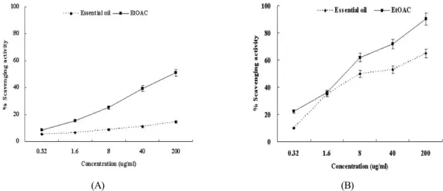
Fig. 1. DPPH free radical (A) and hydroxyl radical (B) scavenging activities of the extracts from Agastache rugosa. The absorbance values were converted into relative values of the positive control without the extracts from Agastache rugosa as scavenging effects (%) and data plotted as the means of replicate scavenging effect (%) values ± 5 S.D. (n=3) against extract concentration in μg extract per ml reaction volume.
of an n-butanol/pyridine mixture (15:1, v/v) was added and the mixture was shaken. After centrifugation at 1000 g for 10 min, the supernatant fraction was isolated and the absorbance was measured spectrophotometrically at 532 nm.
Results and Discussion
DPPH free radical and hydroxyl radical scavenging activities of the extracts from Agastache rugosa
The role of an antioxidant is to remove free radicals. The mechanism for antioxidants to remove free radical involves donating hydrogen to a free radical and hence its reduction to an unreactive species through removing the odd electron fea- ture which is responsible for radical reactivity (Wang et al., 2008). In DPPH radical scavenging assay (Fig. 1A), the EtOAc extracts and the essential oil removed DPPH radical by 2%
and 5.4% at 0.32 µg/ml, 10% and 5.7% at 1.6 µg/ml, 41% and 6.7% at 8 µg/ml, 76% and 11.7% at 40 µg/ml and 91% and 28.4% at 200 µg/ml. And the Fenton reaction (Fe2+ + hydrogen peroxide Fe3+ + hydroxyl radical + OH-) was used as a source of hydroxyl radical. Hydroxyl radical scavenging is an important antioxidant activity because of very high reactivity of hydroxyl radical which enables it to react with a wide range of molecules found in living cells such as sugars, amino acids, lipids and nucleotides (Stohs and Bagchi, 1995). With this
assay (Fig. 1B), the EtOAc extracts and the essential oil sca- venged hydroxyl radical by 14% and 8% at 0.32 µg/ml, 15%
and 25% at 1.6 µg/ml, 26% and 27.6% at 8 µg/ml, 31% and 28.2% at 40 µg/ml and 72% and 56.6% at 200 μg/ml, respec- tively. Epidemiologic studies have shown the effectiveness of antioxidants in reducing the risks of cancer and other diseases (Yang et al., 2000). ROS damage can be reduced by two anti- oxidant factors such as scavenging of radicals formed during reaction and inhibiting the radical generation. The results for scavenging DPPH radical and hydroxyl radical suggest that Agastache rugosa can reduce ROS damage by scavenging generated radical during the reaction.
Hydrogen peroxide scavenging activity and Fe2+ chelating activity of the extracts from Agastache rugosa
Iron and copper are essential transition metal elements in the human body required for the activity of a large range of enzymes and for some proteins involved in cellular respira- tion, O2 transport and redox reactions. Unfortunately, they contain unpaired electrons that enable them to participate in one-electron transfer reactions. Hence, they are powerful cata- lysts of autoxidation reactions (Lioyd et al., 1997). Hydrogen peroxide itself is not very reactive, but it can sometimes be to- xic to cells, since it may give rise to hydroxyl radicals inside the cell (Halliwell and aruoma, 1991). The Fenton reaction
(A) (B)
Fig. 2. Fe2+ chelating (A) activity and hydrogen peroxide (B) scavenging activity of the extracts from Agastache rugosa. The absorbance values were converted into relative values of the positive control without the extracts from Agastache rugosa as scavenging effects (%) and data plotted as the means of replicate scavenging effect (%) values ± 5 S.D. (n=3) against extract concentration in µg extract per ml reaction volume.
between Fe2+ and hydrogen peroxide generates Fe3+, OH- and hydroxyl radical (Halliwell and Gutteridge, 2000). The hyd- roxyl radical generated from the Fenton reaction can cause oxidative DNA damage that has been shown to play a key role in carcinogenesis. In Fe2+ chelating assay and hydrogen pero- xide scavenging assay (Fig. 2A-B), the EtOAc extracts and the essential oil from Agastache rugosa chelated Fe2+ by 8%
and 5.4% at 0.32 μg/ml, 15% and 6.7% at 1.6 μg/ml, 25% and 8.7% at 8 μg/ml, 39% and 11% at 40 μg/ml and 51% and 14.7% at 200 μg/ml, and removed hydrogen peroxide by 22%
and 8% at 0.32 μg/ml, 36% and 25% at 1.6 μg/ml, 62% and 50% at 8 μg/ml, 72% and 53.2% at 40 μg/ml and 90% and 64.9% at 200 μg/ml. These results suggest that Agastache rugosa prevents a mammalian cell from ROS damage by inhibiting the radical generation such as hydroxyl radical.
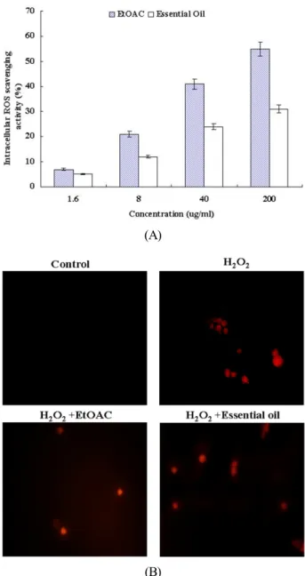
Intracellular ROS scavenging activity and Inhibitory effect of the extracts from Agastache rugosa on oxidative DNA damage
The present investigation also evaluated the ability of the extracts from Agastache rugosa to inhibit oxidative DNA da- mage using φX-174 RF I plasmid DNA and intracellular DNA through intracellular ROS scavenging activity. Excessive ROS (e.g. hydrogen peroxide) can lead to DNA oxidation, cau-
sing cell damage to all cellular constituents. Irreparable DNA damage is involved in carcinogenesis, aging and other dege- nerative diseases (Cozzi et al., 1997). Oxidative DNA damage leads to mutations and is suspected to be a major cause of cancer (Schwarz et al., 1984). Therefore, the inhibition of oxidative DNA damage is important to cancer chemoprevention. The intracellular ROS scavenging activity of the EtOAc extracts and the essential oil was 7% and 5% at 1.6 μg/ml, 21% and 12%
at 8 μg/ml, 41% and 24% at 40 μg/ml and 55% and 31% at 200 μg/ml, respectively (Fig. 2A). As shown in Figure 2B, the treatment of the EtOAc extracts and the essential oil (200 μg/ml) reduced the red fluorescence upon H2O2 treatment alone, which reflects a reduction of ROS generation. The EtOAC ex- tracts (Fig. 3A) and the essential oil (Fig. 3B) from Agastache rugosa inhibited the conversion of supercoiled form to open- circular form induced by hydroxyl radical in φX-174 RF I plasmid DNA by 2.4% and 1.3% at 1.6 μg/ml, 30.1% and 25.8 at 8 μg/ml and 82.4% and 83.1% at 40 μg/ml and 89.6% and 95.4% at 200 μg/ml (Fig. 3). Also it inhibited DNA migration induced by ROS in a does-dependent manner (Fig. 3). DNA migration assay is a sensitive biomarker of the DNA damage.
Together, these data indicate that Agastache rugosa possesses a spectrum of antioxidant and DNA-protective properties com- mon to anti-cancer agents.
(A)
(B) (C)
Fig. 4. Inhibitory effect of the extracts from Agastache rugosa on hydrogen peroxide-induced DNA damage. (A) Oxidative damage of φX-174 RF I plasmid DNA caused by hydrogen peroxide.
Lane 1 and 2 are the normal DNA and treated with 1 mM FeSO4
and 1 mM hydrogen peroxide, respectively. And lane 3~5 were treated with varying concentrations of the extract (1.6, 8, 40, 160 μg/ml) in presence of 1 mM FeSO4 and 1 mM hydrogen peroxide. The plot means % remaining SC form, compared to the negative control (lane 1). Intracellular DNA damage assay of NIH 3T3 cells caused by hydrogen peroxide (Fig. 4, B - EtOAC, Fig. 4, C - Essential oil). Lane 1 and 2 mean the negative control treated with nothing and the positive control treated with 10 mM hydrogen peroxide alone. Lane 3~6 were treated with varying concentrations of the extract (1.6, 8, 40, 200 μg/ml) in presence of 10 mM hydrogen peroxide. (A) The plot shows % remaining supercoiled form against oxidative DNA cleavage. % remaining supercoiled form was quantified using the software Un-SCAN-IT gel Version 5.1 (Silk Scientific, Inc.)
(A)
(B)
Fig. 3. Effect of the extracts from Agastache rugosa on sca- venging intracellular ROS. (A) The intracellular ROS genera- ted was detected by the DCF-DA method. (B) Representative confocal images illustrate the increase in red fluorescence of DCF produced by ROS in hydrogen peroxide-treated cells as compared to the control and of the cells treated with extract from Agastache rugosa in presence of hydrogen peroxide.
Extract Agastache rugosa was treated with 200 μg/ml. The absorbance values were converted into relative values of the positive control without the extracts from Agastache rugosa as scavenging effects (%) and data plotted as the means of replicate scavenging effect (%) values ± 5 S.D. (n=3) against extract concentration in µg extract per ml reaction volume.
(A)
(B)
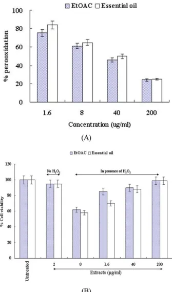
Fig. 5. Inhibitory effect of the extracts from Agastache rugosa on cell death induced by hydrogen peroxide. (A) The effect of the extracts from Agastache rugosa on the inhibition of lipid peroxidation evaluated by measuring the amount of TBARS formation. % peroxidation means the relative values of the positive control without the extracts from Agastache rugosa.
(B) The viability of NIH 3T3 cells on the treatment of hydrogen peroxide was evaluated by a MTT assay. % cell viability means the relative values of the negative control (untreated cells).
Inhibitory effect of the extracts from Agastache rugosa on oxidative cell damage
Inhibitory effect of the EtOAc extracts and the essential oil from Agastache rugosa on oxidative cell damage induced by ROS was examined using MTT assay and lipid peroxidation assay. ROS damage involves injury to cellular membranes.
Measurement of lipid peroxidation is used as an indicator or
membrane damage in mammalian cells. Lipid peroxidation can cause a cascade effect of lipid-derived radicals, thereby causing additional membrane damage. The products of lipid peroxidation, malondialdehyde and other group of aldehyde products may also cause DNA damage (Vaca et al., 1988). It has also been proposed that free radicals derived from lipid peroxidation may function as tumor initiator (Esterbauer et al., 1982). Our result showed that the EtOAc extracts and the essential oil from Agastache rugosa inhibit the lipid peroxi- dation by 25% and 18% at 1.6 μg/ml, 39% and 37% at 8 μg/ml, 54% and 48% at 40 μg/ml and 76% and 77% at 200 μg/ml (Fig. 4A). In MTT assay, the treatment of hydrogen peroxide caused the cell death by about 40% while the EtOAC extracts and the essential oil from Agastache rugosa prevented the cells from the death (Fig. 4B). These results suggest that Agastache rugosa may prevent cell death caused by ROS and have a cancer chemopreventive effect through inhibiting lipid pero- xidation that function as tumor initiator.
In conclusion, the results of the present studies indicate that Agastache rugosa exhibits antioxidant properties, inhibits oxi- dative DNA damage, the cell death and lipid peroxidation caused by ROS through its antioxidant activity. And it is pos- sible to apply to a cancer chemopreventive agent of Agastache rugosa.
Acknowledgement
This work was supported by a support work for training problem solving human resources from Korea research founda- tion and by the second stage of BK21 grants from Ministry of Education and Human Resources Development and GB-Regional Innovation Research Program (2009), Korea
Literature Cited
Aust, S.D., Chignell, C.F., Bray, T.M., Kalyanaraman, B., Mason, R.P., 1993. Free radicals in toxicology. Toxicology and App- lied Pharmacology. 120: 168-178.
Cozzi, R., Ricordy, R., Aglitti, T., Gatta, V., Perticone, P., De Salvia, R., 1997. Ascorbic acid and beta-carotene as modula- tors of oxidative damage. Carcinogenesis. 18: 223-228.
Esterbauer H: In: McBrien, D.C.H., Slater, T.F. (Eds), 1982.
Free Radicals, Lipid Peroxidation and Cancer. Academic Press, New York, pp. 101-128.
Farber E, 1998. Cancer development and its natural history. A cancer prevention perspective. Cance, 62: 1676-1679.
Halliwell, B., Gutteridge JMC, 2000. Oxidative Stress. Free Radicals in Biology and Medicine (3rd ed.), Oxford Univer- sity Press, New York.
Halliwell, B., Aruoma, O.I.,1991. The biological toxicity of free radicals and other reactive species. Free radicals and food additives. 41.
Hus, B., Coupa, I.M., Ng, K.,2006. Antioxidant activity of hot water extract from the fruit of the Doum palm. Hyphaene thebaica. Food Chemistry. 98: 317-328.
Jung, Y., Surh, Y., 2001. Oxidative DNA damage and cytotoxi- city unduced by copper-stimulated redox cycling of salsolinol, a neurotoxic tetrahydroisoquinoline alkaloid. Free Radical Biology and Medicine. 30: 1407-1417.
Kang, K.A., Zhang, R., Piao, M.J., Ko, D.O., Wang, Z.H., Kim, B.J., Park, J.W., Kim, H.S., Kim, D.H., Hyun, J.W., 2008.
Protective effect of irisolidone, a metabolite of kakkalide, against hydrogen peroxide induced cell damage via antioxidant effect. Bioorganic & Medicinal Chemistry. 16: 1133-1141.
Kehrer, J.P., 1993. Free radicals as mediators of tissue injury and disease. Critical Review of Biochemistry and Molecular Biology. 23: 21-48.
Kong, A.N., Yu, R., Hebbar, V., Chen, C., Owuor, R., Hu, R., Mandlekar, S., 2001. Signal transduction events elicited by cancer prevention compounds. Mutation Research. 480/481:
231-241.
Langard, S., 1990. One hundred years of chromium and cancer:
a review of epidemiological evidence and selected case reports.
American Journal of Industrial Medicine. 17: 189-215.
Lee, C.H., Kim, H.N., Kho, Y.E., 2002. Agastinol and Agaste- nol, Novel Ligans from Agastache rugosa and their evaluation in an apoptosis inhibition assay. Journal of Natural Products.
65: 414-416.
Lloyd, R.V., Hanna, P.M., Mason, R.P., 1997. The origin of the hydroxyl radical oxygen in the Fenton reaction. Free Radical Biology and Medicine. 22: 885-888.
Pietraforte, D., Turco, L., Azzini, E., Minetti, M., 2002. On-line EPR study of free radicals induced by peroxidase/H(2)O(2)
in human low-density lipoprotein. Biochimica et Biophysica Acta. 1583: 176-184.
Rosenkranz, A.R., Schmaldienst, S., Stuhlmeier, K.M., Chen, W., Knapp, W., Zlabinger, G.A., 1992. Microplate assay for the detection of oxidative products using 2’,7’-dichloroflu- orescin- diacetate. Journal of Immunological Methods. 156:
39-45.
Salah, N., Miller, N.J., Paganga, G., Tijburg, L., Bolwell, G.P., Rice-Evans, C., 1995. Polyphenolic flavanols as scavengers of aqueous phase radicals and as chain-breaking antioxidants.
Archives of Biochemistry and Biophysics. 322: 339-346.
Shin, S and Kang, C.A., 2003. Antifungal activity of essential oil og agastache rugosa Kuntze and its synergism with keto- conazole. Lett Appl Microbiol. 36: 111-115.
Smirnoff, N., Cumbes, Q.J., 1989. Hydroxyl radical scavenging activity of compatible solutes. Phytochemistry. 28: 1057-1060.
Stohs, S.J., 1995. The role of free radicals in toxicity and disease.
Journal of Basic Clinical Physiology and Pharmacology. 6:
205-228.
Stohs, S.J., Bagchi, D., 1995. Oxidative mechanism in the toxi- city of metal ions. Free Radical Biology and Medicine. 18:
321-336.
Schwarz, S.M., Peres, G., Kunz, W., Furstenberger, G., Kittstein, W., Marks, F., 1984. On the role of superoxide anion radicals in skin tumour promotion. Carcinogenesis. 5: 1663-1670.
Vaca, C.E., Wilhelm, J., Harms-Ringdahl, M., 1988. Interaction of lipid peroxidation products with DNA. A review. Muta- tion Research. 195: 137-149
Vallyathan, V., Shi, X., 1997. The role of oxygen free radicals in occupational and environmental lung diseases. Environmental Health Perspectives. 105: 165-177.
Wang, H., Gao, X.D., Zhou, G.C., Cai, L., YaO, W.B., 2008. In vitro and in vivo antioxidant activity of aqueous extract from Choerospondias axillaris furit. Food Chemistry. 106: 888-895.
Wiseman, H., Halliwell, B., 1996. Damage to DNA by reactive oxygen and nitrogen species: role in inflammatory disease and progression to cancer. Journal of Biochemistry. 313: 17-29.
Yang, C.S., Chung, J.Y., Yang, G., Chhabra, S.K., Lee, M.J., 2000. Tea and tea polyphenols in cancer prevention. Journal of Nutrition. 130: 472-478.
(Received 17 September 2009 ; Accepted 25 Nobember 2009)