online©ML Comm
-
181
- 대한두경부종양학회지제 23 권 제 2 호 2007
하인두암의 보존적 수술과 경부 재발의 술중방사선치료 치험 1예
고려대학교 의과대학 이비인후-두경부외과학교실
조우성·김양수·김무필·백승국·정광윤
= Abstract =
Conservative Surgery of Hypopharyngeal Cancer and Intraoperative Radiation Therapy of Neck Recurrence
Woo-Sung Cho, M.D., Yang Soo Kim, M.D., Moo-Pil Kim, M.D., Seung-Kuk Baek, M.D., Kwang-Yoon Jung, M.D.
Department of Otorhinolaryngology-Head and Neck Surgery, Korea University College of Medicine, Seoul, Korea
Hypopharyngeal cancer usually has invasiveness to adjacent tissue and frequent metastasis to cervical lymph node. In addition, because it often accompanies submucosal extension and second primary malignancy, the sacrifice of larynx and postoperative radiation therapy had been performed in the past. However, it has been reported that conservative surgery of hypopharyngeal cancer show good functional and oncologic outcome according to the development of diagnostic tool and reconstructive technique. We report a case of hypopharyn- geal cancer that received conservative surgery followed radiation therapy and intraoperative radiation therapy for neck recurrence.
KEY WORDS:Hypopharyngeal cancer·Surgery·Radiation therapy.
서 론
하인두암(hypopharyngeal cancer)은 침습적이며, 불량 한 예후를 보이는 대표적인 두경부종양으로 경부로 림프절 전이가 잦고, 이차 중복암이 흔하여 과거로부터 후두를 희 생하고, 술후 방사선 치료를 추가하는 방법이 보편적으로 시행되어 왔으나, 최근 진단 기법의 발달과 더불어 재건술 과 같은 수술적 치료 기법의 발전에 따라 환자의 삶의 질 을 고려한 보존적 술식이 많이 사용되고 있다. 이에 하인두 암에서 보존적 절제술과 재건술을 시행하고 술후 경부 재 발에 대한 술중방사선치료를 시행한 증례에 대해 문헌고찰 과 함께 보고하고자 한다.
증 례
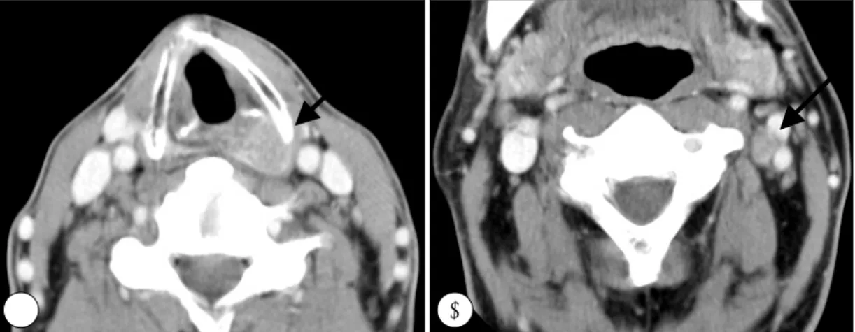
56세 남자 환자가 3개월 된 인후두부 이물감으로 타 병 원에서 시행한 조직 검사상 하인두 편평세포암종으로 진단 받고 본원으로 전원되었다. 후두내시경검사상 좌측 이상와 부의 내측에 2~3cm 가량의 돌출된 병변이 피열연골의 상 부를 침범하였으나 후교련의 중앙을 넘지 않은 소견이었다 (Fig. 1). 전산화단층촬영상 중등도의 조영증강을 보이는 종 물이 갑상연골의 후외측에 인접되어 있었으며 좌측 Level
Ⅱ에 1cm 크기의 전이가 의심되는 림프절이 관찰되었다 (Fig. 2). 원격전이 검사로 시행한 양전자 방출 컴퓨터 단 층 촬영(PET-CT) 검사상 원발부위를 제외한 다른 부위의 특이 소견은 보이지 않았다. T2N1M0인 stage Ⅲ(AJCC 2002)의 경부전이를 동반한 하인두암으로 진단하고, 이에 따라 기관절개술(tracheostomy), 좌측갑상선절제술(hemi- gectomy), 좌측근치적경부절제술(radical neck dissection), 전완피판유리이식술(forearm free flap reconstruction)을 교신저자:정광윤, 136-705 서울 성북구 안암동 5가 126-1
고려대학교 의과대학 이비인후-두경부외과학 교실 전화:(02) 920-5486·전송:(02) 925-5233 E-mail:[email protected]
-
182
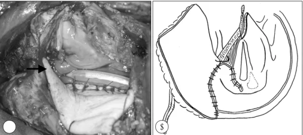
- 이용한 후두측벽재건을 계획하였다. 우선 근치적 경부 절 제술 시행후 설골상 외측 인두 절개술(suprahyoid and la- teral pharyngotomy)통하여 이상와 첨부(pyriform sinus apex)의 병변 유무를 동결절편검사로 확인하였다(Fig. 3).이상와 첨부의 침범이 없음을 확인하고 갑상연골(thyroid cartilage)을 성대 부위까지, 정중선을 따라 절개한 후 동 측 후두개(epiglottis)의 반과 후두실(laryngeal ventricle), 피열 연골(arytenoids cartilage)을 성대 돌기(vocal pro- cess)에서 절개했다. 남아있는 진성대 돌기(true vocal cord process)를 후방 윤상 연골(posterior cricoid cartilage) 에 봉합 하여 정중부에 고정하였다. 노출된 윤상연골과 후 두를 최대한 점막으로 덮기위해 후교련부 부터 노출된 윤 상연골 좌측상부, 고정된 진성대의 후 1/3 부위까지 이상 와 점막에 봉합을 한 후 재건을 위해 전완 피판을 이상와 첨부로부터 시작하여 인두점막에 봉합하였다(Fig. 4). 앞쪽 으로는 갑상연골에 3mm 간격의 구멍을 내어 이에 전완피 판을 고정하였으며 위쪽으로는 설기저부에 피판을 봉합하
여 수술을 마쳤다. 최종 병리조직 소견상 갑상연골과 피열 연골의 침범은 없었으며 경부전이는 level II에서만 확인되었 다. 수술 후 타 대학 병원에서 양측 경부에 2개월에 걸쳐 총
Fig. 1. Preoperative endoscopic view. A huge mass is seen in me- dial aspect of left pyriform sinus(arrows) and vocal cord mobility was normal.
Fig. 2. Preoperative axial CT scan. It shows about 2×3cm enhanced mass in left pyriform sinus(A) and 1×1cm sized mass that is suspected as lymphatic metastasis in level II area(B).
A B
Fig. 3. Intraoperative findings. A:After suprahyoid and lateral pharyngotomy and midline division of epiglottis, hypopharyngeal mass is seen(black arrow), B:Hypopharyngeal mass(black arrow) locate in medial aspect of left pyriform sinus and upper surface of left arytenoid. The broken line shows the range of resection.
A B
-
183
- 량 5220cGy의 방사선 치료를 시행 받았으며, 음성 및 연하 장애는 없이 기능이 잘 유지됨을 추적 관찰할 수 있었다(Fig.5). 방사선 치료 5개월 후에 외래 추적 관찰 중 촉지되는 경 부 종물이 있어(Fig. 6) 시행한 미세침흡인조직검사상 편평세 포암종으로 진단되어 광범위 구제 경부 절제술(extended sal- vage neck dissection)과 술 중 방사선 치료(intraoperative radiation therapy)를 계획하였다. 우선 광범위 구제 경부 절 제술을 시행한 후 1500cGy의 용량을 약 7×7cm 범위에 조 사한 후 수술을 마쳤다(Fig. 7). 현재 약 20개월간 추적 결과 재발소견이나 기능의 이상 없이 술 후 상태가 잘 유지되고 있다.
고 찰
하인두 암은 두경부 영역의 편평세포암종 중에서 예후가 불량한 종양이며, 따라서 후두 전체를 희생하는 공격적인 치 료가 많이 시행되었으나, 술 후 기능 장애 뿐 아니라 보존 적 수술과 비교했을 때 생존률에서도 큰 차이가 없다는 보
고가 있어 최근에는 보존적 술식의 사용이 증가되고 있다1). T1, T2의 초기 병변에서는 술 전 종양의 위치와 도약 병 변의 존재 여부 등을 확인하는 것이 중요하며, 종양의 범위 가 국한되어 있다면, 술 후 기능의 보존을 고려하여 후두의
Fig. 5. Post-operative endoscopic view. It shows well attached forearm free flap without recurrence in post-radiation the-
rapy 5 months. Fig. 6. Post-radiation therapy 5 months axial CT scan. It shows ab- out 2.5×3.5cm sized mass in left level V(Black arrow).
Fig. 7. Intraoperative radiation therapy. After extended salvage neck dissection, radiation therapy was being performed through 7×7cm round cone intraoperatively.
Fig. 4. Intraoperative view of reconstruction using forearm free flap. A:the remnant of left true vocal cord was fixed in midline of posterior commissure and flap was sutured from pyriform sinus apex to posterior pharyngeal wall. B:Schematic figure of re- constructive surgery shows the insetting of forearm free flap and anchoring between flap and thyroid cartilage.
A B
-
184
- 전적출보다는 보존적인 후두 절제술과 함께 술 후 방사선 치료를 시행하는 것이 환자의 삶의 질 향상에 도움이 되며, 이는 또한 공격적인 술식에 비해 생존률과 재발률에도 큰 차이가 없는 것으로 보고되고 있다2). 하인두에는 림프 조 직이 풍부하여 조기에 경부 전이를 일으켜 65~80%에서 진단시 경부 전이를 동반하고 있는 것으로 보고 되고 있으 며, 경부 전이가 없는 환자의 선택적 경부 절제술(selective neck dissection)에서도 30~40%에서 병리 조직학적 경 부 림프절 전이가 있는 것으로 보고되고 있다3)4). 그러나 술 전에 시행한 평가와 달리 병변의 범위가 크다면, 적극적 인 후두 전 적출술을 시행하는 것이 바람직하다. 본 증례에 서는 보존적인 수술을 시행하였고, 술 후 방사선 치료까지 시행 하였으나 재발을 하였다. 그 원인으로는 2가지를 생 각해 볼 수 있다. 첫째, 수술 당시 충분한 경부 절제술(neck dissection)이 되지 않은 점을 꼽을 수 있다. 경부절제술 부위 중에서 횡경동맥이 지나가는 경계 부위의 절제가 충 분하지 않았고, 이에 남아 있는 미세 암종의 재발을 생각해 볼 수 있다. 둘째, 술 후 방사선 치료의 양의 불충분함을 들 수 있다. 술 후 양측 경부에 2개월에 걸쳐 총 5520cGy 의 조사를 받았으나, 후경부 부위의 조사량은 3600cGy로 충분하지 않았고, 이것으로 술 후 남아있는 미세 암종의 치 료가 충분히 되지 않을 가능성이 있다.수술 당시 정확한 경계 부위의 설정 및 충분한 절제 수 술이 필요하며, 술 후 방사선 치료에 있어서도 충분한 조사 범위 및 조사량의 설정이 중요하겠다.
술 중 방사선 치료는 암종의 절제가 불가능하거나, 잔존 암의 가능성이 있거나, 국소 전이의 가능성이 높은 암종의 경우에 치료 적응증이 된다5).
또한 술 중 방사선 치료를 한 후 술 후 방사선 치료를 병합 했을 때, 국소 재발률과 생존률 향상에 도움이 된다고 보고하고 있다6). 그러나 본 증례에서는 술 중 방사선 치료 이후 더 이상의 추가적 방사선 치료가 불가능하여 추가적 방사선 치료를 시행하지 않았고 주기적인 외래 추적관찰로
재발의 여부를 확인하고 있다.
다른 보존적인 치료 방법으로는 수술을 시행하지 않고, 선행항암 요법과 방사선 치료를 시행하는 것이 있으나7) 수 술을 시행 후 방사선 치료를 시행한 군과의 생존률 비교에 있어 성적이 저조한 것으로 보고되고 있다. 따라서 향후 하 인두암의 적정 치료에 대하여 여러가지 연구가 지속되어야 할 것으로 보이며, 병변의 병기가 낮고, 술 전 병변의 범위 등에 대한 정확한 평가가 이루어 질 수 있어 적절한 환자 의 선택이 가능하다면, 환자의 기능 보존을 위하여 보존적 인 후두 절제술 및 술 후 방사선 치료의 병합 요법이 우수 한 치료 방법으로 자리 잡을 수 있다.
중심 단어:
하인두암·보존적 수술·방사선 치료.References
1) Eckel HE, Staar S, Volling P, Sittel C, Damm M, Jungchuclsing M: Surgical treatment for hypopharynx carcinoma: Feasibility, mor- tality, and results. Otolaryngol Head Neck Surg. 2001;124:561-569 2) Czaja JM, Gluckman JL: Surgical management of early-stage
hypopharyngeal carcinoma. AnnOtol Rhino Laryngol. 1997;106: 909-913
3) Shah JP, Shah AR, Spiro RH, Strong EW: Carcinoma of the hypopharynx. Am J Surg. 1976;132:439-443
4) Lefebcre JL, Castelain B, DeLaTorre JC, DelleDeroide A, Van- kemmel B: Lymph node invasion in hypopharynx and lateral epipharynx: A prognostic factor. Head Neck Surg. 1987;10:14-18 5) Kupferman ME, Morrison WH, Santillan AA, et al: The role of
interstitial brachytherapy with salvage surgery for the manage- ment of recurrent head and neck cancers. Cancer. 2007 May 15; 109(10):2052-2057
6) Valentini V, Balducci M, Tortoreto F, Morganti AG, De Giorgi U, Fiorentini G: Intraoperative radiotherapy: current thinking. Euro- pean Journal of Surgical Oncology. 2002;28:180-185
7) Shirinian MH, Weber RS, lippman SM: Larynx preservation by induction chemotherapy plus radiotherapy in locally advanced head and neck cancer. Head Neck. 1994;16:39-44