Received: April 28, 2020 Revised: June 30, 2020 Accepted: June 30, 2020 Trauma and InJury
Correspondence to Jungnam Lee, M.D., Ph.D.
Department of Traumatology, Gil Medical Center, Gachon University College of Medicine, 783 Namdong-daero, Nam- dong-gu, Incheon 21565, Korea Tel: +82-32-460-3010 Fax: +82-32-460-3247 E-mail: [email protected] Co-correspondence to Woo Sung Choi, M.D., Ph.D.
Department of Emergency Medicine, Gil Medical Center, 783 Namdong-daero, Namdong-gu, Incheon 21565, Korea Tel: +82-32-460-3901
Fax: +82-32-460-3019 E-mail: [email protected]
analysis of aspiration risk factors in Severe Trauma Patients: Based on findings of aspiration lung disease in Chest Computed Tomography
Gyu Jin Heo, M.D.
1, Jungnam Lee, M.D., Ph.D.
2, Woo Sung Choi, M.D., Ph.D.
1, Sung Youl Hyun, M.D.,Ph.D.
2, Jin-Seong Cho, M.D., Ph.D.
31
Department of Emergency Medicine, Gil Medical Center, Incheon, Korea
2
Department of Traumatology, Gil Medical Center, Gachon University College of Medicine, Incheon, Korea
3
Department of Emergency Medicine, Gil Medical Center, Gachon University College of Medicine, Incheon, Korea
Purpose: The present study will identify risk factors for aspiration in severe trauma patients by comparing patients who showed a sign of aspiration lung disease on chest computed tomography (CT) and those who did not.
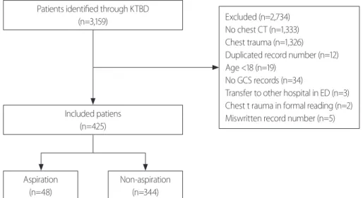
methods: We conducted a retrospective review of the Korean Trauma Data Bank be- tween January 2014 and December 2019 in a single regional trauma center. The inclusion criteria were patients aged ≥18 years with chest CT, and who had an Injury Severity Score ≥16. Patients with Abbreviated Injury Scale (AIS)-chest score ≥1 and lack of medical records were excluded. General characteristics and patient status were analyzed.
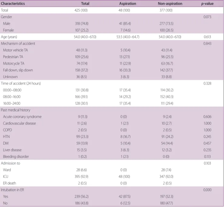
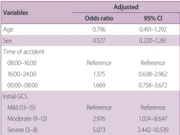
results: 425 patients were included in the final analysis. There were 48 patients show- ing aspiration on CT (11.2%) and 377 patients showing no aspiration (88.7%). Aspira- tion group showed more endotracheal intubation in the ER (p=0.000) and a significant- ly higher proportion of severe Glasgow Coma Scale (GCS) (p=0.000) patients than the non-aspiration group. In AIS as well, the median AIS head score was higher in the aspi- ration group (p=0.046). Median oxygen saturation was significantly lower in the aspi- ration group (p=0.002). In a logistic regression analysis, relative to the GCS mild group, the moderate group showed an odds ratio (OR) for aspiration of 2.976 (CI, 1.024–8.647), and the severe group showed an OR of 5.073 (CI, 2.442–10.539).
Conclusions: Poor mental state and head injury increase the risk of aspiration. To con- firm for aspiration, it would be useful to perform chest CT for severe trauma patients with a head injury.
Keywords: Pneumonia, aspiration; Tomography, X-ray computed; Multiple trauma;
Risk
analysis of aspiration risk factors in Severe Trauma Patients: Based on findings of aspiration lung disease in Chest Computed Tomography
Gyu Jin Heo, M.D.
1, Jungnam Lee, M.D., Ph.D.
2, Woo Sung Choi, M.D., Ph.D.
1, Sung Youl Hyun, M.D.,Ph.D.
2, Jin-Seong Cho, M.D., Ph.D.
31
Department of Emergency Medicine, Gil Medical Center, Incheon, Korea
2
Department of Traumatology, Gil Medical Center, Gachon University College of Medicine, Incheon, Korea
3