□ 연구보고 □
폐암 세포주에서 FHIT 유전자 이입에 의한 Apoptosis의 기전
원자력병원 내과, 실험치료연구실
유정선, 김철현
=Abstract=
Mechanism of FHIT-Induced Apoptosis in Lung Cancer Cell Lines Jung Sun Yoo, Cheol Hyeon Kim, M.D.
Department of Internal Medicine and Laboratory of Experimental Therapeutics, Korea Cancer Center Hospital
Background : The FHIT (fragile histidine triad) gene is a frequent target of deletions associated with abnormal RNA and protein expression in lung cancer. Previous studies have shown FHIT gene transfer into lung cancer cell line lacking FHIT protein expression resulted in inhibition of tumor cell growth attributable to the induction of apoptosis and reversion of tumorigenecity. However, the mechanism of the tumor suppressor activity of the FHIT gene and the cellular pathways associated with its function are not completely understood.
Methods : To gain insight into the biological function of FHIT, we compared the NCI-H358 cell line with its stable FHIT transfectants after treatment with cisplatin or paclitaxel. We investigated the effects of FHIT gene expression on cell proliferation, apoptosis, and activation of caspase system and Bcl-2 family. The induction of apoptosis was evaluated by using DAPI staining and flow cytometry.
Activation of caspases and Bcl-2 members was evaluated by Western blot analysis.
Results : A significantly increased cell death was observed in FHIT transfectants after cisplatin or paclitaxel treatment and this was attributable to the induction of apoptosis. Remarkable changes in caspases and Bcl-2 family were observed in the transfected cells as compared with the control cells after treatment with paclitaxel. Activation of caspase-3 and caspase-7 was markedly increased in cells expressing FHIT. Expression level of Bcl-2 and Bcl-xL protein was significantly decreased and that of Bax and Bad protein was significantly increased in the transfected cells.
Address for correspondence:
Cheol Hyeon Kim, M.D.
Department of Internal Medicine, Korea Cancer Center Hospital 215-4, Gongneung-Dong, Nowon-Gu, Seoul 139-706, Korea
Phone : 02-970-1209 Fax : 02-970-2401 E-mail : [email protected]
Conclusion : FHIT gene delivery into lung cancer cells results in enhanced apoptosis induced by treatment with cisplatin or paclitaxel. The data suggest that apoptosis in FHIT-expressing cells could be related to activation of caspase pathway and Bcl-2 family.(Tuberculosis and Respiratory Diseases 2004, 56:450-464)
Key words : Fragile histidine triad (FHIT), Apoptosis, Lung neoplasms, Paclitaxel, Cisplatin.
서 론
Chromosome 3p의 deletion이 소세포폐암의 90%
이상에서 그리고 비소세포폐암의 80% 이상에서 발견되는, 폐암에서는 가장 흔하게 나타나는 gene
tic alteration이라는 것은 널리 알려진 사실이다1-4. 또한 hyperplasia나 dysplasia 등의 병변과 carci
noma in situ와 같은 발암 단계의 조기에서도 chromosome 3p의 deletion이 잘 관찰되는 것으로 알려져 있다5,6. 따라서 이 위치에 폐암의 암억제유 전자가 존재할 가능성이 매우 높다고 생각되어 과 거부터 이에 대한 연구가 활발하게 진행되어오고 있다7-10. 하지만 현재까지는 몇몇 후보 유전자들이 밝혀져 있을 뿐, 확실한 암억제유전자를 규명해내 지는 못하고 있는 실정이다.
이러한 후보 암억제유전자들 중 최근 Ohta 등이 발표한 3p14.2에 위치하고 있는 FHIT 유전자가 주목을 받고 있다11. 식도, 위, 두경부암 등의 여러 종양에서 이 위치의 homozygous deletion이 보고 된 바 있으며12-15, 서열 분석상 이 유전자가 hu
man genome 중 손상에 가장 취약한 곳 중 하나 인 FRA3B fragile site와 가족성 신세포암에서 잘 발견되는 t(3;8)(p14.2;p24) 유전자 전위의 break
point를 포함하고 있기 때문이다11,16. FHIT 유전자 는 chromosome 3p14.2 위치에 있는 약 1 Mb 정 도의 genomic DNA로서 10개의 exon으로 구성되 어 있으며 이로부터 1.1 kb의 mRNA가 expression 된다11. 10개의 exon중에서도 coding region은 exon 5에서 exon 9까지인데 이로부터 생산되는 16.8 kDa의 단백질은 Schizosaccharomyces pombe
가 생산하는, diadenosine 5',5'''-P1,P4-tetrapho
sphate (Ap4A)를 ATP와 AMP로 분해하는 효소 인, Ap4A asymmetrical hydrolase와 69%의 유사 성을 보이는 것으로 밝혀졌다17,18. 즉, diadenosine triphosphate hydrolase activity를 가지고 있으며, histidine triad (HIT) gene family에 속하기 때문 에 fragile histidine triad gene의 약자로 FHIT gene이라 명명되었다.
폐암 세포주와 조직에 대해서 발표된 기존의 연 구 결과들을 살펴 보면, FHIT 유전자의 hom
ozygous deletion과 이와 관련된 abnormal FHIT mRNA expression이 폐암에서 매우 높은 빈도로 관찰되고 있다19-21. 한 예로 Sozzi 등의 연구에 의 하면 소세포폐암의 80%, 비소세포폐암의 40% 정 도에서 FHIT mRNA transcripts의 이상이 발견되 며 전체의 약 76%에서 FHIT alleles의 loss가 발 견되었다고 한다19. 이후 FHIT 단백질에 대한 항 체를 이용한 면역조직화학염색 실험 결과들이 발 표되었는데 이러한 gene abnormality는 단백질의 발현 결손과 일치함이 밝혀졌다22.
이상의 소견들을 바탕으로 현재 FHIT 단백질이 실제로 세포 내에서 어떠한 기능을 하는가에 대한 연구가 활발히 진행 중에 있다. 일부 연구에 의하 면 FHIT 유전자를 세포 내에 이입시키고 그 변화 를 관찰하였을 때 apoptosis가 유발되었고, 세포 주기의 이상 소견이 관찰되었으며, 나아가 nude mice에서 폐암세포주의 종양형성 능력이 억제됨이 관찰되었다23-25. 하지만 아직까지 FHIT 단백질이 세포내에서 어떠한 기능을 하여 이러한 현상들이 관찰되는지에 대한 지식은 미진한 상태이다.
본 연구에서는 FHIT 유전자를 폐암 세포에 이 입시켰을 때 유발되는 apoptosis의 기전을 규명하 여 FHIT 단백질의 기능을 알아보고자 하였다. 이 를 위하여 FHIT 유전자가 결손된 폐암세포주에 FHIT 유전자를 stable transfection 시킨 후 항암 제를 가하고 apoptosis가 항진되어 나타나는가를 조사해 보았다. 또한 이 과정에서 나타나는 몇몇 apoptosis 관련 단백질들의 변화를 살펴보았다.
대상 및 방법
1. 세포 배양
폐암 세포주 중 FHIT 단백질의 발현이 결손된 것 으로 알려져 있는 NCI-H358 세포를 10% fetal bovine serum (FBS), 100 μg/ml의 streptomycin이 첨가된 RPMI 1640 배지에 37℃, 5% CO2의 조건으 로 배양하였다. NCI-H358 세포에 FHIT gene을 transfection한 세포는 위와 동일한 조건 하에서 700 μg/ml의 G418 (geneticin)을 첨가하여 배양하였다.
2. 시 약
Paclitaxel, cisplatin, 3-(4, 5-dimethyl thiazol-2- yl)- 2, 5-diphenyl tetrazolium bromide (MTT) 용 액, 4',6-diamidino-2-phenylindole dihydrochloride (DAPI)는 Sigma (St. Louis, MO, USA)에서 구입 하였다. Rabbit polyclonal anti-FHIT 항체는 Zymed (South San Francisco, CA, USA)에서, mouse polyclonal anti-PARP 항체, mouse mono
clonal anti-Bcl-2 항체, rabbit polyclonal anti- Bcl-xL 항체, rabbit polyclonal anti-Bad 항체는 Santa Cruz Biotechnology (Santa Cruz, CA, USA) 에서, rabbit polyclonal anti-cleaved caspase-3 (Asp175) 항체, rabbit polyclonal anti-cleaved caspase-7 (Asp178) 항체는 Cell Signaling Te
chnology (Beverly, MA, USA)에서, rabbit poly
clonal anti-Bax 항체는 DakoCytomation (Carpin
teria, CA, USA)에서 각각 구입하여 사용하였다.
ECL Western blotting detection systemTM은 Amersham Biosciences (Piscataway, NJ, USA)의 제품이었고, LipofectamineTM Reagent는 Invitro
gen Life Technologies (Carlsbad, CA, USA)의 제 품이었다. Annexin V-FITC, annexin V binding buffer, propidium iodide, mouse monoclonal anti- caspase-8 항체, rabbit polyclonal anti-caspase-9 항체는 PharMingen (San Diego, CA, USA)에서, Z-VAD-FMK는 R&D Systems (Minneapolis, MN, USA)에서 구입하였다.
3. Eukaryotic expression vector의 제작
먼저 human FHIT cDNA 중 coding region인 exon 5에서 exon 9에 해당하는 cDNA를 얻기위해 정상 조직에서 뽑은 RNA로부터 이 부위를 cover 하는 primer를 이용하여 RT-PCR을 시행하였다.
이때 사용한 primer는 XbaI과 HindIII의 restri
ction site를 가지고 있도록 제작하였다18. Cytome
galovirus (CMV) immediate early gene의 enha
ncer-promotor sequence와 neomycin resistant gene을 포함하고 있는 eukaryotic expression vector인 pRc/CMV plasmid (Invitrogen Life Technologies, Carlsbad, CA, USA)를 구입하였다.
이 vector 역시 XbaI과 HindIII의 restriction site 를 가지고 있다. XbaI/HindIII로 digestion 시킨 pRc/CMV vector에 역시 같은 효소로 digestion 시킨 RT-PCR product를 ligation시켰다. 완성된 plasmid (pRc/CMV-FHIT)는 sequencing을 시행 하여 염기 서열을 확인하였다.
4. 폐암 세포주에 transfection
6-well dish plate에 NCI-H358 세포 1X10⁵개를 분 주한 뒤, CO2 incubator에 37℃에서 18-24시간 정
도 유지하여 세포가 50-80% 정도 confluent해 지 도록 하였다. 한편, 아무 것도 첨가되지 않은 RPMI 1640 배지 100 μl에 FHIT DNA 0.8-1.6 μg 을 첨가하고 또 다른 RPMI 1640 배지 100 μl에 2 mg/ml의 LipofectamineTM Reagent 2-20 μl를 첨 가한 뒤, 두 용액을 부드럽게 혼합하여 실온에 15-45분 정도 유지하였다. 50-80% 정도 confluent 해진 세포를 RPMI 1640 배지로 씻어낸 뒤 0.8 ml 의 배지로 다시 채우고 여기에 위의 혼합액을 상 층으로 가한 뒤 CO2 incubator에 37℃에서 5시간 동안 유지하였고, 5시간 후 20% FBS가 첨가된 RPMI 1640 배지 1 ml을 첨가하였다. 48시간이 지 난 후에 G418 700 μg/ml이 첨가된 10% FBS RPMI 1640 배지로 교환한 뒤 selection을 시작하 였다. 2주간 G418로 selection을 시행한 후 G418- resistant colony를 isolation하여 배양하였다. 형성 된 colony는 rabbit polyclonal anti-FHIT 항체로 Western blotting을 시행하여 FHIT 발현을 확인 하였다.
5. Paclitaxel, cisplatin의 처리
폐암 세포주에서 FHIT transfection이 paclitaxel 이나 cisplatin에 의해 유발되는 apoptosis에 미치 는 영향을 알아보고자 하였다. FHIT가 transfec
tion된 세포와 그렇지 않은 세포에 각각 paclitaxel 0-5 μg/ml 혹은 cisplatin 0-30 μg/ml을 가하고, 48-72시간 후 세포 생존율을 분석하여 비교해 보 았다.
6. MTT assay에 의한 세포 생존율의 측정
3-(4, 5-dimethyl thiazol-2-yl)-2, 5-diphenyl tetra
zolium bromide (MTT) assay를 이용하여 세포 생존율을 측정하였다. 96-well plate에 well당 1×
104개의 세포를 분주하고 4-5시간 뒤 항암제를 처 리하여 정해진 시간 동안 배양하였다. 배양된 세포
에 5 mg/ml의 MTT 용액을 15 μl 첨가하고 CO2
incubator에 37℃에서 5시간 동안 배양하였다. 배 양액을 제거한 후 50 μl의 DMSO를 첨가하여 녹 이고 분광광도계를 이용하여 590 nm 파장에서 흡 광도를 측정하였다. 4개 well의 성적을 분석하였다.
7. DAPI staining
6-well plate에 well당 1×10⁶개의 세포를 분주하고 4-5시간 뒤에 cisplatin을 5 μg/ml로, paclitaxel을 0.1 μg/ml로 처리한 뒤 CO2 incubator에서 37℃로 48시간 동안 배양하였다. 1200 rpm으로 3분간 원 심분리를 하여 pellet을 만들고 이 pellet에 me
thanol 100-200 μl를 혼합하여 세포를 고정시켰다.
이 상태로 4℃에서 30분 정도 보관한 후 슬라이드 에 고정된 세포를 약 1-2방울 떨어뜨리고 건조시 켰다. 여기에 4',6-diamidino-2-phenylindole dihy
drochloride (DAPI)를 1 μg/ml 농도로 가한 후 30 분 동안 염색하였다. 3번의 세척 후 형광현미경으 로 관찰하였다.
8. Annexin V/propidium iodide staining and flow cytometry (FACS)
6-well plate에 well당 1×10⁶개의 세포를 분주하고 4-5시간 뒤에 cisplatin (5 μg/ml), paclitaxel (0.1 μ g/ml)을 처리한 뒤 CO2 incubator에서 37℃로 48 시간 동안 배양하였다. Tripsin/EDTA를 처리하여 세포를 수확한 후 차가운 phosphate-buffered saline (PBS)으로 세척한 뒤 1X annexin V bin
ding buffer에 세포를 다시 부유 시켰다. 100 μl의 부유액을 5 ml culture tube에 옮긴 뒤 5 μl의 annexin V-FITC와 5 μl의 propidium iodide를 첨 가하였다. 세포를 vortex하고 어두운 실온에 15분 간 놔둔 뒤 1X annexin V binding buffer 400 μl 를 첨가하여 한 시간 이내에 flow cytometry 분석 을 시행하였다.
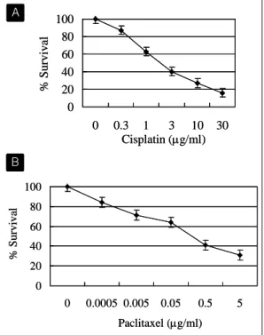
Fig. 1. Cisplatin and paclitaxel decrease cell survival in lung cancer cells. (A) Dose- dependent effect of cisplatin on the cell survival. NCI-H358 cells were incubated with 0, 0.3, 1, 3, 10, 30 μg/ml of cisplatin for 72 hours. (B) Dose-dependent effect of paclitaxel on the cell survival. NCI-H358 cells were incubated with 0, 0.0005, 0.005, 0.05, 0.5, 5 μg/ml of paclitaxel for 48 hours. Cell viability was assayed by MTT assay. Data are shown as mean percentage of control ± standard deviation.
% Survival
Paclitaxel (μg/ml) 0
20 40 60 80 100
0 0.0005 0.005 0.05 0.5 5
% Survival
Paclitaxel (μg/ml) 0
20 40 60 80 100
0 0.0005 0.005 0.05 0.5 5 0
20 40 60 80 100
0 0.3 1 3 10 30
Cisplatin (μg/ml)
% Survival
0 20 40 60 80 100
0 0.3 1 3 10 30
Cisplatin (μg/ml)
% Survival
9. Western blot 분석
RIPA buffer (1% Nonidet P-40, 0.1% SDS, 0.5%
Deoxycholic acid, 50 mM Tris-Hcl (pH 7.5), 150 mM NaCl, 0.5 mM PMSF, 5 μg/ml Leupeptin, 5 μg/ml Aprotinin, 1 mM Na3VO4, 3 mM DTT)를 이용하여 총세포 단백을 추출하였다. 50 μg의 세포 단백을 10% SDS-polyacrylamide gel에서 전기영 동 시켰다. 1시간 30분 동안 180 mA의 일정한 전
류로 단백질들을 nitrocellulose membrane으로 transfer시키고, 이 membrane을 blocking solution (5% skin milk in 1X PBS/Tween20)으로 30분 동 안 block 시킨 후 일차 항체와 12시간 동안 반응 시켰다. 일차 항체들의 반응 농도는 rabbit poly
clonal anti-FHIT 항체가 1:500, mouse polyclonal anti-PARP 항체가 1:1000, rabbit polyclonal anti- cleaved caspase-3 (Asp175) 항체가 1:1000, rabbit polyclonal anti-cleaved caspase-7 (Asp178) 항체 가 1:1000, mouse monoclonal anti-caspase-8 항체 가 1:500, rabbit polyclonal anti-caspase-9 항체가 1:1000, mouse monoclonal anti-Bcl-2 항체가 1:
1000, rabbit polyclonal anti-Bcl-xL 항체가 1:500, rabbit polyclonal anti-Bax 항체가 1:500, rabbit polyclonal anti-Bad 항체가 1:200 이었다. 세척 후 이차 항체를 1:2000-1:10000으로 첨가하여 반응시 킨 후 면역신호의 검출은 ECL Western blotting detection systemTM을 이용하였다.
10. 통계 분석
농도별 세포 생존율의 차이는 SPSS for Windows Release 11.0 프로그램의 Mann-Whitney U-test를 이용하여 분석하였다.
결 과
1. 항암제 투여에 따른 폐암 세포의 생존율
NCI-H358 폐암 세포주에 cisplatin을 72시간 동안, paclitaxel을 48시간 동안 농도별로 투여하고 세포 의 생존율을 MTT assay로 조사하였다. Cisplatin 을 0-30 μg/ml 농도까지, paclitaxel을 0-5 μg/ml 농도까지 투여하였을 때, 농도를 증가시킴에 따라 세포의 생존율은 지속적으로 감소함을 관찰할 수 있었다(Fig. 1).
A
B
Fig. 2. Expression of FHIT increases during cell death induced by cisplatin or paclitaxel.
(A) NCI-H1573 and A549 cells were treated with cisplatin (3 μg/ml) for the indicated times. (B) NCI-H1573 and A549 cells were incubated with paclitaxel (0.1 μ g/ml) for the indicated times. Total cellular extracts were separated by 10% SDS- PAGE and FHIT was detected by Western blot analysis.
H1573
A549
Cisplatin 0 24 48 72 (hr)
Actin
Actin FHIT
FHIT H1573
A549
Cisplatin 0 24 48 72 (hr)
Actin
Actin FHIT
FHIT
Actin FHIT H1573
Paclitaxel 0 24 48 72 (hr)
Actin FHIT A549
Actin FHIT H1573
Paclitaxel 0 24 48 72 (hr)
Actin FHIT A549
Fig. 3. Western blot analysis showing FHIT ex pression in stably transfected NCI-H358 cells. Signals due to anti-FHIT, anti-β -actin antibody used as a protein loading control are indicated. Lanes 1, untran sfeced; 2, transfected with pRc/CMV pla smid; 3, transfected with recombinant pRc/
CMV-FHIT.
1 2 3
Actin FHIT
1 2 3
Actin FHIT
2. 항암제 투여시 발생하는 세포 사멸에서 FHIT 단백질의 발현 증가
항암제를 투여하였을 때 나타나는 세포 사멸 과정 중 혹시 FHIT 단백질의 발현에 어떠한 변화가 나 타나는지를 알아보기 위해, FHIT가 결손되지 않은 세포주인 NCI-H1573과 A549 세포주에 항암제를 투여하고 FHIT 발현을 Western blotting으로 관찰
하였다(Fig. 2). 먼저 cisplatin 3 μg/ml을 투여하고 시간별로 관찰하였을 때 NCI-H1573과 A549 세포 모두에서 시간 경과에 따른 FHIT 단백질의 발현 증가를 관찰할 수 있었다. Paclitaxel 0.1 μg/ml을 투여하고 관찰하였을 때도 같은 결과를 얻을 수 있 었다. 이러한 소견은 항암제에 의한 세포 사멸 과정 에 FHIT가 관여할 수도 있음을 시사하고 있다.
3. 폐암 세포주에 FHIT 유전자를 stable tran
sfection 시킨 후 세포 생존율의 관찰
FHIT 유전자의 발현이 항암제에 의한 세포의 사 멸에 미치는 영향을 알아보기 위해 FHIT가 결손 된 폐암 세포주인 NCI-H358 세포에 FHIT 유전 자를 stable transfection 시킨 후 항암제에 대한 반응의 변화를 살펴보고자 하였다. FHIT 유전자 를 삽입한 plasmid인 pRc/CMV-FHIT를 tran
sfection 시켰을 때, transfection을 시키지 않은 세 포나 FHIT 유전자를 삽입하지 않은 plasmid인 pRc/CMV를 transfection 시킨 세포에서는 관찰되 지 않는 FHIT 단백질의 발현을 확인 할 수 있었 다(Fig. 3). 이렇게 FHIT가 stable transfection된 세포에서 항암제를 투여하기 전 기저 상태에서 생 A
B
Fig. 4. The effects of pRc/CMV-FHIT stable tran sfection on the growth of NCI-H358 cells.
The stable transfection of pRc/CMV-FHIT shows no significant effect on the cell gro wth in NCI-H358 cells, as compared with untransfected and pRc/CMV transfected cells.
0 20 40 60 80 100
H358 H358-
pRc/CMV
H358- pRc/CMV-
FHIT
% Survival
0 20 40 60 80 100
H358 H358-
pRc/CMV
H358- pRc/CMV-
FHIT
% Survival
0 2 0 4 0 6 0 8 0 1 0 0
0 0 .3 1 3 1 0 3 0
H 3 5 8 H 3 5 8 - p R c /C M V H 3 5 8 - p R c /C M V- F H I T
Cisplatin (μg/ml)
% Survival
* *
*
0 2 0 4 0 6 0 8 0 1 0 0
0 0 .3 1 3 1 0 3 0
H 3 5 8 H 3 5 8 - p R c /C M V H 3 5 8 - p R c /C M V- F H I T
Cisplatin (μg/ml)
% Survival
* *
*
Cisplatin (μg/ml)
% Survival
* *
*
0 2 0 4 0 6 0 8 0 1 0 0
0 0 .0 0 0 5 0 .0 0 5 0 .0 5 0 .5 5
H 3 5 8 H 3 5 8 - p R c /C M V H 3 5 8 - p R c /C M V-F H I T
Paclitaxel (μg/ml)
% Survival *
* * *
0 2 0 4 0 6 0 8 0 1 0 0
0 0 .0 0 0 5 0 .0 0 5 0 .0 5 0 .5 5
H 3 5 8 H 3 5 8 - p R c /C M V H 3 5 8 - p R c /C M V-F H I T
Paclitaxel (μg/ml)
% Survival *
* * *
Paclitaxel (μg/ml)
% Survival *
* * *
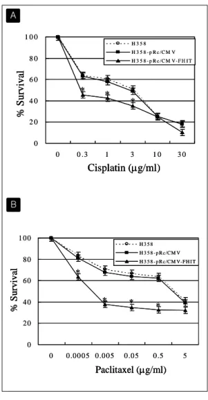
Fig. 5. Transfection of FHIT gene increases the cell death induced by cisplatin and pa clitaxel. (A) Cells were treated with 0, 0.3, 1, 3, 10, 30 μg/ml of cisplatin for 72 hours.
In NCI-H358, pRc/CMV-FHIT tranfection enhanced the decrease of cell survival as compared with control cells. (B) Cells were incubated with 0, 0.0005, 0.005, 0.05, 0.5, 5 μg/ml of paclitaxel for 48 hours. In NCI-H358, pRc/CMV-FHIT tranfection en hanced the decrease of cell survival as compared with control cells. Cell viability was assayed by MTT assay. Data are shown as mean percentage of control±
standard deviation. *: p<0.05 compared to control.
존율에 차이가 있는지를 MTT assay로 분석해 보 았다. Transfection을 시키지 않은 NCI-H358 세포 의 생존율을 100%로 하였을 때 FHIT 유전자를 삽입하지 않은 plasmid인 pRc/CMV를 transfection 시킨 세포 (H358-pRc/CMV)와 FHIT 유전자를 삽입한 plasmid인 pRc/CMV-FHIT를 transfection 시킨 세포 (H358-pRc/CMV-FHIT)에서 모두 차 이를 보이지 않았다(Fig. 4).
4. FHIT 유전자가 stable transfection된 세포에 서 항암제 투여에 의한 apoptosis의 변화
위의 H358, H358-pRc/CMV, H358-pRc/CMV- FHIT 세 가지 세포에 각각 항암제를 가하고 이 때 발생하는 apoptosis에 차이가 나타나는가를 조 사해 보았다. Cisplatin을 0-30 μg/ml 농도로 72 시 간 동안 투여하였을 때 H358-pRc/CMV-FHIT 세 포는 나머지 두 세포에 비해 통계적으로 유의하게 생존율이 감소하였다 (p<0.05). 예를 들어 cisplatin 농도 1 μg/ml에서 H358은 61%, H358-pRc/CMV
A
B
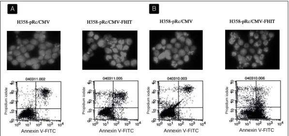
Fig. 6. Cisplatin and paclitaxel induce markedly increased apoptosis in cells transfected with FHIT gene. H358-pRc/CMV and H358-pRc/CMV-FHIT cells were treated with 3 μg/ml of cisplatin for 48 hours (A) or with 0.1 μg/ml of paclitaxel for 24 hours (B). Apoptosis was analysed by DAPI staining and FACS analysis by using both annexin V and propidium iodide. Comparison of fluorescent staining profiles of H358-pRc/CMV with that of H358-pRc/CMV-FHIT cells indicated a higher percentage of apoptotic H358-pRc/CMV-FHIT with condensed and fragmented nuclei. The apoptotic rate of H358-pRc/CMV-FHIT and H358-pRc/CMV cells was 23.4% and 12.7% respectively after cisplatin treatment, and 58.1% and 7.8% respectively after paclitaxel treatment.
H358-pRc/CMV H358-pRc/CMV-FHIT
Annexin V-FITC Annexin V-FITC
Propidiumiodide Propidiumiodide
H358-pRc/CMV H358-pRc/CMV-FHIT
Annexin V-FITC Annexin V-FITC
Propidiumiodide Propidiumiodide
H358-pRc/CMV H358-pRc/CMV-FHIT
Propidiumiodide Propidiumiodide
Annexin V-FITC Annexin V-FITC
H358-pRc/CMV H358-pRc/CMV-FHIT
Propidiumiodide Propidiumiodide
Annexin V-FITC Annexin V-FITC
는 58%의 생존율을 보인 반면, H358-pRc/CMV- FHIT 세포는 43%로 생존율이 뚜렷이 낮게 나타 났다. 이러한 현상은 paclitaxel을 투여하였을 때 더욱 뚜렷이 나타났다. Paclitaxel 농도 0.05 μg/
ml에서 각각 H358은 67%, H358-pRc/CMV는 64%, H358-pRc/CMV-FHIT 세포는 35%의 생존 율을 보였고, IC50는 H358이 1.7 μg/ml, H358-pRc/
CMV가 1.4 μg/ml, H358-pRc/CMV-FHIT가 현저 히 낮은 0.002 μg/ml 였다(Fig. 5). 이러한 세포 생 존율의 감소가 apoptosis에 기인한 것인지를 확인 하기 위해 H358-pRc/CMV 세포와 H358-pRc/
CMV-FHIT 세포에 항암제를 투여한 후 DAPI staining과 FACS 분석을 시행하였다. Cisplatin을 투여하고 48시간 후 관찰하였을 때, H358-pRc/
CMV-FHIT 세포는 H358-pRc/CMV 세포에 비해 DAPI staining상 apoptosis가 유의하게 증가되어
있었으며 FACS 결과에서도 apoptosis 분획이 증가 되어 있음을 확인할 수 있었다 (각각 23.4%, 12.7%).
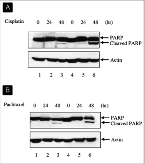
Paclitaxel을 투여하고 24시간 후에 관찰하였을 때도 같은 현상을 관찰할 수 있었는데 FACS 분석상 apoptosis 분획은 각각 58.1%와 7.8%로 더욱 큰 차 이를 보였다(Fig. 6). 한편, apoptosis의 특징적 소견 인 poly (ADP-ribose) polymerase (PARP)의 분해 를 시간 경과에 따라 Western blotting으로 조사해 보았다. H358-pRc/CMV와 H358-pRc/CMV-FHIT 세포에 3 μg/ml의 cisplatin 혹은 0.1 μg/ml의 paclitaxel을 투여하고 48시간까지 PARP의 분해를 관찰하였을 때, PARP의 분해시 나타나는 86 kDa 분절은 48시간째에 나타났는데 band의 density를 비교해 보았을 때 H358-pRc/CMV-FHIT 세포에 서 훨씬 뚜렷이 나타남이 확인되었고(Fig. 7), 이는 MTT assay 결과와 같은 양상이라고 할 수 있다.
A B
Fig. 7. Cisplatin and paclitaxel induce markedly increased poly(ADP-ribose) polymerase (PARP) cleavage in cells transfected with FHIT gene. H358-pRc/CMV (Lanes 1, 2, and 3) and H358-pRc/CMV-FHIT cells (Lanes 4, 5, and 6) were treated with 3 μ g/ml of cisplatin for 0, 24, and 48 hours (A) or with 0.1 μg/ml of paclitaxel for 0, 24, and 48 hours (B). Total cellular extracts were separated by 10% SDS- PAGE and PARP was detected by Western blot analysis.
Cisplatin 0 24 48 0 24 48 (hr)
1 2 3 4 5 6
PARP Cleaved PARP
Actin
Cisplatin 0 24 48 0 24 48 (hr)
1 2 3 4 5 6
PARP Cleaved PARP
Actin
Paclitaxel 0 24 48 0 24 48 (hr)
1 2 3 4 5 6
PARP Cleaved PARP
Actin Paclitaxel 0 24 48 0 24 48 (hr)
1 2 3 4 5 6
PARP Cleaved PARP
Actin
이상의 결과는 cisplatin이나 paclitaxel 투여에 의 한 세포 생존율의 감소는 apoptosis에 의하며, FHIT가 발현된 세포에서는 그렇지 않은 세포에 비해 항암제에 의한 apoptosis가 증가함을 보여주 고 있다.
5. FHIT 유전자가 transfection된 세포에서 ca
spase system의 변화
활성화된 caspase에 의한 단백 분해는 apoptosis에 서 관찰되는 특징적인 소견으로서, DNA의 분절화
와 함께 세포 사멸 과정을 비가역적인 과정이 되 게 한다. 위에서 관찰된 FHIT 단백질 발현에 의 해 유발되는 apoptosis의 증가 과정에 caspase sys
tem이 관여하는지를 평가하기 위하여 H358-pRc/
CMV 세포와 H358-pRc/CMV-FHIT 세포에 pacli
taxel을 투여한 후 caspase-3, -7, -8, -9의 활성을 Western blot 분석으로 조사하였다(Fig. 8). 32 kDa의 caspase-3는 12 kDa 분절로 분해되어 활성 화됨이 알려져 있다. 0.1 μg/ml의 paclitaxel을 투여 하고 0, 24, 48, 72 시간 후에 cleaved caspase-3를 관찰하였을 때, H358-pRc/CMV, H358-pRc/CMV- FHIT 세포 모두에서 48시간째에 cleaved ca
spase-3의 발현이 나타났으나 H358-pRc/CMV- FHIT에서 훨씬 강하게 발현되었다. Cleaved ca
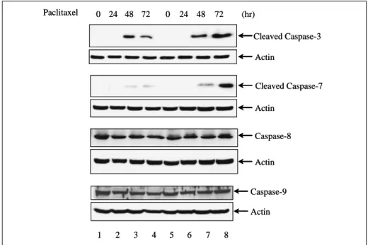
spase-7 (20 kDa)에 있어서도 같은 소견을 관찰할 수 있었다. 하지만 caspase-8, -9에 있어서는 두 세포간에 차이를 관찰할 수 없었다. 나아가 이러한 caspase-3. -7 활성화의 증가가 apoptosis의 증가 와 직접 관련되어 있는지를 확인하기 위하여, pan-caspase inhibitor인 Z-VAD-FMK를 10 μM로 1시간동안 전처치한 후 paclitaxel을 투여하고 PARP의 분해와 세포 생존율의 변화를 관찰해 보 았다(Fig. 9). H358-pRc/CMV, H358-pRc/CMV- FHIT 세포 모두에서 paclitaxel 투여 48시간째에 관찰되던 PARP의 분해가 Z-VAD-FMK의 투여 에 의해 소실됨이 확인되었다(Fig. 9A). MTT 분 석 결과를 살펴보면 Z-VAD-FMK를 투여했을 때 H358-pRc/CMV, H358-pRc/CMV-FHIT 세 포 모두에서 생존율이 증가되었는데 paclitaxel 만 을 투여했을 때 관찰되던 두 세포간의 유의한 차 이가 소실된 것을 관찰할 수 있었다(Fig. 9B). 이 결과는 NCI-H358 세포에서 FHIT 단백질 발현 에 의해 항암제 투여 후 나타나는 apoptosis가 증 가되는 경로에 caspase-3과 caspase-7의 활성화 가 관련되어 있음을 보여주는 소견이라 할 수 있 다.
A
B
Fig. 8. Modulation of caspases in cells transfected with pRc/CMV-FHIT. Restored FHIT expression induces markedly increased activation of caspase-3 and -7. On the contrary, caspase-8 and -9 expression is not significantly affected by exogenous FHIT expression. H358-pRc/CMV (Lanes 1, 2, 3, and 4) and H358-pRc/CMV-FHIT cells (Lanes 5, 6, 7, and 8) were treated with 0.1 μg/ml of paclitaxel for 0, 24, 48, and 72 hours. Total cellular extracts were sepa rated by 10% SDS-PAGE and caspases were detected by Western blot analysis.
Paclitaxel 0 24 48 0 24 48 (hr)
1 2 3 4 5 6
Cleaved Caspase-7 Actin
72 72
7 8
Cleaved Caspase-3 Actin
Caspase-9 Actin
Caspase-8
Actin
Paclitaxel 0 24 48 0 24 48 (hr)
1 2 3 4 5 6
Cleaved Caspase-7 Actin
72 72
7 8
Cleaved Caspase-3 Actin
Caspase-9 Actin
Caspase-8
Actin
6. FHIT 유전자가 stable transfection된 세포에 서 Bcl-2 family의 변화
한편, FHIT 단백질 발현에 의해 유발되는 apopto
sis의 증가 과정에 Bcl-2 family가 관여하는지를 평가하기 위하여 H358-pRc/CMV 세포와 H358- pRc/CMV-FHIT 세포에 paclitaxel을 투여한 후 Bcl-2, Bcl-xL, Bax, Bad의 활성을 Western blot 분석으로 조사하였다(Fig. 10). Bcl-2 family는 미 토콘드리아의 투과성과 cytochrome c의 분비를 조 절함으로써 apoptosis를 제어함이 잘 알려져 있다.
여기에는 apoptosis를 억제하는 단백질과 항진시키 는 단백질이 모두 포함되어 있는데, Bcl-2, Bcl-xL 등은 미토콘드리아의 외벽에 존재하면서 cyto
chrome c의 분비를 막아 apoptosis를 억제하며,
Bax, Bad 등은 세포질 내에 존재하다가 사멸 신호 가 전달되면 미토콘드리아로 이동하여 cytochrome c의 분비를 촉진함으로써 apoptosis를 항진시키게 된다. H358-pRc/CMV, H358-pRc/CMV-FHIT 두 세포에 0.1 μg/ml의 paclitaxel을 투여하고 0, 48 시 간 후에 관찰하였을 때, H358-pRc/CMV-FHIT 세 포에서는 paclitaxel을 투여하고 48 시간 후 H358- pRc/CMV 세포에 비해 Bcl-2와 Bcl-xL는 유의하 게 감소하고 Bax와 Bad는 유의하게 증가함을 관찰 할 수 있었다. 즉, apoptosis 억제 단백질의 감소와 apoptosis 유발 단백질의 증가를 관찰할 수 있었다.
이 결과는 NCI-H358 세포에서 FHIT 단백질 발현 에 의해 유발되는 항암제 투여 후 apoptosis의 증 가 과정이 미토콘드리아 의존성 기전에 의한 것임 을 시사하는 소견이라 할 수 있다.
Fig. 9. Z-VAD-FMK prevents enhanced apoptosis induced by paclitaxel in FHIT-transfected cells.
(A) H358-pRc/CMV (Lanes 1, 2, 3, and 4) and H358-pRc/CMV-FHIT cells (Lanes 5, 6, 7, and 8) were treated with 0.1 μg/ml of paclitaxel for 48 hours with or without Z-VAD-FMK preincubation (10 μM). Total cellular extracts were separated by 10% SDS-PAGE and PARP was detected by Western blot analysis. (B) Cells were incubated with 0, 0.0005, 0.005, 0.05, 0.5, 5 μg/ml of paclitaxel for 48 hours with or without Z-VAD-FMK preincubation (10 μM).
Z-VAD-FMK significantly inhibited paclitaxel-induced cell death in both H358-pRc/CMV and H358-pRc/CMV-FHIT cells and prevented enhanced apoptosis in FHIT-transfected cells. Cell viability was assayed by MTT assay. Data are shown as mean percentage of control ± standard deviation.
1 2 3 4 5 6 7 8 Paclitaxel - - + +
Z-VAD-fmk - + - +
- - + + - + - +
Cleaved PARPPARP
Actin 1 2 3 4 5 6 7 8
Paclitaxel - - + + Z-VAD-fmk - + - +
- - + + - + - +
Cleaved PARPPARP
Actin
0 2 0 4 0 6 0 8 0 1 0 0
0 0 .0 0 0 5 0 .0 0 5 0 .0 5 0 .5 5
H 3 5 8 -p R c /C M V
H 3 5 8 -p R c /C M V- F H IT
H 3 5 8 -p R c /C M V, Z -VA D
H 3 5 8 -p R c /C M V- F H IT , Z - VA D
Paclitaxel (μg/ml)
% Survival
0 2 0 4 0 6 0 8 0 1 0 0
0 0 .0 0 0 5 0 .0 0 5 0 .0 5 0 .5 5
H 3 5 8 -p R c /C M V
H 3 5 8 -p R c /C M V- F H IT
H 3 5 8 -p R c /C M V, Z -VA D
H 3 5 8 -p R c /C M V- F H IT , Z - VA D
Paclitaxel (μg/ml)
% Survival
고 찰
일부 연구에 의하면 FHIT 유전자를 폐암 세포 내 에 이입시키고 그 변화를 관찰하였을 때 apoptosis 가 유발되었으며, 세포 주기 분석에서도 이상 소견 이 관찰되었다23-25. 나아가 nude mice에서 폐암세 포주의 종양형성 능력이 억제됨도 관찰되었다24,25. 하지만 아직까지 FHIT 단백질이 세포내에서 어떠 한 경로를 거쳐 apoptosis를 유발하는지에 대한 지 식은 매우 부족한 상태이다. 현재까지 보고된 몇몇
apoptosis 관련 단백질들에 대한 연구 결과를 보면 caspase system의 활성화가 관련되어 있음이 알려 지고 있다. Adenoviral vector를 이용하여 FHIT 유전자를 transduction시켰을 때, 식도암 세포에서는 caspase-8, -9이26, 췌장암 세포에서는 caspase-3, -8, -9이27, 폐암세포에서는 caspase-8이25, 유방암 세포에서는 caspase-2, -9이 활성화 되었다28. 본 연구에서도 caspase system의 활성화를 관찰 할 수 있었다. FHIT가 발현된 세포에서는 PARP 의 바로 위 단계에 있는 caspase-7과 그 위 단계 A
B
Fig. 10. Activation of proapoptotic proteins and inactivation of anti-apoptotic proteins of Bcl-2 family in cells transfected with pRc/CMV-FHIT. H358-pRc/CMV (‘FHIT (-)’) and H358-pRc/CMV-FHIT cells (‘FHIT (+)’) were treated with 0.1 μg/ml of paclitaxel for 0 (‘paclitaxel (-)’) and 48 hours (‘paclitaxel (+)’). Total cellular ex tracts were separated by 10% SDS- PAGE and caspases were detected by Western blot analysis.
Bcl-2
Bcl-xL
Bax
Bad
Actin Paclitaxel - - + +
FHIT - + - +
Bcl-2
Bcl-xL
Bax
Bad
Actin Paclitaxel - - + +
FHIT - + - +
의 caspase-3 활성화가 유의하게 증가되는 것이 관찰되었다. 하지만 그 보다 위 단계에 있는 caspase-8, -9의 차이는 관찰할 수 없었다. 또한, 이러한 caspase 활성화가 apoptosis 유발에 직접적 으로 관련되어 있음을 Z-VAD-FMK를 이용한 억 제 실험으로 확인하였다. 폐암 세포에 대한 기존의 연구에서는 caspase-8의 활성화만이 관찰되었으나25, 이는 NCI-H460 세포에 대한 실험 결과로서 본 연 구와는 차이를 보이는 것으로 생각된다.
한편, 본 연구에서는 기존에 발표된 연구 결과에 서는 확실치 않았던 Bcl-2 family의 활성화가 FHIT 단백질이 발현된 세포에서 관찰되었다. 본 실험 결과를 보면, FHIT가 발현된 세포에서는 apoptosis 억제 단백질인 Bcl-2, Bcl-xL의 활성이
감소하고 apoptosis 유발 단백질인 Bax, Bad의 활 성은 증가함이 관찰되어 apoptosis를 유발하는 방 향으로 변화되어 있음을 알 수 있다. FHIT 발현에 의한 apoptosis 과정에 미토콘드리아가 관련되어 있 을 가능성을 시사하는 소견들이 발표돤 바 있는데, 췌장암 세포에서 미토콘드리아에 대한 형광 염색을 시행하였을 때 FHIT가 발현된 세포에서는 정상 세 포에 비해 미토콘드리아의 형광 염색이 변화되어 관 찰되었고27, 위암 세포에서 미토콘드리아 내막 전위 (inner mitochondrial transmembrane potential)을 synchronous luminescence spectroscopy를 이용하 여 측정하였을 때, FHIT가 발현된 세포에서는 정상 세포에 비해 막전위가 파괴되어 관찰되었다는 보고 가 있다29.
본 연구의 제한점은 FHIT 발현에 상대적으로 resistant한 폐암 세포를 대상으로 한 연구라는 점 이다. NCI-H358 세포에 FHIT 유전자를 stable transfection 시킨 후 시행한 실험이므로, selection 과정에서 FHIT 발현 자체에 의해 apoptosis가 일 어난 세포, 즉 FHIT 발현에 상대적으로 sensitive 한 세포는 이미 제거되었을 것이다. 따라서 이러한 세포에 대해서도 결과를 얻기 위해 transient tran
sfection을 시키고 실험을 진행하였으나, transfec
tion 효율이 충분하지 못하여 결과를 얻을 수 없었 다. 따라서 이러한 세포군에 대한 연구를 위해서는 향후 효율이 높은 viral vector를 이용한 실험이나 inducible promotor를 가진 plasmid vector를 이용 한 실험이 필요할 것으로 생각된다. 또한, FHIT 유전자를 transfection 시킨 후 관찰된 이상의 실험 결과들이 FHIT 발현과 직접적인 관련이 있는지를 확인하기 위한 FHIT의 억제 실험들이 뒤따라야 할 것이다.
유력한 폐암억제유전자인 FHIT가 apoptosis를 유발하는 기전을 규명한다면, 이에 대한 이해를 바 탕으로 유전자 치료 등의 새로운 치료 방법의 소재 로서 중요한 기여를 할 수 있을 것으로 생각된다.
요 약 연구배경 :
FHIT 유전자의 homozygous deletion과 이와 관 련된 mRNA 발현 이상, 단백질의 발현 결손은 폐 암에서 매우 높은 빈도로 관찰되고 있다. 일부 연 구에 의하면 FHIT 유전자를 폐암 세포 내에 이입 시켰을 때 apoptosis가 유발되었고, 세포 주기의 이상 소견이 관찰되었으며, 종양형성 능력이 억제 됨이 관찰되었다. 하지만 아직까지 FHIT 단백질의 기능에 대한 지식은 미진한 상태이다. 본 연구에서 는 FHIT 유전자를 폐암 세포에 이입시켰을 때 유 발되는 apoptosis의 기전을 규명하고자 하였다.
방 법 :
FHIT 유전자가 결손된 NCI-H358 세포주에 FHIT 유전자를 stable transfection 시킨 후, cisplatin 혹은 paclitaxel을 가하고 apoptosis가 항 진되어 나타나는가를 DAPI staining과 flow cytometry로 관찰해 보았다. 또한 이 과정에서 나 타나는 caspase system의 변화와 Bcl-2 family의 변화를 Western blotting으로 조사해 보았다.
결 과 :
FHIT를 발현시킨 세포에서는 cisplatin 혹은 pacli
taxel을 투여하였을 때 유의하게 생존율이 감소하 였으며, 이는 apoptosis 증가에 의한 것으로 확인 되었다. 이 과정에서 FHIT가 발현된 세포는 ca
spase-3, caspase-7의 활성화가 유의하게 증가되 었으며, Bcl-2와 Bcl-xL 발현은 유의하게 감소하 고 Bax와 Bad 발현은 유의하게 증가하였다.
결 론 :
FHIT가 발현된 폐암 세포에 항암제를 투여하였을 때 유의하게 증가한 apoptosis는 caspase system 과 Bcl-2 family의 활성화와 관련되어 있다.
참 고 문 헌
1. Brauch H, Johnson B, Hovis J, Yano T,
Gazdar A, Pettengill OS, Graziano S, Sore
nson GD, Poiesz BJ, Minna J, Linehan N, Zbar B. Molecular analysis of the short arm of chromosome 3 in small-cell and non- small-cell carcinoma of the lung. N Engl J Med 1987;317:1109-13.
2. Naylor SL, Johnson BE, Minna JD, Saka
guchi AY. Loss of heterozygosity of chro
mosome 3p markers in small-cell lung cancer. Nature(London) 1987;329:451-4.
3. Yokota J, Wada M, Shimosato Y, Terada M, Sugimura T. Loss of heterozygosity on chro
mosome 3, 13, 17 in small cell carcinoma and on chromosome 3 in adenocarcinoma of the lung. Proc Natl Acad Sci USA 1987;84:9252- 6.
4. Hibi K, Takahashi T, Yamakawa K, Ueda R, Sekido Y, Ariyoshi Y, Suyama M, Takagi H, Nakamura Y, Takahashi T. Three distinct regions involved in 3p deletions in human lung cancer. Oncogene 1992;7:445-9.
5. Sundaresan V, Ganly P, Hasleton P, Rudd R, Sinha G, Bleehen NM, Rabbitts P. p53 and chromosome 3 abnormalities, characteristic of malignant lung tumors, are detectable in preinvasive lesions of the bronchus. Onco
gene 1992;7:1989-97.
6. Hung J, Kishimoto Y, Sugio K, Virmani A, McIntire DD, Minna JD, Gazdar AF. Allele- specific chromosome 3p deletions occur at an early stage in the pathogenesis of lung carcinoma. J Am Med Assoc 1995;273:558-63.
7. Killary AM, Wolf ME, Giambernardi TA, Naylor SL. Definition of a tumor suppressor locus within human chromosome 3p21-p22.
Proc Natl Acad Sci USA 1992;89:10877-81.
8. Daly MC, Xiang RH, Buchhagen D, Hensel
CH, Garcia DK, Killary AM, Minna JD, Naylor SL. A homozygous deletion on chro
mosome 3 in a small cell lung cancer cell line correlates with a region of tumor sup
pressor activity. Oncogene 1993;8:1721-9.
9. Kok K, Hosfra R, Pilz A, van den Berg A, Terpstra P, Buys CH, Carrit B. A gene in the chromosomal region 3p21 with greatly reduced expression in lung cancer is similar to the gene for ubiquitin activation enzyme.
Proc Natl Acad Sci USA 1993;90:6071-5.
10. Sekido Y, Bader S, Latif F, Gnarra JR, Gadzar AF, Linehan M, Zbar B, Lerman MI, Minna JD. Molecular analysis of the von Hippel-Lindau disease tumor suppressor gene in human lung cancer cell lines. Oncogene 1994;9:1599-604.
11. Ohta M, Inoue H, Cotticelli MG, Kastury K, Baffa R, Palazzo J, Siprashvili Z, Mori M, McCue P, Druck T, Croce CM, Heubner K.
The FHIT gene, spanning the chromosome 3p14.2 fragile site and renal carcinoma- associated t(3;8) breakpoint, is abnormal in digestive tract cancers. Cell 1996;84:587-97.
12. Kastury K, Baffa R, Druck T, Ohta M, Cotticelli MG, Inoue H, Negrini M, Rugge M, Huang D, Croce CM, Palazzo J, Huebner K.
Potential gastrointestinal tumor suppressor locus at the 3p14.2 FRA3B site identified by homozygous deletions in tumor cell lines.
Cancer Res 1996;56:978-83.
13. Sozzi G, Alder H, Tornielli S, Corletto V, Baffa R, Veronese ML, Negrini M, Pilotti S, Pierotti MA, Huebner K, Croce CM. Aber
rant FHIT transcripts in Merkel cell car
cinoma. Cancer Res 1996;56:2472-4.
14. Negrini M, Monaco C, Vorechovsky I, Ohta M, Druck T, Baffa R, Huebner K, Croce CM.
The FHIT gene at 3p14.2 is abnormal in breast carcinomas. Cancer Res 1996;56:3173-9.
15. Virgilio L, Shuster M, Gollin SM, Veronese ML, Ohta M, Huebner K, Croce CM. FHIT gene alterations in head and neck squamous cell carcinomas. Proc Natl Acad Sci USA 1996;93:9770-5.
16. Smeets DF, Scheres JM, Hustinx TW. The most common fragile site in man is 3p14.
Hum Genet 1986;72:215-20.
17. Huang Y, Garrison PN, Barnes LD. Cloning of the Schizosaccharomyces pombe gene encoding diadenosine 5'5'''-P1,P4-tetrapho
sphate (Ap4A) asymmetrical hydrolase: seque
nce similarity with histidine triad (HIT) family. Biochem J 1995;312:925-32.
18. Barnes LD, Garrison PN, Siprashvili Z, Gura
nowski A, Robinson AK, Ingram SW, Croce CM, Otha M, Huebner K. Fhit, a putative tumor suppressor in humans, is a dinu
cleoside 5', 5'''-P1,P3 triphosphate hydrolase.
Biochemistry 1996;35:11529-35.
19. Sozzi G, Veronese ML, Negrini M, Baffa R, Cotticelli MG, Inoue H, Tornielli S, Pilotti S, Gregorio LD, Pastorino U, Pierotti MA, Ohta M, Heubner K, Croce CM. The FHIT gene at 3p14.2 is abnormal in lung cancer. Cell 1996;85:17-26.
20. Yanagisawa K, Kondo M, Osada H, Uchida K, Takagi K, Masuda A, Takahashi T, Taka
hashi T. Molecular analysis of the FHIT gene at 3p14.2 in lung cancer cell lines.
Cancer Res 1996;56:5579-82.
21. Fong KM, Biesterveld EJ, Virmani A,
Wistuba I, Sekido Y, Bader SA, Ahmadian M, Ong ST, Rassool FV, Zimmerman PV, Giaccone G, Gazdar AF, Minna JD. FHIT and FRA3B 3p14.2 allele loss are common in lung cancer and preneoplastic bronchial le
sions and are associated with cancer-related FHIT cDNA splicing aberrations. Cancer Res 1997;57:2256-67.
22. Sozzi G, Tornielli S, Tagliabue E. Absence of Fhit protein in primary lung tumors and cell lines with FHIT gene abnormalities.
Cancer Res 1997;57:5207-12.
23. Sard L, Accornero P, Tornielli S, Delia D, Bunone G, Campiglio M, Colombo MP, Gramegna M, Croce CM. The tumor-sup
pressor gene FHIT is involved in the regula
tion of apoptosis and in cell cycle control.
Proc Natl Acad Sci USA 1999;96:8489-92.
24. Ji L, Fang B, Yen N, Fong K, Minna JD, Roth JA. Induction of apoptosis and inhi
bition of tumorigenicity and tumor growth by adenovirus vector-mediated fragile histi
dine triad (FHIT) gene overexpression. Can
cer Res 1999;59:3333-9.
25. Roz L, Gramegna M, Ishii H, Croce CM, Sozzi G. Restoration of fragile histidine triad (FHIT) expression induces apoptosis and suppresses tumorigenicity in lung and cer
vical cancer cell lines. Proc Natl Acad Sci USA 2002;99:3615-20.
26. Ishii H, Dumon KR, Vecchione A, Trapasso F, Mimori K, Alder H, Mori M, Sozzi G, Baffa R, Huebner K, Croce CM. Effect of adenoviral transduction of the fragile histi
dine triad gene into esophageal cancer cells.
Cancer Res 2001;61:1578-84.
27. Dumon KR, Ishii H, Vecchione A, Trapasso F, Baldassarre G, Chakrani F, Druck T, Rosato EF, Williams NN, Baffa R, During MJ, Huebner K, Croce CM. Fragile histidine triad expression delays tumor development and induces apoptosis in human pancreatic cancer. Cancer Res 2001;61:4827-36.
28. Sevignani C, Calin GA, Cesari R, Sarti M, Ishii H, Yendamuri S, Vecchione A, Trapas
so F, Croce CM. Restoration of fragile histidine triad (FHIT) expression induces apoptosis and suppresses tumorigenicity in breast cancer cell lines. Cancer Res 2003;
63:1183-7.
29. Askari MD, Vo-Dinh T. Implication of mito
chondrial involvement in apoptotic activity of fragile histidine triad gene: application of synchronous luminescence spectroscopy. Bio
polymers 2004;73:510-23.