Abstract (J. Kor. Oral Maxillofac. Surg. 2008;34:525-531)
Ⅰ . 서 론
세포는 인접 다른 세포들 또는 세포외 기질(extracelluar matrix) 등과 결합하여 자신의 형태를 유지, 고정하고 인접 구조들과
경계를 가지게 되는데 이를 세포 부착(cell adhesion)이라고 한 다. 세포막 표면에는 세포 부착 물질(cell adhesion molecules)이 존재하며, 이들은 인접 세포 또는 세포외 기질과 상호 작용을 통해 세포 부착을 조절 한다1)
.
세포 부착 물질은 intracellular domain, transmembrane domain 및
extracellular domain 3개의 부분으로 구성되어 있으며 외부환경
과 상호작용하는 부위는 transmembrane, extracelluar domain이다.인체 내 세포 부착 물질은 크게 4개의 종류가 있는데, Ig
(immunoglobulin) superfamily, integrins, selectins 및 cadherins 등이
있다1,2).
구강 편평상피암종에서 CDH-13 유전자의 promoter methylation에 대한 연구
이문주∙한세진∙김경욱
단국대학교 치과대학 구강악안면외과학교실
김 경 욱
330-714 충남 천안시 안서동29
단국대학교 치과대학 부속병원 구강악안면외과 Kyung-Wook Kim
Dept. of OMFS. College of Dentistry, Dankook University 29 Anseodong, Choenan, Chungnam, 330-714, Korea Tel: 82-41-550-1991~3 Fax: 82-41-551-8988 E-mail: [email protected]
PROMOTER METHYLATION OF THE CDH-13 GENE IN THE ORAL SQUAMOUS CELL CARCINOMA
Kyung-Wook Kim, Moon-Joo Lee, Se-Jin Han
Department of Oral and Maxillofacial Surgery, College of Dentistry, Dankook University
CDH-13(T-cadherin), which is one of a kind among the 20 cadherins, can be found mainly in wall of aorta, neuron, spleen, blood vessel etc. It is also called H-cadherin. This structural difference can explain that CDH-13 is thought to play a key role in maintaining mutual relation between extra and intra-cellular environment rather than in cell adhesion. The main function of CDH-13 is to participate in blood vessel function. Additionally, it is known to regulate cell growth and cell contact inhibition.
When cells are proliferating, cell surface perceives other cells so that substance such as CDH-13 can inhibit their growth or proliferation resulting in homeostasis without endless proliferation or invasion of connective tissue boundaries. However, tumor cell itself appears to be different from normal cells’
growth, invasion or transmission. Therefore, it can be diagnosed that these characteristics are closely related to expression of CDH-13 in tumor cells.
This study is to investigate expression of CDH-13 in SCC and its correlation with promoter methylation.
20 of tissue species for the study are excised and gathered from 20 patients who are diagnosed as SCC in department of OMS, dental hospital, dankook university. To find development of CDH-13 in each tissue samples, immunohistochemical staining, RT-PCR gene analysis and methylation specific PCR are processed. The results are as follows.
1.Immunohistochemical staining: In normal oral squamous epithelial tissue, strong expression of CDH-13 was found in cell plasma membrane of basal cell layer. On the other hand, in case of low-differentiated oral SCC, development of CDH-13 was hardly seen.
2.The development of CDH-13 gene: In 9 of samples, expression of CDH-13 gene could be seen and 2 of them showed low expression compared to the others. And rest of the 11 samples showed no expression of CDH-13 gene.
3.Methylation of CDH-13 gene: Among 9 samples which expressed CDH-13 gene, 7 of them showed unmethylation. In addition, among 11 sam- ples without CDH-13 gene expression, 10 showed methylation.
According to the results stated above, promoter methylation were found in 13 samples(65%) among 20 of oral SCC samples. In low-differentiated SCC, suppression of gene expression could be seen accompanying promoter methylation. These phenomenon of gene expression was proved by immunohistochemical investigation.
Finally, for development of oral SCC, conclusions can be made that suppression of CDH-13 played a main role and suppression of gene expression was originated from promoter methylation. Considering this, it is expected that suppression of CDH-13 from promoter methylation to be utilized as a good diagnostic marker of oral SCC.
Key words
:
CDH-13 gene, Promoter methylation, Oral squamous cell carcinomaCadherin은 세포 부착 물질로서 type-1 transmembrane proteins group을 총칭하며 칼슘이온의 결합 형태에 따라 그 기능 및 명
칭이 달라지기도 하는데 가장 중요한 역할은 역시 cell adhesion 이다. 현재까지 알려진 cadherin은 총 20여 종이며, 종류에 따라 신체 내에서 주로 발견되는 위치가 다르다2,3).
Cadherin 중 대표적인 CDH-1(E-cadherin)은 현재까지 가장 잘
밝혀진 종류 중 하나로서 주로 구강 상피를 포함하는 상피 조 직에서 발견되며, 이러한 이유로 구강편평상피세포암종과의 연관성에 대한 연구가 많이 시행되었고 연구 결과들에 따르 면, 저분화 구강편평상피세포암종에서 그 발현이 감소 또는 소실한다고 하였다. 또한 구강편평상피세포암종에서 뿐만 아 니라 유방, 전립선, 간, 갑상선에서 생기는 종양에서도 그 발현 양상의 감소가 연관이 있다고 한다4-10).
CDH-1의 발현 감소 또는 기능의 상실은 조직내 세포 부착력
의 감소를 의미하며 이는 세포의 동요도를 증가시키게 되고 암종의 경우, 종양 세포가 상피 조직의 기저부를 관통하여 주 변 결합조직 등으로 침습하는 것을 허락한다4,11,12).
또 다른 cadherin 중 하나인 CDH-13(T-cadherin)은 최근에 활발 히 그 연구가 진행되고 있으며, 주로 대동맥벽, 뉴론, 비장 또는 다른 장기들의 작은 혈관 등에서 발견되며 H-cadherin이라고도 한다. 하지만, 몇몇 연구에서 구강 및 피부 상피조직의 basal cell
layer에도 존재한다고 한다
13-15). 아직 CDH-13의 기능이 완전히
밝혀지지 않았지만 현재까지 알려진 바에 의하면 다른 cad-herin과는 달리 intracellular domain이 없으며 이런 구조상의 차이
점은 cell adhesion 보다는 세포와 외부 환경과의 상호 관계를 조 절하는데 더 중요한 역할을 한다는 증거이다16).
CDH-13
의 주요한 작용은 혈관 기능 및 혈관 손상 시 재형성에 관여하는 것이며 동맥경화 같은 혈관 질환과의 연관성이 밝혀졌다. 또한 세포 성장의 negative growth regulator 및 세포 접 촉 저지작용(cell contact inhibition) 등을 조절한다고 한다17,18)
.
정상 세포가 증식하는 경우, 세포 표면에서 다른 세포를 인 식하여 그 운동이나 증식을 제한하는 CDH-13 같은 물질이 있 어 무한 증식이나 결합조직과의 경계를 침범하지 않고 항상성 을 유지하게 된다.
하지만, 종양 세포는 그 성장 및 침습, 전이 과정에서 정상 세 포와는 다른 양상을 나타내는데 특히, 접촉 저지 작용의 상실 로 인해 무한 증식하며, 결합 조직내로 침습, 전이하게 된다4,5)
.
이런 특성은 종양 세포에서 CDH-13의 발현과 그 연관성이 있 다고 추론할 수 있다. 현재까지 여러 연구들은 non-small celllung carcinoma(NSCLC), ovarian cancer, pancreatic cancer, colorectal cancer 등에서 CDH-13 발현이 감소 또는 소실되었다고 보고하
고 있다19-23)
. 하지만 구강편평상피세포암종의 CDH-13유전자
발현 감소에 대한 연구는 많이 시행되지 않았으며,이에 본 연 구는 구강편평상피세포암종에서 CDH-13 유전자의 발현양상 을 검사하고 그것이 CDH-13 유전자의 promoter methylation과 관련이 있는지 알아보고자 하였다.
Ⅱ. 연구 재료 및 방법 1. 연구 재료
실험에 사용된 조직편은 단국대학교 치과대학 부속 치과병 원 구강악안면외과에서 구강편평상피세포암으로 최종 진단 받은 환자 20명의 수술 후 절제된 조직 20편을 사용하였다.
절제된 조직은 10% neutral buffered formalin으로 8-12시간 고 정 후 통상적인 방법으로 paraffin block으로 만들어졌다.
2. 연구 방법
1) 면역조직화학적 염색
절취한 조직을 고정한 후 Poly-L-Lysine으로 처리된 슬라이드 에 4㎛ 파라핀 절편을 제작하였다. 통상적인 방법으로 탈 파라 핀 후 antigen retrieval을 위하여 0.01M Citrate buffer (pH 6.0)로
pressure cooker로 15분 처리 한 후 endogenous peroxidase와 non- specific binding을 막기 위하여 20% 과산화수소용액/methanol에 15분 처리 후 normal goat serum에 20분 처리 하였다.
CDH-13에 대한 polyclonal antibody(Sigma Chemical Co, USA)를 1:1000으로 희석 후 조직에 얹어 4℃에서 8시간 이상 incubation
하였다.그 후 PBS(phosphate buffered saline, pH 7.0)으로 3회 수세 후 lab
Vision Kit 에 있는 일차항체 enhancer 에 20분간 incubation 하였
고 PBS로 3회 수세한 후, Polymer로 40분간 실온에서 incubation 하였다. 역시 3회 PBS로 수세 후 DAB (Diaminobenzidine)으로 발색하여 hematoxylin으로 대조 염색하였다. 맹검법에 의해 병 리의사가 광학현미경으로 관찰하여 세포막의 염색 정도에 따 라 70% 이상일 경우 정상적인 발현으로 70% 이하일 경우 발현 의 감소로 그 양상을 기록하였다(Table 1).2) CDH-13 유전자 RT-PCR 분석
탈 파라핀한 조직 절편에서 종양조직을 취하여 total RNA를
RNA Tissue Kit(inTron, Korea)로 추출한 후 cDNA를 역시 RT- PCR Kit(inTron, Korea)로 합성 후 PCR을 시행하였다. 대조군으
로 house keeping gene인 GAPDH의 발현을 보았다.PCR은 2분동안 95℃에서 initial denaturation을 시행하였고 95
℃에서 30초 동안, 55℃에서 30초 동안, 72℃에서 30초 동안
35cycle 처리하였다. PCR pro- duct는 capilary electrophoresis 기계 (eGene INC., USA)로 분석하였으며, 각각의 primer sequence와 PCR 조건은 다음과 같았다(Table 2).
3) Methylation Specific PCR 분석
탈 파라핀 조직절편에서 종양조직을 현미경하에서 채취한 후 그 종양조직에서 QuickGene DNA Tissue Kit(Fujifilm, Japan)를 이용하여 제조사의 manual에 따라 DNA를 추출하였다. 추출된
DNA를 EpiTech Bisulfite Kit (Qiagen, Germany)로 처리한 후 PCR
을 시행하였다.Fig. 1. Immunohistochemical stainings for T- cadherin(CDH-13) of normal oral squamous cells (×250)
Fig. 2. Immunohistochemical stainings for T- cadherin(CDH-13) of normal secretory ductal epithelial cells (×250)
Fig. 3. Immunohistochemical stainings for T- cadherin(CDH-13) of moderate differentiated oral squamous cell carcinoma (×300)
Fig. 4. Immunohistochemical stainings for T- cadherin(CDH-13) of poor differentiated and invasive oral squamous cell carcinoma (×300) Table 1.Specifications of Antibody ; Polyclonal anti-CDH13 Ab
Antibody Dilution Manufacturer
Polyclonal anti-CDH13 Ab 1:1000 Sigma Chemical Co, USA
Table 2.Sequences of Primers for CDH-13 Gene RT-PCR
Primer Primer sequence Product size AT
CDH13 RT-PCR-forward 5’- TTCAGCAGAAAGTGTTCCATAT-3’
203 bp 55℃
CDH13 RT-PCR-reverse 5’- GTGCATGGACGAACAGAGT-3’
GAPDH RT-PCR-forward 5’- CATGGGGTGTGAACCATGAGA-3’
166 bp 55℃
GAPDH RT-PCR-reverse 5’- GTCTTCTGGGTGGCAGTGAT-3’
Table 3.Sequences of Primers for CDH-13 Gene RT-PCR
Primer Primer sequence Product size AT
MSP-forward 5’- TCGCGGGGTTCGTTTTTCGC-3’
243 bp 60℃
MSP-reverse 5’- GACGTTTTCATTCATACACGCG-3’
USP-forward 5’- TTGTGGGGTTTGTTTTTTGT-3’
242 bp 052℃
USP-reverse 5’- AACATTTTCATTCATACACACA-3’
PCR
은 2분동안 94℃에서 initial denaturation을 시행하였고 94℃에서 30초 동안, 60℃에서 30초동안, 72℃에서 30초 동안 35
cycle 처리하였다.
Primer sequence와 PCR 조건은 다음과 같았으며 PCR product
는 2% agarose gel에서 전기영동기로 분석하였으며, 필름에 노 출 후 현상하였다(Table 3).4) 통계 분석
CDH-13 유전자 발현과 promoter methylation과의 관련성을 검
증하기 위해 chi-square test를 사용하였다. 유의성은 5%이하로 하였다.Ⅲ. 연구 결과 1. 면역조직화학적 염색 소견
정상적인 구강편평상피조직에서 CDH-13은 basal cell layer의
cell plasma membrane에서 붉은 오렌지 색으로 진하게 염색되어
강한 발현을 나타나는 것이 관찰되었다. 또한 구강선조직의 분비 도관 이장세포(normal secretory ductal epithelial cells)에서도CDH-13이 진하게 염색되어 있는 것을 관찰할 수 있었다(Fig. 1, 2).
반면에 병리조직학적으로 중등도 분화된 구강편평상피세포 암종에서도 CDH-13의 발현이 나타났지만, 정상 세포군과 비 교 시 그 염색 정도가 덜하였다. 특히, 저등도로 분화된 구강편 평상피세포 암종의 경우 CDH-13 발현이 거의 관찰되지 않았 다(Fig 3, 4).
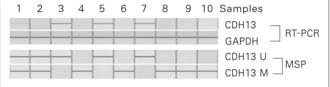
Fig. 5. T-cadherin(CDH-13) gene expression after RT-PCR & promoter methylation specific PCR of #1-#10 samples(U: unmethylation, M: methylation, MSP: methylation specific PCR)
CDH13 GAPDH CDH13 U CDH13 M
RT-PCR
MSP 1 2 3 4 5 6 7 8 9 10 Samples
Fig. 6. T-cadherin(CDH-13) gene expression after RT-PCR & promoter methylation specific PCR of #11-#20 samples(U: unmethylation, M: methylation, MSP: methylation specific PCR)
CDH13
GAPDH RT-PCR
MSP 11 12 13 14 15 16 17 18 19 20 Samples
Table 4.Relation of CDH-13 Gene Expression and Promoter Methylation
CDH-13 Gene Expression No Expression of CDH-13 Gene
Promoter Unmethylation 7 samples 0 sample
Promoter Methylation 0 sample 10 samples
Unmethylation+Methylation 2 samples 1 sample
Total 9 samples 11 samples
chi-square test, p<0.05
CDH13 U
CDH13 M
2. CDH-13 유전자의 발현
실험에 사용된 총 20개의 조직편 중 9개의 샘플에서 CDH-13 유전자의 발현을 관찰할 수 있었으며, 그 중 2개의 샘플에서는
CDH-13 유전자의 발현이 다른 것과 비교하여 다소 약하게 관
찰되었다. 또한 나머지 11개의 샘플에서는 CDH-13 유전자의 발현을 관찰할 수 없었다(Fig. 5, 6).3. CDH-13 유전자의 Methylation
CDH-13 유전자가 발현되었던 9개의 샘플 중 7개에서 CDH- 13 유전자의 promoter unmethylation이 관찰되었으며 2개에서는 promoter unmethylation과 methylation이 동시에 나타났다. 또한 CDH-13 유전자 발현이 나타나지 않았던 11개의 샘플 중 10개
에서 CDH-13 유전자 promoter methylation이 관찰되었으며 1개 에서는 promoter unmethylation과 methylation이 동시에 관찰되었 다(chi-square test, p<0.05), (Table 4 & Fig. 5, 6).Ⅳ. 총괄 및 고찰
Cadherin은 칼슘 의존성 세포 간 접착분자로 막당단백질로
구성되어 있으며, 정상 조직 구조의 세포막 형성과 유지에 매 우 중요한 역할을 담당하고 있다. 이들은 여러 종류가 있는데 종류마다 주로 발견되는 위치가 다르다. CDH-1(E-cadherin)은 상피 조직에서 발견되며 CDH-2(N-Cadherin)은 신경세포에서 발견되고 CDH-13(T-cadherin)은 심혈관조직에서 주로 발견되 었다. 그 중에서 E-cadherin이 상피조직에 주로 존재하고 그 기 능상의 이유로 암조직과의 관련성에 대한 연구가 많이 이루어 졌다2,24).
CDH-1은 세포질에 있는 α -catenin, β -catenin, γ -catenin 등과 결
합하여 E-cadherin/catenin 복합체를 형성하고 있다25-27). 또한 catenin들은 CDH-1과 세포내 골격을 이루는 actin filament와 세
포막 사이의 연결 역할을 하여 세포 구조를 유지하도록 한다.이러한 복합체에 생기는 변이는 세포 간 결합력을 저하시키고 암의 전이 및 침윤을 일으킨다28-30)
.
많은 연구들에서 CDH-1의 발현 감소가 식도암, 위암, 대장 암, 폐암, 유방암, 자궁경부, 두경부상피세포암 등 여러 암조직 의 분화 정도와 침윤, 전이, 예후와 밀접하게 연관되어있다고
하였다31-34)
. 또한 CDH-1의 발현 감소는 CDH-1 promoter의 과메
칠화(hypermetylation)와 관련이 있으며 이런 현상은 인간의 위 암 뿐만 아니라 두경부암에서도 관찰되었다35-38)
.
CDH-13
은 CDH-1에 비해 아직 많은 것이 명확히 밝혀지지 않았지만, 여러 암세포와의 관련성에 대해 주목받고 있다. CDH-
13은 대동맥벽, 뉴론, 비장 또는 다른 장기들의 작은 혈관 등에
서 높은 발현을 나타내며, 특이하게 동맥경화 같은 병리적 상 태 또는 혈관 손상 시에 그 발현이 증가하였다. 이런 증거들은CDH-13이 혈관세포들의 기능과 재형성을 조절하는데 관여한
다는 것을 의심케 한다39).
Zhou 등
15)은 CDH-13이 심혈관 조직뿐만 아니라 쥐와 인간의 상피 조직의 기저세포층(basal cell layer)에도 존재한다고 하였 다. 또한 다른 연구에서 CDH-13은 기존의 E-cadherin 또는 P-cad-herin과는 상이한 구조를 하고 있는데 가장 특징적인 것이 세포
내 골격 구조를 담당하는 actin 또는 catenin과 상호 작용하는intracelluar domain이 없이 세포막 표면에 부착한다는 것이다
16).
Takeuchi 등
17,18)은 앞서 기술한 CDH-13과 다른 cadherin과의 구 조적 차이에 의해 CDH-13이 세포 부착 보다는 세포내 신호 전 달 체계에 관여하며 그에 따른 기능적 차이가 있다고 하였다.CDH-13에 대한 신호 동반자 또는 adaptor protein이 앞으로 명확
히 밝혀져야 한다는 조건이 있겠지만, 현재까지 CDH-13은 세 포의 이주 증가 및 표현형(phenotype)을 변화시키는 저밀도 지 질단백 수용체(low density lipoprotein)로 생각 된다. 또한 CDH-13이 세포접촉저지현상(cell contact inhibition)에 의해 세포의 성
장과 증식을 조절하는 negative 성장 인자로 작용한다고 하 였 다40).
Hepatocellular carcinoma나 폐전이 Lewis carcinoma 같은 몇몇
종양의 경우에 CDH-13의 발현이 종양내부의 신생혈관부에서 증가된 것을 관찰 할 수 있는데 이는 CDH-13이 결합조직으로 부터 신생혈관이 종양 조직 내로 성장하도록 유도하는 역할을 하기 때문으로 추정되며, 그 외 다른 Non-small cell lung carcino-ma(NSCLC), ovarian cancer, pancreatic cancer, colorectal cancer 등
여러 암조직에서 CDH-13의 발현이 감소 또는 소실된 것이 관찰되었다19-23)
.
이러한 점은 종양세포의 성장 및 증식에 있어 CDH-13의 발 현 감소가 중요한 역할을 하며 CDH-13이 종양 억제 인자일 가 능성을 추측해 볼 수 있다. 이는 CDH-13의 발현이 없는 신경아 세포종에서 CDH-13의 첨가 시 EGF(epidermal growth factor)에 의한 성장 자극 반응의 소실이 관찰된 실험적 모델에서 증명 되었으며, 피하 부위에 종양을 이식한 누드마우스 실험에서
CDH-13을 발현시켰을 때 종양의 성장이 억제된 것을 관찰 할
수 있었다17).
또한 Takeuchi 등41)은 실제 인간의 피부 기저부 세포 암종에 서 CDH-13 발현의 감소 또는 소실을 관찰 보고하였고 Toyooka 등42)은 colorectal cancer와 adenoma에서의 연구를 통해 종양 조직 에서 CDH-13의 발현 감소를 관찰 하였으며 또한 CDH-13 유전 자의 promoter methylation specific PCR 분석을 통해 CDH-13의 발 현 감소와의 연관성을 보고하였다.
일반적으로 DNA에서 염기의 특정 부위에 메칠화기 되는 것 을 흔히 볼 수 있는데, DNA 메칠화는 그 가닥 중 A와 C가 G와
T보다 쉽게 메칠화되며 특정한 염기 서열 또는 영역내에서 일
어나고 DNA 메칠화 효소(DNA methylase) 모두가 메틸기의 공 여체로 아데노실 메티오닌(adenocyl-methionine)을 사용한다는 특징이 있다. DNA 분자에서 이와 같이 변형된 또는 이례적인 염기는 유전 정보의 발현을 조절한다든지 혹은 유전 정보를 여러 가지 파기요인으로 부터 방어하기 위한 특이한 신호로 이해되고 있다. 한편, 종양 조직 등 병적 상태에서 유전자 pro-moter 메칠화(methylation)는 유전자 발현이 억제되는 가장 중요
한 메카니즘 중 하나로 알려져 있다43)
.
본 연구에서 CDH-13 발현을 알아보기 위한 면역조직화학적 검사 결과 정상적인 구강편평상피조직의 경우, 상피 전층에서 발현되지 않고 기저 세포층(basal cell layer)에서만 발현되는 양 상은 CDH-13의 세포 성장 및 증식 조절 인자일 가능성과 관련 이 있다고 생각된다. 특히, 구강 선조직의 분비도관상피세포 의 세포질 내 CDH-13의 발현양상은 세포 접착이라는 일반 cad-
herin의 주요 기능과는 다른 CDH-13만의 역할이 있을 수 있다
고 추측 된다.다른 연구에서처럼 정상조직세포들에 비해 구강편평상피세 포암종에서 CDH-13의 발현이 감소 또는 소실 된 것을 관찰 할 수 있었는데, 특이한 점은 조직병리학적 조직 소견 상 중등도 로 분화되고 침습성이 덜한 구강편평상피세포암종에서는
CDH-13의 발현이 정상 세포에 비해 중간 정도로 감소하였으
나 더 공격적인 분화도가 낮은 구강편평상피세포암종에서는CDH-13의 발현이 거의 나타나지 않았다는 것이다. 이는 CDH- 13의 발현 정도가 암종의 분화도 또는 침습성과 상관관계가 있
다고 생각해 볼 수 있다.면역조직화학적 검사 소견을 증명하기 위한 CDH-13 유전자
RT-PCR 분석에서 20편의 샘플 중 11개의 샘플(55%)에서 유전
자 발현이 관찰되지 않았다. 이런 결과는 Tamotsu 등44)이 피부 편평상피세포암종의 56 샘플 중 50 샘플(89%)에서 유전자 발 현이 소실되었다는 연구 보고와는 차이가 있는데 아마도 실험 에 사용된 암종의 분화도와 침습성의 차이에 따른 것이라 생 각된다.CDH-13 유전자의 발현과 유전자 promoter 메칠화의 상관관
계를 알아보기 위한 methylation specific PCR 분석에서 primer와primer sequence는 Sato 등
21)이 보고했던 것을 사용하였으며, 10 개의 샘플(50%)에서 promoter의 메칠화(methylaton)가 관찰되었 다. 구강편평상피암종에서의 CDH-13 유전자 메칠화에 대한 연구가 거의 없기 때문에 다른 부위의 암종 연구들의 결과와 비교하였는데 Maruyama 등45)의 방광암 CDH-13 유전자 메칠화 에 대한 연구에서는 전체 조직 중 29%에서 메칠화가 관찰되었 다고 보고하였으며, Kim 등46)의 원발성 nonsmall cell lung carci-noma에 대한 연구에서는 43%의 조직 샘플에서의 CDH-13 유
전자 메칠화를 보고하였고 이는 비록 암종의 종류 및 부위가 다르기는 하지만 본 연구의 결과와 비슷한 비율이었다.또한 주목할 점은 CDH-13 유전자가 발현되지 않은 11개의 샘플 중 10개에서 promoter의 메칠화(methylaton)가 관찰되었다 는 것으로 유전자 발현이 감소 또는 소실이 promoter 메칠화와 밀접한 상관관계가 있다는 증거가 된다.
Ⅴ. 결 론
CDH-13의 주요한 작용은 혈관 기능 및 재형성에 관여하는
것이며 동맥경화 같은 혈관 질환과의 연관성이 밝혀졌다. 또 한 세포 성장 및 세포 접촉 저지작용(cell contact inhibition) 등을 조절한다고 한다.정상 세포가 증식하는 경우, 세포 표면에서 다른 세포를 인 식하여 그 운동이나 증식을 제한하는 CDH-13 같은 물질이 있 어 무한 증식이나 결합조직과의 경계를 침범하지 않고 항상성 을 유지하게 된다. 하지만, 종양 세포는 그 성장 및 침습, 전이 과정에서 정상 세포와는 다른 양상을 나타내는데 특히, 접촉 저지 작용의 상실로 인해 무한 증식하며, 결합 조직내로 침습, 전이하게 된다. 이런 특성은 종양 세포에서 CDH-13의 발현과 그 연관성이 있다고 추론할 수 있다.
본 실험의 결과에서 보면, 20개의 구강편평상피암종 조직편 중 promoter methylation은 13예(65%)에서 관찰 되었으며 promot-
er methylation이 관찰된 예에서는 RT-PCR 분석에서 유전자의
발현이 관찰 되지 않은 반면 unmethylation이 동반된 예에서는 유전자 발현이 관찰되었다. 또한 분화도가 낮은 편평상피암종 에서promoter methylation과 함께 유전자 발현이 억제된 것이 관 찰 되었다. 이러한 유전자 발현 현상은 면역조직학적 검사에 서 증명 되었다.따라서 구강편평상피세포암종 발생에 CDH-13 발현 억제는 중요한 역할을 하며 유전자 발현 억제는 promoter methylation에 의한 것임을 알 수 있었으며 이러한 결과로 향후 CDH-13 유전 자의 promoter methylation에 의한 발현 억제가 구강편평상피세 포암종의 좋은 진단적 marker로 활용 될 수 있을 것으로 기대 된다.
참고문헌
1. Cooper, Hausman : The Cell ; A molecular approach. Sinaur Associates Inc. 2007;4.
2. Takeichi M. : Cadherin cell adhesion receptors as a morphogenetic regulator. Science 1991; 251:1451-55.
3. 표성운, 김영실, 박지영, 김창현, 이 원, 박민규 : 구강 편평세포 암에서 E-cadherin 유전자의 과메틸화 J. Kor. Oral Maxillofac.
Surg. 2008; 34:135-140.
4. Behrens J. : The role of cell adhesion molecules in cancer invasion and metastasis. Breast Cancer Res Treat 1994;24:175-84.
5. Ziober BL, Silverman SS, Kramer RH : Adhesive mechanisms regu- lating invasion and metastasis in oral cancer, Crit. Rev. Oral Biol.
Med 2001;12:499-510.
6. Oka H, Shiozaki H, Kobayashi K : Expression of E-cadherin cell adhesion molecules in human breast cancer tissues and its relation- ship to metastasis. Cancer Res 1993;53:1696-1701.
7. Umbas R, Isaacs WB, Bringuier PP : Decreased E-cadherin expres- sion is associated with poor prognosis in patients with prostate can- cer, Cancer Res 1994;54:3929-33.
8. Umbas R, Schalken JA, Aalders TW : Expression of the cellular ad- hesion molecule E-cadherin is reduced or absent in high-grade prostate cancer, Cancer Res 1992;52:5104-9.
9. Jiao W, Miyazaki K, Kitajima Y : Inverse correlation between Ecadherin and Snail expression in hepatocellular carcinoma cell lines in vitro and in vivo, Br. J. Cancer 2002;86:98-101.
10. Graff JR, Greenberg VE, Herman JG : Distinct patterns of E-cadherin CpG island methylation in papillary, follicular, Hurthle’s cell, and poorly differentiated human thyroid carcinoma, Cancer Res 1998;58:2063-6.
11. Schipper JH, Frixen UH, Behrens J : E-cadherin expression in squa- mous cell carcinomas of head and neck: inverse correlation with tumor dedifferentiation and lymph node metastasis. Cancer Res 1991;51:6328-37.
12. Rodrigo JP, Dominguez F, Alvarez C : Expression of E-cadherin in squamous cell carcinomas of the supraglottic larynx with correlations to clinicopathological features. Eur. J. Cancer 2002;38:1059-64.
13. Tkachuk VA, Bochkov VN, Philippova MP : Identification of an atypical lipoprotein-binding protein from human aortic smooth mus- cle as T-cadherin. FEBS Lett. 1998;421:208-12.
14. Philippova MP, Bochkov VN, Stambolsky DV : T-cadherin and sig- nal-transducing molecules co-localize in caveolin-rich membrane domains of vascular smooth muscle cells. FEBS Lett 1998;429:207- 10.
15. Zhou S, Liang SB, Miyachi Y : Expression of T-cadherin in basal keratinocytes of skin. J Invest Dermatol 2002;118:1080-4.
16. Ranscht B and Zimmermann D : T-cadherin, a novel cadherin adhe- sion molecule in the nervous system, lacks the conserved cytoplasmic region. Neuron 1991;7:391-402.
17. Takeuchi T, Misaki A, Liang SB : Expression T-cadherin in human brain and its characteristics as a negative growth rgulator of EGF in neuroblastoma cells. J Neurochem 2000;74:1489-97.
18. Takeuchi T and Ohtsuki Y : Recent progress in T-cadherin (CDH13, H-cadherin) research. Histol Histopathol 2001;16:1287-93.
19. Lee SW : H-cadherin, a novel cadherin with growth inhibitory func- tions and diminished expression in human breast cancer. Nat Med 1996;2:776-82
20. Lee SW, Reimer CL, Campbell DB : H-cadherin expression inhibits in vitro invasiveness and tumor formation in vivo. Carcinogenesis 1998;19:1157-9.
21. Sato M, Mori Y, Sakurada A, Fujimura S : The Hcadherin(CDH13)gene is inactivated in human lung cancer. Hum Genet 1998;103:96-101.
22. Toyooka KO, Toyooka S, Virmani AK : Loss of expression andaber- rant methylation of the CDH13 (H-cadherin) gene in breast and lung carcinomas. Cancer Res 2001;61:4556-60.
23. Zhong Y, Delgado Y, Gomez J : Loss of H-cadherin protein expres- sion in human non-small cell lung cancer is associated with tumori- genicity. Clin Cancer Res 2001;7:1683-7.
24. Hatta K, Okada TS, Yakeichi M : A monoclonal antibody disrupting calcium-dependent cell-cell adhesion of brain tissues: possible role of its target antigen in animal pattern formation. Proc Natl Acad Sci USA 1985;82:2789-93.
25. Boller K, Vestweber D, Kemler R : Cell-adhesion molecule uvo- morulin is localized in the intermediate junctions of adult intestinal epithelial cells. J Cell Biol 1985;100:327-32.
26. Ozawa M and Kemler R : Molecular organization of the uvomorulin- catenin complex. J Cell Biol 1992;116:989-96.
27. Rubinfeld B, Souza B, Alvert I : Association of the APC gene prod- uct with beta-catenin. Science 1993;262:1731-4.
28. Breen E, Steele G, Mercurio AM : Role of E-cadherin/α-catenin com- plex in modulating cell-cell and cell-matrix adhesive properties of invasive colon carcinoma cells. Ann Surg Oncol 1995;2:378-85.
29. Caca K, Kolligs FT, Ji X : β-catenin and γ-catenin mutations, but not E-cadherin inactivation, underlie T-cell factor/lymphoid enhancer factor transcriptional deregulation in gastric and pancreatic cancer.
Cell Growth Differ 1999;10:369-376.
30. Mareel M, Boterberg T, Noe V : E-cadherin/catenin/cytoskeleton complex : a regulator of cancer invasion. J Cell Physiol 1997;173:271-4.
31. Takayama T, Shiozaki H, Shibamoto S : β-Catenin expression in human cancers. Am J Pathol 1996;148:39-46.
32. Shimoyama Y, Nagafuchi A, Fujita S : Cadherin dysfunction in a human cancer cell line:possible involvement of loss of α-catenin expression in reduced cell-cell adhesiveness. Cancer Res 1992;52:5770-4.
33. Oyama T, Kanai Y, Ochiai A : A truncated β-Catenin disrupts the interaction between E-caherin and α-catenin: A cause of loss of inter- cellular adhesiveness in human cancer cell lines. Cancer Res 1994;54:6282-7.
34. Jawhari A, Jordan S, Poole S : Abnormal immunoreactivity of the E- cadherin-catenin complex in gastric carcinoma: relationship with patient survival. Gastroenterology 1997;112:46-54.
35. Chang HW, Chow V, Lam KY : Loss of Ecadherin expression result- ing from promoter hypermethylation in oral tongue carcinoma and its prognostic significance, Cancer 2002;94;386-92.
36. Melki JR, Vincent PC, Brown RD : Hypermethylation of E-cadherin in leukemia, Blood 2000;95:3208?13.
37. Nakayama S, Sasaki A, Mese H : The E-cadherin gene is silenced by CpG methylation in human oral squamous cell carcinomas, Int. J.
Cancer 2001;93:667-73.
38. Saito Y, Takazawa H, Uzawa K : Reduced expression of E-cadherin in oral squamous cell carcinoma: relationship with DNA methylation of 50 CpG island, Int. J. Oncol 1998;12;293-8.
39. Ivanov D, Philippova M, Antropova J : Expression of cell adhesion molecule T-cadherin in the human vasculature. Histochem. Cell Biol 2001;115:231-42.
40. Resink TJ, Kuzmenko YS, Kern F : LDL binds to surface-expressed human T-cadherin in transfected HEK293 cells and influences homophilic adhesive interactions. FEBS Lett 2000;463:29-34.
41. Takeuchi T, Liang SB, Ohtsuki Y : Downregulation of expression of a novel cadherin molecule, T-cadherin, in basal cell carcinoma of the skin. Mol. Carcinog 2003;35:173-9.
42. Toyooka S, Toyooka KO, Harada K : “Aberrant methylation of the CDH13 (H-cadherin) promoter region in colorectal cancers and ade- nomas.”. Cancer Res 2002;62:3382-6.
43. Esteller M, Corn PG, Baylin SB, Herman JG : A gene hypermethyla- tion profile of human cancer, Cancer Res 2001;61;3225-9.
44. Tamotsu T, Sheng-Ben, Hiroshi S : Loss of T-cadherin (CDH13, H- cadherin) expression in cutaneous squamous cell carcinoma.
Laboratory Investigation 2002;82:1023-9.
45. Maruyama R, Toyooka KO, Harada K : “Aberrant promoter methyla- tion profile of bladder cancer and its relationship to clinicopathologi- cal features.”. Cancer Res 2001;61:8659-63.
46. Kim JS , Han J, Shim YM : Aberrant Methylation of H-Cadherin (CDH13) Promoter Is Associated with Tumor Progression in Primary Nonsmall Cell Lung Carcinoma. CANCER 2005;104;1825-32.