Cyclosporine A eyedrops with self-
nanoemulsifying drug delivery systems have improved physicochemical properties and efficacy against dry eye disease in a murine dry eye model
Seung Pil Bang1,2, Chang Yeor Yeon3, Nirpesh AdhikariID3, Sanjiv NeupaneID3,
Harim Kim1, Dong Cheol Lee1, Myeong Jin Son1, Hyun Gyo Lee1, Jae-Young Kim3, Jong Hwa JunID1*
1 Department of Ophthalmology, Keimyung University School of Medicine, Dongsan Medical Centre, Daegu, Republic of Korea, 2 Department of Biomedical Engineering, University of Rochester, Rochester, New York, United States of America, 3 Department of Biochemistry, School of Dentistry, IHBR, Kyungpook National University, Daegu, Republic of Korea
Abstract
Purpose
We aimed to compare the physicochemical properties and in vivo efficacy of commercially available nanoemulsion cyclosporine A (CsA) eyedrops in benzalkonium chloride (BAC)- induced dry eye disease (DED).
Methods
Particle size analysis was performed on conventional 0.05% CsA (Restasis, C-CsA) and two new types of 0.05% CsA eyedrops based on a self-nanoemulsifying drug delivery sys- tem (SNEDDS, SNEDDS-N and -T). Turbidometry, pH measurements and instability indices of each CsA solution were measured. DED was induced with BAC, and animals were treated with vehicle or CsA preparations. Tear volume and fluorescein staining scores were evaluated on days 7 and 14. Eyes were enucleated and subjected to IHC, TUNEL staining, periodic acid-Schiff (PAS) staining, real-time PCR and western blotting.
Results
Both SNEDDSs had lower and more uniform particle size distribution than C-CsA, and a similar optical density to phosphate-buffered saline and stable pH, in contrast to the high tur- bidity and unstable pH of C-CsA. Aqueous tear volume and fluorescein staining scores were improved in C-CsA- and SNEDDS-treated mice. Numbers of PAS-positive goblet cells and levels of inflammatory mediators were decreased by both C-CsA and SNEDDS, although SNEDDS resolved inflammation more effectively than C-CsA.
a1111111111 a1111111111 a1111111111 a1111111111 a1111111111
OPEN ACCESS
Citation: Bang SP, Yeon CY, Adhikari N, Neupane S, Kim H, Lee DC, et al. (2019) Cyclosporine A eyedrops with self-nanoemulsifying drug delivery systems have improved physicochemical properties and efficacy against dry eye disease in a murine dry eye model. PLoS ONE 14(11):
e0224805.https://doi.org/10.1371/journal.
pone.0224805
Editor: Collynn F. Woeller, University of Rochester School of Medicine and Dentistry, UNITED STATES
Received: July 18, 2019 Accepted: October 22, 2019 Published: November 18, 2019
Copyright:© 2019 Bang et al. This is an open access article distributed under the terms of the Creative Commons Attribution License, which permits unrestricted use, distribution, and reproduction in any medium, provided the original author and source are credited.
Data Availability Statement: All relevant data are within the manuscript and its Supporting Information files.
Funding: This work was supported by the National Research Foundation of Korea (NRF) Grant funded by the Korea Government (MSIP)
(No.2014R1A5A2010008) and NRF-
2018R1D1A1B07045362. This funding body had no role in the design of the study, in the collection,
Conclusions
Cyclosporine A eyedrops with SNEDDS have improved physicochemical properties and treatment efficacy in BAC-induced DED.
Introduction
Dry eye disease (DED) is a multifactorial disorder of the ocular surface defined by dysfunc- tional tear film homeostasis [1]. Hyperosmolarity, tear film instability, neurosensory abnor- malities, ocular surface inflammation and tissue damage contribute to the undesirable symptoms and aetiology of DED [1]. Ocular surface inflammation plays a significant role in DED pathology, and is primarily mediated by CD4+T cells [2,3]. Numerous inflammatory cytokines associated with conjunctival T cells are elevated in the tear film of DED patients [4].
Induction of DED-associated inflammation occurs due to a diverse array of pathologies. Sys- temic autoimmune diseases such as Sjo¨gren syndrome lead to destruction of the lacrimal gland, causing aqueous deficient dry eye [1,5]. Contrastingly, meibomian gland dysfunction diminishes tear film lipid content, resulting in evaporative dry eye [1,5]. However, regardless of the cause, the downstream result is loss of tear film integrity and stability, promoting ocular surface inflammation and damage to the corneal epithelium. Therefore, ophthalmic anti- inflammatory agents are important treatments for all forms of DED.
Cyclosporine A (CsA) is the anti-inflammatory treatment of choice for DED, as it can be utilised long-term without adverse side effects associated with long-term use of other anti- inflammatory drugs such as corticosteroids [6]. In addition, unlike steroids, the specific and reversible effects of CsA on T cells make CsA desirable for extended use [7]. However, CsA has a large molecular weight and hydrophobicity (Log P = 1.4–3.0; solvent-dependent), resulting in poor aqueous solubility (6.6–106 lg/mL; temperature-dependent) [8]. Therefore, CsA is challenging to administer with conventional topical ophthalmic delivery systems. Thus, it is important to improve drug delivery strategies for CsA to maximise its ocular bioavailability.
Over the past several decades, various drug delivery tactics have been developed to improve the ophthalmic bioavailability of CsA after topical administration to alleviate DED without the systemic side effects associated with oral CsA treatment [9]. These delivery strategies include hydrogels,in situ gelling systems, liposomes, nanoparticles and micelles [10]. Restasis (CsA ophthalmic emulsion 0.05%; Allergan, Irvine, CA, USA) is the most authorised and widely used CsA worldwide, and is a preservative-free, anionic oil-in-water emulsion, in which CsA is enclosed and dissolved in castor oil with the emulsifying agent polysorbate 80 [11]. However, Restasis is often associated with adverse effects such as visual disturbances due to its turbidity, ocular discomfort and conjunctival hyperaemia. Restasis is an anisotropic complex emulsion, which can break down when applied to the tear film, causing release of free surfactants that may irritate the ocular surface [12]. Also, in the dispersed phase, a wide range of particle sizes and distribution can cause creaming and flocculation of the preparation, decreasing its long- term physicochemical stability [13,14]. Furthermore, Restasis is a complex emulsion, so CsA exists in various phases that may have differential biological effects, which may restrict suffi- cient delivery of bioactive CsA to the tissue [12].
On the other hand, 0.05% CsA nanoemulsion products based on the self-nanoemulsifying drug delivery system (SNEDDS) [15], such as Cyporin-N (SNEDDS-N; Taejoon Pharma, Seoul, South Korea) and T-sporin (SNEDDS-T; Hanlim Pharma, Seoul, South Korea), are cur- rently available. In addition to cyclosporine, a significant number of other drugs have poor
analysis and interpretation of data, or in the preparation of the manuscript.
Competing interests: The authors have declared that no competing interests exist.
water solubility and permeability, leading to poor bioavailability and variable drug efficacy.
SNEDDSs are anhydrous homogenous mixtures formed by mixing an oil, a drug, a surfactant, and a co-surfactant. Gentle dilution with water results in optically transparent emulsions, in which the sizes of the particles are a few hundreds of nanometers in diameter. SNEDDS result in better chemical and physical stability of drugs and higher solubility of water insoluble drugs, such as cyclosporine A, than conventional nanoemulsions [16] In one previous clinical study, the SNEDDS eyedrop had comparable effects to Restasis, with more rapid improvement of temporal conjunctival surface damage and tear film stability than Restasis [13].
These studies suggest that enhancing the bioavailability of CsA to ocular tissues is necessary to maximise the clinical potency of CsA for DED [9]. We hypothesised that the physicochemi- cal stability provided by the homogenous and smaller particle size in SNEDDS eyedrops might increase the bioavailability of CsA to ocular tissues, especially the cornea and conjunctiva. In the present study, we compared the physicochemical characteristics andin vivo anti-inflam- matory efficacy of three commercially available ocular CsA preparations, conventional 0.05%
nanoemulsion CsA (Restasis, C-CsA) and two 0.05% nanoemulsion CsA preparations (SNEDDS-N and -T), in the murine benzalkonium chloride (BAC)-induced experimental DED model.
Materials and methods
Size distribution of CsA emulsions
In a pilot experiment, a significant discrepancy in particle size was identified between C-CsA and SNEDDS preparations. C-CsA preparations have highly variable particle sizes, with size ranging from several nanometers to several tens of micrometers. Therefore, two different types of particle analysers with different thresholds were used to accommodate each preparation. To evaluate all three CsA preparations, a liquid particle size analyser (Cilas 1064; Cilas, Orle´ans, France) was used to accommodate the wide range of size distribution in C-CsA. A laser nano- particle analyser (Zetasizer Nano; Malvern Panalytical Ltd., Worcestershire, England) was also used to analyse SNEDDS-N and -T for a more sensitive analysis. Particle size analysis was per- formed at 25˚C.
Scanning electron microscope (SEM) evaluation of CsA emulsions
A drop of each 0.05% CsA emulsion was applied to an electron microscope (EM) sample stand and dried in an EM oven at 37˚C for 12 h. The sample surface was coated with Platinum-Palla- dium (10 nm thickness) to reduce fluorescence. Each CsA emulsion was evaluated by SEM (Hitachi S-4200; SEMTech Solutions, North Billerica, MA, USA) at 20.0 kV.
Turbidity of CsA emulsions
To evaluate CsA solubility in each preparation, light scatter was analysed at specific wave- lengths using a microplate absorbance reader (iMark; Bio-Rad Laboratories, Hercules, CA, USA). After brief vortexing, 50μL of each emulsion was placed in a 96-well plate. Absorbance was measured at 415, 450, 490, 560, 595, 655 and 750 nm. Phosphate-buffered saline (PBS), ultrapure water and balanced salt solution were used as references. Each measurement was repeated in triplicate and performed at 25˚C.
Instability index of CsA emulsions
Because the C-CsA emulsion is an unstable oil-in-water emulsion, phase separation may occur gradually over time. Therefore, the instability index was evaluated and compared over time for
each CsA preparation. During a 2400 min experiment, the instability of each emulsion was detected by centrifugation at 2300g at 25˚C or 35˚C, and subsequent evaluation with a par- ticle analyser (LUMiSizer; LUM GmbH, Berlin, Germany). Each value was detected every 3 min.
pH instability of CsA emulsions
Since a changing pH value indicates unstable dissolution of the solute in the solvent, the sta- bility of the emulsions can be indirectly determined by performing pH measurements.
Unstable pH can also result in inconsistent drug efficacy and discomfort to the patient during administration of the drug. [17] Therefore, drug pH was monitored 30 min after brief vortexing at 25˚C and 35˚C using a benchtop pH meter (Orion Star A211; Thermo Fisher Scientific, Waltham, MA, USA). The pH value of each CsA emulsion was read every minute.
Static and kinematic viscosity
Because eyedrop viscosity affects drug efficacy and blurring after instillation, and nanoemulsi- fication decreases viscosity, we measured dynamic (absolute) viscosity using a Brookfield type viscometer (Brookfield DV2T, SC4-18, 5 rpm, 25˚C), and kinetic viscosity using the following equation: Kinematic viscosity = K (0.08390) x t (mean value of three repeated measures using a capillary tube viscometer).
Induction of murine BAC-induced DED model and application of CsA emulsion eyedrops
All study procedures were conducted in accordance with the ARVO Statement for the Use of Animals in Ophthalmic and Vision Research. The animal protocol was approved by the Kei- myung University Institutional Animal Care and Use Committee (KM-2018-11R).
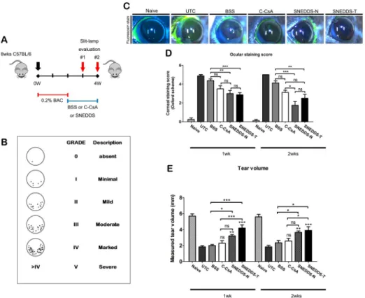
Ninety 8-week-old male C57BL6J mice were purchased from Hyochang Science (Daegu, South Korea). Mice were kept in a facility with a standard environment (temperature, 25˚C± 1˚C; relative humidity, 60% ± 5%; and 12 h light/12 h dark cycles [9 AM to 9 PM]), and provided food and waterad libitum. Experimental dry eye was induced by twice-daily (9 AM and 6 PM) application of 5μL of 0.2% BAC (Sigma-Aldrich) for 2 weeks. After BAC administration was complete and DED was successfully induced, mice were randomly divided into six groups (15 mice per group): Naïve control, non-treated control, BSS-treated control, C-CsA, SNEDDS-N and SNEDDS-T groups. Fifteen naïve control mice without BAC-induced DED were used as a normal control.
After group allocations, the naïve control was maintained for a 4 week experimental period (untreated for 2 week DED induction period and 2 week DED treatment period), the
untreated control group was maintained without application of eyedrops for 2 weeks after the 2 week BAC induction period, the solvent-treated control was treated with balanced salt solu- tion (BSS; Alcon Laboratories, Fort Worth, TX, USA) twice daily for 2 weeks after the 2 week DED induction period and the three 0.05% CsA treatment groups were treated twice daily (9 AM/6 PM) with the specified CsA preparation for 2 weeks after the 2 week DED induction period (Fig 3A). At 1 and 2 weeks of dry eye treatment, 2, 2, 2-tribromoethanol (Avertin) was injected intraperitoneally at a 250 mg/kg dose for evaluations of the corneal fluorescein stain- ing and tear meniscometry. Two weeks after dry eye treatment, mice were sacrificed through cervical dislocation and the eyeballs were enucleated and analyzed.
Corneal fluorescein staining and tear meniscometry
At both 1 and 2 weeks after beginning treatment of DED animals, corneal fluorescein staining scoring and tear volume measurement were performed. After application of fluorescein dye using fluorescein paper (Haag-Streit AG, Koeniz, Switzerland) into the lower conjunctival sac, corneal staining scores were calculated under cobalt blue light using a portable slit lamp (SL- 17; Kowa Optimed, Torrance, CA, USA). The corneal staining score was calculated by a blinded investigator (C.Y.Y.) as follows: absent, 0; slightly punctuate staining with fewer than 30 spots, 1; non-diffuse punctuate staining with more than 30 spots, 2; severe diffuse staining with no positive plaques, 3; and positive fluorescein plaques, 4. The scores from four quadrants were averaged to a final grade (ranging from 0 to 16 points) (Fig 3B). Tear volume was mea- sured 1 and 2 weeks after beginning CsA treatment using strip meniscometry (SM Tube;
EchoElectricity Co., Ltd., Fukushima, Japan) from the lower conjunctival sac. After anaesthe- sia, the tear meniscometry tip was introduced to the lower tear lake for 5 s. The length of wet- ted thread was measured per manufacturer’s instructions.
Immunohistochemistry of central and peripheral cornea
The eye and ocular adnexa were excised, fixed in 4% paraformaldehyde for 3 days and embed- ded in optimal cutting temperature (OCT) compound at -80˚C. A 7μm section was cut and air dried at room temperature. Sectioned tissues were permeabilised with 0.4% (v/v) Triton X-100 (Structure Probe, West Chester, PA, USA) for 15 min and blocked with 1% (w/v) bovine serum albumin in 0.1% (v/v) Tween 20 (AMRESCO, Solon, OH, USA) in PBS for 30 min. Sec- tions were then incubated with anti-IL-1β, IL-6, TNF-α, CK-10 and Ki-67 primary antibodies (BioLegend, San Diego, CA, USA) overnight at 4˚C, and subsequently with Alexa Fluor 594-conjugated secondary antibodies (Molecular Probes, Eugene, OR, USA) for 2 h at room temperature. After several rinses, nuclei were stained with Vectashield mounting solution (Vector Laboratories, Burlingame, CA, USA) containing 4’,6-diamidino-2-phenylindole (DAPI). Each slide was observed under an upright fluorescence microscope (Leica Microsys- tems GmbH, Wetzlar, Germany), and images were obtained using an inverted fluorescence microscope (Ti-U; Nikon Instruments, Melville, NY, USA), and captured using NIS-Elements software (Nikon Instruments). Ki-67-positive cells at the basal area of the corneal epithelium were counted with ImageJ (http://imagej.nih.gov/ij/; provided in the public domain by the National Institutes of Health, Bethesda, MD, USA) at five different locations by a blinded investigator (C.Y.Y.). All experiments were performed in triplicate.
TUNEL assay
To compare the treatment efficacy of each CsA preparation, TUNEL staining was performed on corneal cryosections using anin situ cell death detection kit (Merck, Kenil Worth, NJ, USA) per the manufacturer’s instructions. Each section was counterstained and mounted with Vectashield mounting medium (Vector Laboratories). Each slide was examined, and images were captured as described for immunohistochemistry.
PAS staining
Eyeballs and adnexa were surgically excised and fixed with 4% paraformaldehyde (Biosesang, Seongnam-si, Gyeonggi-do, South Korea) for 3 days at 4˚C followed by a 3 day PBS (Wel- gene, Gyeongsan-si, Gyeongsangbuk-do, South Korea) rinse, cryoprotected using a sucrose gradient and embedded in OCT compound (Tissue-Tek; Sakura Finetek, Torrance, CA, USA) at -80˚C. A 7μm section was cut and dried at room temperature. The sectioned tissue
was stained with periodic acid-Schiff (PAS; Sigma-Aldrich) for 15 min, with haematoxylin counterstaining (Muto Pure Chemicals, Tokyo, Japan) for 5 min. Each slide was examined and photographed under a microscope (Ti-U; Nikon Instruments), and images were cap- tured using NIS-Elements software (Nikon Instruments). PAS-positive goblet cells were counted with ImageJ on three individual slides for each treatment by a blinded investigator (C.Y.Y.).
RNA isolation and real-time polymerase chain reaction
Total corneal and conjunctival RNA was extracted using a NucleoSpin RNA Plus kit (Macherey-Nagel GmbH & Co. KG, Du¨ren, Germany), and cDNA was synthesised using ReverTraAce qPCR RT Master Mix (FSQ-201; TOYOBO, Osaka, Japan). Real-time PCR was performed using a CFX96 real-time system (Bio-Rad Laboratories) using SsoAdvanced Uni- versal SYBR Green Supermix (172–5271; Bio-Rad Laboratories). Primer sequences are shown inTable 1. The amplification program included an initial denaturation step at 95˚C for 30 s, followed by 40 cycles of 95˚C for 15 s and 60˚C for 30 s, after which a melt curve analysis was conducted to check amplification specificity. Results were analysed by the comparative thresh- old cycle (Ct) method, normalised with HPRT as an endogenous reference and calibrated against the normal control group.
Western immunoblot analysis
Cornea and conjunctival tissues were excised. Frozen cornea and conjunctival tissues were crushed with stainless beads in a bead mill homogeniser (Bullet blender; Next Advance, Troy, NY, USA). Samples were then lysed in radioimmunoprecipitate buffer (Elpis Biotech, Daejeon, South Korea). Lysate protein concentration was measured using a protein bicinchoninic acid assay kit (Thermo Fisher Scientific). Equal quantities of total protein samples were loaded onto 8–15% polyacrylamide gels for sodium dodecyl sulphate polyacrylamide gel electrophore- sis (Bio-Rad Laboratories). Resolved proteins were electroblotted onto nitrocellulose blotting membranes (Sigma-Aldrich) and blocked with 5% skim milk solution (Honeywell Interna- tional, Morris Plains, NJ, USA) for 2 h. After overnight incubation with the appropriate pri- mary antibodies, the membranes were incubated with secondary antibodies for 1 h. The fluorescent band of a stain-free gel (Mini-PROTEAN TGX stain-free gel; Bio-Rad Laborato- ries) was used as a loading control for band densitometry.
Table 1. Primer sequences for semi-quantitative real-time PCR.
Gene Accession no. Sequence (5’!3’) Product size (bp)
ICAM-1 NM_010493.3 Forward: TTCTCATGCCGCACAGAACT 73
Reverse: TCCTGGCCTCGGAGACATTA
VCAM-1 NM_011693.3 Forward: CTGGGAAGCTGGAACGAAGT 115
Reverse: GCCAAACACTTGACCGTGAC
F4/80 NM_010130.4 Forward: GTGCCATCATTGCGGGATTC 79
Reverse: AAGAGCATCACTGCCTCCAC
MMP-2 NM_008610.3 Forward: CTGATGGCCCCGATCTACAC 74
Reverse: AGCTCCTGGATCCCCTTGAT
HPRT NM_013556.2 Forward: CCTAAGATGAGCGCAAGTTG 86
Reverse: CACAGGACTAGAACACCTGCTAA https://doi.org/10.1371/journal.pone.0224805.t001
Statistical analysis
Representative data obtained from experiments conducted in triplicate are presented as mean± standard deviation. Data were evaluated using independent t-tests for two groups, or one-way analysis of variance (ANOVA) for three or more groups and Tukey’s post hoc com- parison, to identify statistically significant differences. A p-value <0.05 was considered statisti- cally significant.
Results
SNEDDS preparations reduced particle size, particle size variability, preparation turbidity, and viscosity
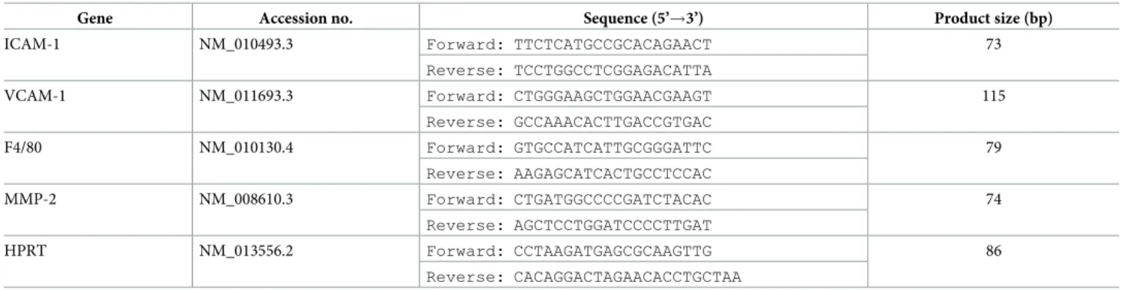
Based on particle size analysis, self-nanoemulsified CsA particles were distributed evenly and in a single size. Particle size analysis of C-CsA identified two larger size peaks at approximately 1000 and 10,000 nanometres with a wider range (6–13,000 nm) (Fig 1A). However, both SNEDDS preparations had a single peak in particle distribution (SNEDDS-N: 42.67± 20.59 nm, SNEDDS-T: 26.49± 8.32 nm) (Fig 1B and 1C). SEM analysis revealed variable particle sizes in C-CsA (Fig 1D); however, particles of the SNEDDS preparations were not identifiable by the general sample preparation technique.
Fig 1. Size distribution (percent) of CsA emulsions by intensity: (A) Restasis (C-CsA), (B) T-sporin (SNEDDS-T) and (C) Cyporin-N (SNEDDS-N). (D) Scanning electron microscope (SEM) evaluation of C-CsA. (E) Turbidity (optical density) of CsA emulsions.
https://doi.org/10.1371/journal.pone.0224805.g001
C-CsA is a milky colour emulsion due to a mixture of unevenly distributed, variably sized oil-in-water emulsions. Contrastingly, an evenly distributed and single-size emulsion would have improved transparency. Therefore, we evaluated the effects of self-nanoemulsification on CsA preparation turbidity. C-CsA had much higher turbidity than PBS or BSS at every wave- length (Fig 1E). However, both SNEDDS preparations had extremely low turbidity, which was similar to that of ultrapure water, PBS and BSS (Fig 1E). Both SNEDDS-T and SNEDDS-N showed significantly lower turbidity than C-CsA (Fig 1F and 1G).
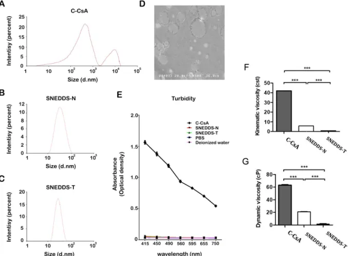
SNEDDS preparations had lower instability indices and stable pH
Because C-CsA is manufactured using a high-speed homogeniser, the drug itself is unstable, and is likely to be denatured over time due to flocculation and creaming. Contrastingly, the SNEDDS preparations are likely to be more stable, as these preparations are self-emulsifying.
Therefore, we analysed the instability indices of the three CsA preparations. Both SNEDDS preparations were remarkably stable until 2400 min, but C-CsA exhibited a gradual increase in instability index at 25˚C and 35˚C (Fig 2A–2D). In addition, consistent with the instability results, the pH of each preparation exhibited a similar pattern. After brief vortexing of each preparation, C-CsA exhibited a gradual decrease in pH over 30 min, while the pH of the SNEDDS preparations remained stable at 25˚C and 35˚C (Fig 2E–2H).
SNEDDS preparations conferred earlier recovery of corneal fluorescein stain and tear volume in the murine DED model
After 2-week induction of experimental dry eye using 0.2% BAC, we monitored alterations in corneal fluorescein staining scores at 1 and 2 weeks after treatment with each preparation.
Both SNEDDS preparations conferred accelerated recovery of corneal staining relative to the BSS and untreated control groups (Fig 3C and 3D). Although all three CsA eyedrops improved the staining score compared with the BSS or untreated control group at 1 week after initiation of treatment, SNEDDS preparations had similar or higher treatment efficacy than C-CsA (Fig 3C and 3D). C-CsA did not significantly improve tear volume at 1 and 2 weeks after treatment,
Fig 2. (A) Instability index along time elapse (2400 min) and (B) mean instability index of CsA emulsions (n = 30). (C) pH instability along time elapse after vortexing (30 min) and (D) mean pH values of CsA emulsions (n = 30). Data are expressed as mean± SD.�P < 0.05,��P < 0.01,���P < 0.001 by independent t-test.
https://doi.org/10.1371/journal.pone.0224805.g002
but the SNEDDS preparations significantly improved tear volume at both 1 and 2 weeks (Fig 3E).
SNEDDS preparations ameliorate corneal epithelial inflammation and squamous metaplasia
After 2 week application of each eyedrop, we enucleated the eyes of all animals, and measured levels of inflammatory mediators in both the central and peripheral cornea using IHC. Both SNEDDS preparations decreased levels of IL-1β, IL-6 and TNF-α relative to both the BSS and C-CsA groups (Fig 4). In addition, we assessed corneal surface metaplasia of each group using anti-CK-10 immunostaining. Corneal squamous metaplasia was almost completely recovered in SNEDDS-treated animals, while metaplasia was only partially recovered by C-CsA treat- ment (Fig 5).
SNEDDS preparations increased Ki-67 staining and decreased numbers of apoptotic cells in the corneal epithelium
We evaluated the effect of each CsA preparation on corneal epithelial proliferation and apo- ptosis. Expression of Ki-67 was increased in both the central and peripheral cornea in all CsA groups relative to the BSS-treated control, which was suggestive of increased cellular
Fig 3. (A) Schematic diagram of experimental protocol of the murine benzalkonium chloride (BAC)-induced DED model and subgroups. (B) Schematic diagram of corneal fluorescein staining scoring system. (C) Representative images of corneal fluorescein staining in the untreated control (UTC), balanced salt solution (BSS)-treated, C-CsA- treated, SNEDDS-N-treated and SNEDDS-T-treated subgroups on week 2. (D, E)In vivo evaluations of dry eye murine model by (D) corneal fluorescein staining (n = 8) and (E) tear volume (n = 8) on weeks 1 and 2. Data are expressed as mean± SD.�P < 0.05,��P < 0.01,���P < 0.001 by independent t-test or one-way ANOVA.
https://doi.org/10.1371/journal.pone.0224805.g003
Fig 4. Representative images for immunofluorescent staining of inflammatory cytokines including (A) IL-1β, (B) IL-6 and (C) TNF-α in central and peripheral cornea in the UTC, BSS-treated, C-CsA-treated, SNEDDS-N- treated and SNEDDS-T-treated subgroups.
https://doi.org/10.1371/journal.pone.0224805.g004
proliferation (Fig 6A). However, SNEDDS-treated animals had more Ki-67+cells than did C-CsA-treated animals (Fig 6A). Additionally, we utilised a TUNEL assay to investigate DED- induced corneal epithelial apoptosis. C-CsA decreased apoptotic superficial corneal epithelial cells relative to the BSS control, but both SNEDDS preparations further decreased apoptosis relative to C-CsA, with very few apoptotic cells detected (Fig 6B).
Fig 5. Representative images for immunofluorescent staining of CK-10 in central cornea to assess corneal epithelial squamous metaplasia in the untreated control, BSS-treated, C-CsA-treated, SNEDDS-N-treated and SNEDDS-T-treated subgroups.
https://doi.org/10.1371/journal.pone.0224805.g005
Effect of SNEDDS preparations on goblet cell density
We quantified goblet cell density in DED animals treated with each CsA preparation using PAS staining. Both SNEDDS preparations increased goblet cell density at the 2 week timepoint with greater efficacy than either C-CsA or the BSS control, while C-CsA did not show signifi- cant superiority in efficacy to the BSS control (Fig 7).
Effect of SNEDDS preparations on corneal and conjunctival surface inflammation
To compare and verify the regulatory effects of topical CsA, we measured mRNA and protein levels of inflammatory mediators in the corneoconjunctival tissue. Both SNEDDS prepara- tions significantly decreased ICAM-1 and VCAM-1 mRNA levels relative to the BSS control, suggesting that they decreased inflammation more effectively (Fig 8A). In addition, both SNEDDS preparations decreased NF-κB activation and protein levels of 1L-1β, IL-6 and TNF-α relative to the BSS control and C-CsA, as demonstrated by the results of western blot analysis (Fig 8B).
Fig 6. Representative images for (A) immunofluorescent staining of Ki-67 in central (n = 5) and peripheral cornea to assess corneal epithelial cell proliferation, and (B) TUNEL assay to evaluate corneal epithelial cell apoptosis in the untreated control, BSS-treated, C-CsA-treated, SNEDDS-N-treated and SNEDDS-T-treated subgroups. Data are expressed as mean± SD.�P < 0.05,��P < 0.01,���P < 0.001 by independent t-test.
https://doi.org/10.1371/journal.pone.0224805.g006
Fig 7. Mucin-containing goblet cells along the conjunctival fornices were visualised by PAS staining in the untreated control, BSS-treated, C-CsA-treated, SNEDDS-N-treated and SNEDDS-T-treated subgroups, and mean numbers of PAS+cells in each subgroup were quantified (n = 5). Data are expressed as mean± SD.�P < 0.05,
��P < 0.01,���P < 0.001 by independent t-test.
https://doi.org/10.1371/journal.pone.0224805.g007
Fig 8. (A) Real-time PCR was used to quantify corneal mRNA expression ofIcam-1, Vcam-1, F4/80 and Mmp-2 in each subgroup (n = 5). (B) Corneal NF-κB activation and IL-1β, IL-6 and TNF-α expression were assessed by western blot analysis (n = 5). Data are expressed as mean± SD.�P < 0.05,��P < 0.01,���P < 0.001 by independent t-test.
https://doi.org/10.1371/journal.pone.0224805.g008
Discussion
An emulsion is a liquid-liquid dispersion system in which at least one liquid is dispersed in another liquid with which it is immiscible. Most emulsions have a particle size distribution ranging from 0.1 to several tens of micrometers [18]. Microemulsions are thermodynamically unstable, and ultimately separate through various processes, including flocculation, sedimenta- tion, creaming, Ostwald ripening, and coalescence [19]. If the sizes of dispersed emulsion par- ticles are reduced to the nanometer range, emulsion stability is greatly enhanced due to Brownian motion between particles [20]. C-CsA is a nanoemulsion with a particle size of the dispersed phase in the nanometer range.
To prepare C-CsA as a nanoemulsion, a high-pressure homogeniser may be used to apply high physical power to the emulsion [21], or a high-speed stirring or shearing machine, such as a microfluidiser, may also be used [22]. These preparation methods are costly and require large preparation equipment. Further, these methods apply high energy to the emulsion, sig- nificantly increasing temperature during emulsification, which is detrimental to emulsion components vulnerable to heat, such as CsA, which is a phospholipid [23]. In addition, Resta- sis has non-uniform particle size, increasing flocculation and creaming, which makes long- term storage prohibitive [13]. Further, because the distribution of particles in the dispersed phase is relatively wide, it is difficult to ensure uniform product quality in every preparation lot [24]. On the other hand, SNEDDS is formulated without the use of high-pressure homoge- nisation, and has enhanced nanoemulsion stability due to homogeneity of particle size and distribution.
Due to the discrepancies in physiochemical properties between the C-CsA and SNEDDS preparations, we evaluated particle size distribution of each preparation with two different methods, including a laser particle size analyser and an ultrasound liquid particle analyser.
This analysis demonstrated that the particle sizes of both SNEDDS preparations were distrib- uted in a narrower range, while C-CsA exhibited an extremely wide size distribution. C-CsA is an anisotropic complex emulsion existing in multiple phases, which may cause the wider range of particle size [12]. CsA emulsified in C-CsA may have distinct bioactivities in each phase, and only a fractional portion of the CsA may penetrate corneal and conjunctival tissues to suppress inflammation.
In contrast to the conglomerate emulsions in C-CsA, SNEDDS is an isotropic liquid formu- lation of oil, water and surfactants. This Winsor type IV nanoemulsion consists of a single phase, such that the activity of CsA is consistent throughout the nanoemulsion. In the present study, these preparations were optically transparent with extremely low turbidity, and were thermodynamically and chemically stable, as demonstrated by low instability indices and con- sistent pH values over time. Unlike the high-energy preparation method for C-CsA, SNEDDS is formulated using a low-energy method [25]. The mixture of surfactant and cosurfactant at the appropriate ratio of oil and water cause a spontaneous formulation of the nanoemulsion, with a homogeneous droplet size retaining the internal phase. We detected different particle size distribution in the two SNEDDS preparations, which may be due to a differential compo- sition of surfactant/cosurfactant.
A previous clinical comparative study demonstrated that another SNEDDS CsA prepara- tion, Clacier, more rapidly improved tear breakup time scores than did C-CsA [13]. Additional prior studies demonstrated that the effects of topical C-CsA continue to improve with use, and that long-term topical CsA therapy alleviates disease progression [11,26,27]. We hypothesised that SNEDDS is a faster-acting agent due to increased CsA bioavailability afforded by the smaller homogenous particle size. In the present study, the SNEDDS preparations may have enhanced anti-inflammatory efficacy due in part to the relatively short course of treatment in
the murine DED model. While there was a significant difference in tear film stability between the C-CsA and SNEDDS groups after 4 weeks of treatment in a previous clinical study [13], we demonstrated improved anti-inflammatory activity of SNEDDS even after 2 weeks of treat- ment. This suggests that enhancing the physicochemical properties of topical CsA by reducing particle size and improving homogeneity and stability can reduce the induction time of the agent.
BAC-induced dry eye causes pathologic changes of the corneal epithelium, including squa- mous metaplasia and apoptosis, which are closely associated with inflammatory progression [28]. We used this murine DED model to demonstrate that all three topical CsA preparations significantly decreased corneal fluorescein staining in DED, which suggests repair of ocular surface damage and improved corneal epithelium integrity, consistent with the results of a pre- vious clinical study [13]. Interestingly, at 2 weeks of treatment, there was no significant differ- ence in fluorescein staining between the BSS group and C-CsA group. This result indicates that even simple hydration of the cornea with BSS promotes corneal epithelial wound healing, potentially due to surface protection and humidity preservation, independent of anti-inflam- matory effects.
The Schirmer tear test is a standard test for quantitative assessment of tear production, but is difficult to perform in rodent models because the test takes a long time (60 s for veterinary application) and causes irritation that is intolerable to mice. Strip meniscometry is a simple and fast method (5 s) for quantitative evaluation of tear volume [29], and can easily be con- ducted in rodent models. To evaluate the efficacy of topical CsA treatments, it is preferable to assess both lacrimal gland function and ocular surface integrity. Tear volume was increased by both SNEDDS preparations relative to BSS and C-CsA, consistent with previous reports [13, 30]. Together with the corneal staining result, this suggests that topical CsA treatment, espe- cially in SNEDDS preparations, improves the epithelial quality of the corneal surface by ame- liorating lacrimal gland inflammation. These improvements of the ocular surface and lacrimal gland secretion were likely related to tear film stability.
A previous study demonstrated that, in the murine BAC-induced DED model, corneal expression of the proinflammatory cytokines IL-1β, IL-6 and TNF-α and the chemokine MIP- 2 was increased, suggesting inflammatory pathologies similar to those of human DED [31].
We demonstrated that all three topical CsA preparations, especially SNEDDSs, reduced expression of proinflammatory cytokines such as IL-1β, TNF-α and IL-6 and chemokines such as ICAM-1, VCAM-1. ICAM-1, synthesized by conjunctival epithelium cells, serves as a potent signalling molecule to induce ocular surface inflammation and lymphocytic infiltration of the lacrimal gland. In addition, the ocular surface adhesion molecule, VCAM, may induce ocular surface damage and recruit leukocytes along with ICAM-1 and E-selectin from the vascular compartment of the ocular surface. These results are consistent with those of previous studies, which identified CsA as an anti-inflammatory agent [32,33].
Previous studies have reported that DED triggers p38 MAPK activation, which increases activity of NF-κB and levels of inflammatory mediators, including cytokines and MMPs [34–
36]. CsA was reported to suppress p38 MAPK activation in an inflammatory model of DED [37]. The present study demonstrated that BAC induction of DED provoked NF-κB activation, which was markedly suppressed by topical CsA, consistent with decreased expression of inflammatory mediators. We suggest that CsA suppression of NF-κB signalling may down- regulate expression of inflammatory molecules, including cytokines such as IL-1β [38–40], TNF-α [40,41] and IL-6 [40,42,43], and chemokines [44–46] such as ICAM-1 and VCAM-1.
Squamous metaplasia, in which corneal epithelial cells are transformed into keratinised cells, is associated with numerous ocular surface diseases, including DED [47]. Proinflamma- tory cytokines contribute to squamous metaplasia [48], and the severity of DED and intensity
of squamous metaplasia are correlated in DED patients [49]. Cytokeratin-10 (CK-10) expres- sion in the corneal epithelial layer is indicative of squamous metaplasia. In the present study, we found that CK-10 expression was increased after BAC induction of DED, and decreased by topical CsA treatment. This suggests that topical CsA treatment successfully suppresses scar formation after superficial punctate keratitis related to DED.
Ki-67 is indicative of cell cycle status, as Ki-67 is expressed during the G1, S, G2 and M phases, but not in the G0 phase, of the cell cycle [50]. Therefore, we measured Ki-67 expres- sion in corneal epithelial cells to evaluate the effect of topical CsA on the corneal epithelial proliferation, and found that both SNEDDS preparations significantly increased proliferation.
A previous study identified that BAC causes DNA damage in corneal epithelial cells, and decreases Ki-67 protein expression in human corneal cells [51]. Other previous studies using the murine BAC-induced DED model identified that few Ki-67+cells were detected in the superonasal corneal epithelium in both untreated control and solvent-treated control groups, similar to our results [31,52]. CsA has been reported to protect human conjunctival epithelial cells from cell death by preventing both extrinsic and intrinsic apoptosis pathways, consistent with the TUNEL staining results in the present study [53]. Together, our findings demonstrate that topical CsA, especially in SNEDDS preparations, decreases corneal epithe- lial cell death and increases cellular proliferation to restore damaged cells in BAC-induced DED.
Goblet cells lining the surface of the conjunctiva secrete MUC5AC, the gel-forming mucous element of the tear film [54]. We observed significantly fewer PAS+goblet cells in murine DED, which were significantly increased by topical CsA treatment, consistent with a previous study [54]. Our fluorescein staining score results suggest that these goblet cells secreted suffi- cient mucins to stabilise the tear film. In DED patients with or without Sjo¨gren syndrome, T cells infiltrate the conjunctiva [55,56]. Proinflammatory cytokines released by activated T cells, such as interferon-γ, change the differentiation pattern of the mucosal epithelia, includ- ing conjunctival goblet cells [57,58]. By suppressing T cell activation, CsA may prevent pro- duction and release of various cytokines and other mediators, including interferon-γ, restoring the integrity of goblet cells.
The current study has some limitations. Since we could not obtain the three cyclosporine vehicles, we were unable to compare and exclude completely vehicle effects. Therefore, in this study, we compared the physiochemical characteristics of three different cyclosporins. In addi- tion, in the BAC induced dry eye murine model, some signs of dry eye disease, such as decreased tear production, low goblet cell density, and pro-inflammatory cytokine induction were observed, but have not yet been validated for CD4 + T cell or IL-2 introduction. There- fore, it was not clear whether SNEDDS-preparations are better at inhibiting T cell responses and IL-2 expression than C-CsA. This issue should be addressed in future studies.
The present study demonstrated that topical SNEDDS CsA eyedrops had improved phar- macochemical properties andin vivo clinical efficacy relative to C-CsA. Further, the SNEDDS CsA preparations protected the corneal epithelium and conjunctival goblet cells by suppress- ing inflammation more efficiently than C-CsA in this acute model and course of treatment.
This information is relevant to clinical management of DED, as topical SNEDDS CsA prepara- tions have enhanced physiochemical properties that decrease the induction period of CsA therapy.
Supporting information
S1 Fig. Gel and western blot images. Raw gel and blot images ofFig 8B.
(TIF)
S1 File. The raw excel file of Ki-67 positive cells. Raw excel file ofFig 6A.
(XLSX)
S2 File. The raw excel file of Ocular staining score. Raw excel file ofFig 3D.
(XLSX)
S3 File. The raw excel file of PAS-positive cells. Raw excel file ofFig 7.
(XLSX)
S4 File. The raw excel file of Tear volume measurements. Raw excel file ofFig 3E.
(XLSX)
Author Contributions
Conceptualization: Jong Hwa Jun.Data curation: Dong Cheol Lee.
Funding acquisition: Jong Hwa Jun.
Investigation: Chang Yeor Yeon, Nirpesh Adhikari, Sanjiv Neupane, Harim Kim, Dong Cheol Lee, Myeong Jin Son, Hyun Gyo Lee.
Methodology: Harim Kim, Dong Cheol Lee, Jong Hwa Jun.
Project administration: Jong Hwa Jun.
Resources: Nirpesh Adhikari, Sanjiv Neupane, Hyun Gyo Lee, Jae-Young Kim, Jong Hwa Jun.
Supervision: Jae-Young Kim, Jong Hwa Jun.
Validation: Seung Pil Bang.
Visualization: Seung Pil Bang, Harim Kim, Jong Hwa Jun.
Writing – original draft: Seung Pil Bang.
Writing – review & editing: Dong Cheol Lee, Jong Hwa Jun.
References
1. Craig JP, Nichols KK, Akpek EK, Caffery B, Dua HS, Joo CK, et al. TFOS DEWS II Definition and Clas- sification Report. Ocul Surf. 2017; 15(3):276–83. Epub 2017/07/25.https://doi.org/10.1016/j.jtos.2017.
05.008PMID:28736335.
2. Niederkorn JY, Stern ME, Pflugfelder SC, De Paiva CS, Corrales RM, Gao J, et al. Desiccating stress induces T cell-mediated Sjogren’s Syndrome-like lacrimal keratoconjunctivitis. J Immunol. 2006;
176(7):3950–7. Epub 2006/03/21.https://doi.org/10.4049/jimmunol.176.7.3950PMID:16547229.
3. Zhang X, Chen W, De Paiva CS, Volpe EA, Gandhi NB, Farley WJ, et al. Desiccating stress induces CD4+ T-cell-mediated Sjogren’s syndrome-like corneal epithelial apoptosis via activation of the extrinsic apoptotic pathway by interferon-gamma. Am J Pathol. 2011; 179(4):1807–14. Epub 2011/08/17.https://
doi.org/10.1016/j.ajpath.2011.06.030PMID:21843497.
4. Lee SY, Han SJ, Nam SM, Yoon SC, Ahn JM, Kim TI, et al. Analysis of tear cytokines and clinical corre- lations in Sjogren syndrome dry eye patients and non-Sjogren syndrome dry eye patients. Am J Ophthalmol. 2013; 156(2):247–53.e1. Epub 2013/06/12.https://doi.org/10.1016/j.ajo.2013.04.003 PMID:23752063.
5. Bron AJ, de Paiva CS, Chauhan SK, Bonini S, Gabison EE, Jain S, et al. TFOS DEWS II pathophysiol- ogy report. Ocul Surf. 2017; 15(3):438–510. Epub 2017/07/25.https://doi.org/10.1016/j.jtos.2017.05.
011PMID:28736340.
6. Utine CA, Stern M, Akpek EK. Clinical review: topical ophthalmic use of cyclosporin A. Ocul Immunol Inflamm. 2010; 18(5):352–61. Epub 2010/08/26.https://doi.org/10.3109/09273948.2010.498657 PMID:20735287.
7. Power WJ, Mullaney P, Farrell M, Collum LM. Effect of topical cyclosporin A on conjunctival T cells in patients with secondary Sjogren’s syndrome. Cornea. 1993; 12(6):507–11. Epub 1993/11/01.https://
doi.org/10.1097/00003226-199311000-00008PMID:8261782.
8. Lallemand F, Felt-Baeyens O, Besseghir K, Behar-Cohen F, Gurny R. Cyclosporine A delivery to the eye: a pharmaceutical challenge. Eur J Pharm Biopharm. 2003; 56(3):307–18. Epub 2003/11/07.
https://doi.org/10.1016/s0939-6411(03)00138-3PMID:14602172.
9. Lallemand F, Schmitt M, Bourges JL, Gurny R, Benita S, Garrigue JS. Cyclosporine A delivery to the eye: A comprehensive review of academic and industrial efforts. Eur J Pharm Biopharm. 2017; 117:14–
28. Epub 2017/03/21.https://doi.org/10.1016/j.ejpb.2017.03.006PMID:28315447.
10. Agarwal P, Rupenthal ID. Modern approaches to the ocular delivery of cyclosporine A. Drug Discov Today. 2016; 21(6):977–88. Epub 2016/04/16.https://doi.org/10.1016/j.drudis.2016.04.002PMID:
27080149.
11. Sall K, Stevenson OD, Mundorf TK, Reis BL. Two multicenter, randomized studies of the efficacy and safety of cyclosporine ophthalmic emulsion in moderate to severe dry eye disease. CsA Phase 3 Study Group. Ophthalmology. 2000; 107(4):631–9. Epub 2000/04/18.https://doi.org/10.1016/s0161-6420(99) 00176-1PMID:10768324.
12. Coursey TG, Wassel RA, Quiambao AB, Farjo RA. Once-Daily Cyclosporine-A-MiDROPS for Treat- ment of Dry Eye Disease. Transl Vis Sci Technol. 2018; 7(5):24. Epub 2018/10/17.https://doi.org/10.
1167/tvst.7.5.24PMID:30323997.
13. Kim HS, Kim TI, Kim JH, Yoon KC, Hyon JY, Shin KU, et al. Evaluation of Clinical Efficacy and Safety of a Novel Cyclosporin A Nanoemulsion in the Treatment of Dry Eye Syndrome. J Ocul Pharmacol Ther.
2017; 33(7):530–8. Epub 2017/08/02.https://doi.org/10.1089/jop.2016.0164PMID:28759302.
14. Tadros T. Application of rheology for assessment and prediction of the long-term physical stability of emulsions. Adv Colloid Interface Sci. 2004; 108–109:227–58. Epub 2004/04/10.https://doi.org/10.
1016/j.cis.2003.10.025PMID:15072944.
15. Rehman FU, Shah KU, Shah SU, Khan IU, Khan GM, Khan A. From nanoemulsions to self-nanoemul- sions, with recent advances in self-nanoemulsifying drug delivery systems (SNEDDS). Expert Opin Drug Deliv. 2017; 14(11):1325–40. Epub 2016/08/04.https://doi.org/10.1080/17425247.2016.1218462 PMID:27485144.
16. Shahba AA, Mohsin K, Alanazi FK. Novel self-nanoemulsifying drug delivery systems (SNEDDS) for oral delivery of cinnarizine: design, optimization, and in-vitro assessment. AAPS PharmSciTech. 2012;
13(3):967–77.https://doi.org/10.1208/s12249-012-9821-4PMID:22760454.
17. Farkouh A, Frigo P, Czejka M. Systemic side effects of eye drops: a pharmacokinetic perspective. Clin Ophthalmol. 2016; 10:2433–41.https://doi.org/10.2147/OPTH.S118409PMID:27994437.
18. Mehta RC, Head LF, Hazrati AM, Parr M, Rapp RP, DeLuca PP. Fat emulsion particle-size distribution in total nutrient admixtures. Am J Hosp Pharm. 1992; 49(11):2749–55. Epub 1992/11/01. PMID:
1471641.
19. Tadros T, Izquierdo P, Esquena J, Solans C. Formation and stability of nano-emulsions. Adv Colloid Interface Sci. 2004; 108–109:303–18. Epub 2004/04/10.https://doi.org/10.1016/j.cis.2003.10.023 PMID:15072948.
20. Einhorn-Stoll U, Weiss M, Kunzek H. Influence of the emulsion components and preparation method on the laboratory-scale preparation of o/w emulsions containing different types of dispersed phases and/or emulsifiers. Nahrung. 2002; 46(4):294–301. Epub 2002/09/13.https://doi.org/10.1002/1521-3803 (20020701)46:4<294::AID-FOOD294>3.0.CO;2-2PMID:12224428.
21. Saheki A, Seki J, Nakanishi T, Tamai I. Effect of back pressure on emulsification of lipid nanodisper- sions in a high-pressure homogenizer. Int J Pharm. 2012; 422(1–2):489–94. Epub 2011/11/24.https://
doi.org/10.1016/j.ijpharm.2011.10.060PMID:22108638.
22. Tang SY, Shridharan P, Sivakumar M. Impact of process parameters in the generation of novel aspirin nanoemulsions—comparative studies between ultrasound cavitation and microfluidizer. Ultrason Sono- chem. 2013; 20(1):485–97. Epub 2012/05/29.https://doi.org/10.1016/j.ultsonch.2012.04.005PMID:
22633626.
23. Sadeghpour Galooyak S, Dabir B. Three-factor response surface optimization of nano-emulsion forma- tion using a microfluidizer. J Food Sci Technol. 2015; 52(5):2558–71. Epub 2015/04/22.https://doi.org/
10.1007/s13197-014-1363-1PMID:25892755.
24. Barnadas-Rodriguez R, Sabes M. Factors involved in the production of liposomes with a high-pressure homogenizer. Int J Pharm. 2001; 213(1–2):175–86. Epub 2001/02/13.https://doi.org/10.1016/s0378- 5173(00)00661-xPMID:11165105.
25. Anton N, Vandamme TF. Nano-emulsions and micro-emulsions: clarifications of the critical differences.
Pharm Res. 2011; 28(5):978–85. Epub 2010/11/09.https://doi.org/10.1007/s11095-010-0309-1PMID:
21057856.
26. Rao SN. Reversibility of dry eye deceleration after topical cyclosporine 0.05% withdrawal. J Ocul Phar- macol Ther. 2011; 27(6):603–9. Epub 2011/10/18.https://doi.org/10.1089/jop.2011.0073PMID:
21999340.
27. Mah F, Milner M, Yiu S, Donnenfeld E, Conway TM, Hollander DA. PERSIST: Physician’s Evaluation of Restasis((R)) Satisfaction in Second Trial of topical cyclosporine ophthalmic emulsion 0.05% for dry eye: a retrospective review. Clin Ophthalmol. 2012; 6:1971–6. Epub 2012/12/12.https://doi.org/10.
2147/OPTH.S30261PMID:23226002.
28. Lin Z, Liu X, Zhou T, Wang Y, Bai L, He H, et al. A mouse dry eye model induced by topical administra- tion of benzalkonium chloride. Mol Vis. 2011; 17:257–64. Epub 2011/02/02. PMID:21283525.
29. Dogru M, Ishida K, Matsumoto Y, Goto E, Ishioka M, Kojima T, et al. Strip meniscometry: a new and simple method of tear meniscus evaluation. Invest Ophthalmol Vis Sci. 2006; 47(5):1895–901. Epub 2006/04/28.https://doi.org/10.1167/iovs.05-0802PMID:16638996.
30. Wan KH, Chen LJ, Young AL. Efficacy and Safety of Topical 0.05% Cyclosporine Eye Drops in the Treatment of Dry Eye Syndrome: A Systematic Review and Meta-analysis. Ocul Surf. 2015; 13(3):213–
25. Epub 2015/06/06.https://doi.org/10.1016/j.jtos.2014.12.006PMID:26045239.
31. Zhang Z, Yang WZ, Zhu ZZ, Hu QQ, Chen YF, He H, et al. Therapeutic effects of topical doxycycline in a benzalkonium chloride-induced mouse dry eye model. Invest Ophthalmol Vis Sci. 2014; 55(5):2963–
74. Epub 2014/04/12.https://doi.org/10.1167/iovs.13-13577PMID:24722696.
32. Turner K, Pflugfelder SC, Ji Z, Feuer WJ, Stern M, Reis BL. Interleukin-6 levels in the conjunctival epi- thelium of patients with dry eye disease treated with cyclosporine ophthalmic emulsion. Cornea. 2000;
19(4):492–6. Epub 2000/08/06.https://doi.org/10.1097/00003226-200007000-00018PMID:10928765.
33. Shetty R, Ghosh A, Lim RR, Subramani M, Mihir K, Reshma AR, et al. Elevated expression of matrix metalloproteinase-9 and inflammatory cytokines in keratoconus patients is inhibited by cyclosporine A.
Invest Ophthalmol Vis Sci. 2015; 56(2):738–50. Epub 2015/02/05.https://doi.org/10.1167/iovs.14- 14831PMID:25648341.
34. Luo L, Li DQ, Doshi A, Farley W, Corrales RM, Pflugfelder SC. Experimental dry eye stimulates produc- tion of inflammatory cytokines and MMP-9 and activates MAPK signaling pathways on the ocular sur- face. Invest Ophthalmol Vis Sci. 2004; 45(12):4293–301. Epub 2004/11/24.https://doi.org/10.1167/
iovs.03-1145PMID:15557435.
35. Seo MJ, Kim JM, Lee MJ, Sohn YS, Kang KK, Yoo M. The therapeutic effect of DA-6034 on ocular inflammation via suppression of MMP-9 and inflammatory cytokines and activation of the MAPK signal- ing pathway in an experimental dry eye model. Curr Eye Res. 2010; 35(2):165–75. Epub 2010/02/09.
https://doi.org/10.3109/02713680903453494PMID:20136427.
36. Chen M, Hu DN, Pan Z, Lu CW, Xue CY, Aass I. Curcumin protects against hyperosmoticity-induced IL-1beta elevation in human corneal epithelial cell via MAPK pathways. Exp Eye Res. 2010; 90(3):437–
43. Epub 2009/12/23.https://doi.org/10.1016/j.exer.2009.12.004PMID:20026325.
37. Warcoin E, Baudouin C, Gard C, Brignole-Baudouin F. In Vitro Inhibition of NFAT5-Mediated Induction of CCL2 in Hyperosmotic Conditions by Cyclosporine and Dexamethasone on Human HeLa-Modified Conjunctiva-Derived Cells. PLoS One. 2016; 11(8):e0159983. Epub 2016/08/04.https://doi.org/10.
1371/journal.pone.0159983PMID:27486749.
38. Martinon F, Burns K, Tschopp J. The inflammasome: a molecular platform triggering activation of inflammatory caspases and processing of proIL-beta. Mol Cell. 2002; 10(2):417–26. Epub 2002/08/23.
https://doi.org/10.1016/s1097-2765(02)00599-3PMID:12191486.
39. Chen M, Wang H, Chen W, Meng G. Regulation of adaptive immunity by the NLRP3 inflammasome. Int Immunopharmacol. 2011; 11(5):549–54. Epub 2010/12/02.https://doi.org/10.1016/j.intimp.2010.11.
025PMID:21118671.
40. Xie YF, Shu R, Jiang SY, Song ZC, Guo QM, Dong JC, et al. miRNA-146 negatively regulates the pro- duction of pro-inflammatory cytokines via NF-kappaB signalling in human gingival fibroblasts. J Inflamm (Lond). 2014; 11(1):38. Epub 2015/01/20.https://doi.org/10.1186/s12950-014-0038-zPMID:
25598707.
41. Zhang W, Shao M, He X, Wang B, Li Y, Guo X. Overexpression of microRNA-146 protects against oxy- gen-glucose deprivation/recovery-induced cardiomyocyte apoptosis by inhibiting the NF-kappaB/TNF- alpha signaling pathway. Mol Med Rep. 2018; 17(1):1913–8. Epub 2017/12/20.https://doi.org/10.3892/
mmr.2017.8073PMID:29257202.
42. Liu X, Ye F, Xiong H, Hu DN, Limb GA, Xie T, et al. IL-1beta induces IL-6 production in retinal Muller cells predominantly through the activation of p38 MAPK/NF-kappaB signaling pathway. Exp Cell Res.
2015; 331(1):223–31. Epub 2014/09/23.https://doi.org/10.1016/j.yexcr.2014.08.040PMID:25239226.
43. Wang Z, Yang Y, Yang H, Capo-Aponte JE, Tachado SD, Wolosin JM, et al. NF-kappaB feedback con- trol of JNK1 activation modulates TRPV1-induced increases in IL-6 and IL-8 release by human corneal epithelial cells. Mol Vis. 2011; 17:3137–46. Epub 2011/12/16. PMID:22171160.
44. Zhu S, Xu X, Liu K, Gu Q, Wei F, Jin H. Peptide GC31 inhibits chemokines and ICAM-1 expression in corneal fibroblasts exposed to LPS or poly(I:C) by blocking the NF-kappaB and MAPK pathways. Exp Eye Res. 2017; 164:109–17. Epub 2017/08/06.https://doi.org/10.1016/j.exer.2017.07.017PMID:
28778400.
45. Liu Y, Kimura K, Yanai R, Chikama T, Nishida T. Cytokine, chemokine, and adhesion molecule expres- sion mediated by MAPKs in human corneal fibroblasts exposed to poly(I:C). Invest Ophthalmol Vis Sci.
2008; 49(8):3336–44. Epub 2008/07/29.https://doi.org/10.1167/iovs.07-0972PMID:18660424.
46. Orita T, Kimura K, Zhou HY, Nishida T. Poly(I:C)-induced adhesion molecule expression mediated by NF-{kappa}B and phosphoinositide 3-kinase-Akt signaling pathways in human corneal fibroblasts.
Invest Ophthalmol Vis Sci. 2010; 51(11):5556–60. Epub 2010/06/25.https://doi.org/10.1167/iovs.09- 4909PMID:20574012.
47. Murube J, Rivas L. Impression cytology on conjunctiva and cornea in dry eye patients establishes a correlation between squamous metaplasia and dry eye clinical severity. Eur J Ophthalmol. 2003;
13(2):115–27. Epub 2003/04/17.https://doi.org/10.1177/112067210301300201PMID:12696629.
48. Li S, Nikulina K, DeVoss J, Wu AJ, Strauss EC, Anderson MS, et al. Small proline-rich protein 1B (SPRR1B) is a biomarker for squamous metaplasia in dry eye disease. Invest Ophthalmol Vis Sci.
2008; 49(1):34–41. Epub 2008/01/04.https://doi.org/10.1167/iovs.07-0685PMID:18172072.
49. Chen YT, Li S, Nikulina K, Porco T, Gallup M, McNamara N. Immune profile of squamous metaplasia development in autoimmune regulator-deficient dry eye. Mol Vis. 2009; 15:563–76. Epub 2009/04/15.
PMID:19365590.
50. Scholzen T, Gerdes J. The Ki-67 protein: from the known and the unknown. J Cell Physiol. 2000;
182(3):311–22. Epub 2000/02/01.https://doi.org/10.1002/(SICI)1097-4652(200003)182:3<311::AID- JCP1>3.0.CO;2-9PMID:10653597.
51. Wu H, Zhang H, Wang C, Wu Y, Xie J, Jin X, et al. Genoprotective effect of hyaluronic acid against ben- zalkonium chloride-induced DNA damage in human corneal epithelial cells. Mol Vis. 2011; 17:3364–70.
Epub 2012/01/06. PMID:22219631.
52. Xiao X, He H, Lin Z, Luo P, He H, Zhou T, et al. Therapeutic effects of epidermal growth factor on ben- zalkonium chloride-induced dry eye in a mouse model. Invest Ophthalmol Vis Sci. 2012; 53(1):191–7.
Epub 2011/12/14.https://doi.org/10.1167/iovs.11-8553PMID:22159022.
53. Gao J, Sana R, Calder V, Calonge M, Lee W, Wheeler LA, et al. Mitochondrial permeability transition pore in inflammatory apoptosis of human conjunctival epithelial cells and T cells: effect of cyclosporin A.
Invest Ophthalmol Vis Sci. 2013; 54(7):4717–33. Epub 2013/06/20.https://doi.org/10.1167/iovs.13- 11681PMID:23778874.
54. Pflugfelder SC, De Paiva CS, Villarreal AL, Stern ME. Effects of sequential artificial tear and cyclospor- ine emulsion therapy on conjunctival goblet cell density and transforming growth factor-beta2 produc- tion. Cornea. 2008; 27(1):64–9. Epub 2008/02/05.https://doi.org/10.1097/ICO.0b013e318158f6dc PMID:18245969.
55. Kunert KS, Tisdale AS, Stern ME, Smith JA, Gipson IK. Analysis of topical cyclosporine treatment of patients with dry eye syndrome: effect on conjunctival lymphocytes. Arch Ophthalmol. 2000; 118 (11):1489–96. Epub 2000/11/14.https://doi.org/10.1001/archopht.118.11.1489PMID:11074805.
56. Stern ME, Gao J, Schwalb TA, Ngo M, Tieu DD, Chan CC, et al. Conjunctival T-cell subpopulations in Sjogren’s and non-Sjogren’s patients with dry eye. Invest Ophthalmol Vis Sci. 2002; 43(8):2609–14.
Epub 2002/07/31. PMID:12147592.
57. Pflugfelder SC, De Paiva CS, Moore QL, Volpe EA, Li DQ, Gumus K, et al. Aqueous Tear Deficiency Increases Conjunctival Interferon-gamma (IFN-gamma) Expression and Goblet Cell Loss. Invest Ophthalmol Vis Sci. 2015; 56(12):7545–50. Epub 2015/12/01.https://doi.org/10.1167/iovs.15-17627 PMID:26618646.
58. Barbosa FL, Xiao Y, Bian F, Coursey TG, Ko BY, Clevers H, et al. Goblet Cells Contribute to Ocular Sur- face Immune Tolerance-Implications for Dry Eye Disease. Int J Mol Sci. 2017; 18(5). Epub 2017/05/06.
https://doi.org/10.3390/ijms18050978PMID:28475124.