Journal of Geriatric Cardiology (2018) 15: 574584
©2018 JGC All rights reserved; www.jgc301.com
http://www.jgc301.com; [email protected] | Journal of Geriatric Cardiology
Research Article
Open Access
Incremental age-related one-year MACCE after acute myocardial infarction in the drug-eluting stent era (from KAMIR-NIH registry)
Dae-Won Kim
1, Sung-Ho Her
1, Ha Wook Park
1, Kiyuk Chang
2, Wook Sung Chung
2, Ki Bae Seung
2, Myung Ho Jeong
3, Hyo-Soo Kim
4, Hyeon Cheol Gwon
5, In Whan Seong
6, Kyung Kuk Hwang
7, Shung Chull Chae
8, Kwon-Bae Kim
9, Young Jo Kim
10, Kwang Soo Cha
11, Seok Kyu Oh
12, Jei Keon Chae
13, Ji-Hoon Jung
14; on behalf of all KAMIR-NIH Investigators
1Division of Cardiology, Daejeon St. Mary’s Hospital, College of Medicine, the Catholic University of Korea, Seoul, South Korea
2Division of Cardiology, Seoul St. Mary’s Hospital, College of Medicine, the Catholic University of Korea, Seoul, South Korea
3Chonnam National University Hospital, Gwangju, South Korea
4Seoul National University Hospital, Seoul, South Korea
5Sungkyunkwan Universtiy Samsung Medical Center, Seoul, South Korea
6Chungnam National University Hospital, Daejeon, South Korea
7Chungbuk National University Hospital, Cheongju, South Korea
8Kyungpook National University Hospital, Daegu, South Korea
9Keimyung University Dongsan Medical Center, Daegu, South Korea
10Yeungnam University Hospital, Daegu, South Korea
11Pusan National University Hospital, Busan, South Korea
12Wonkwang University Hospital, Iksan, South Korea
13Chonbuk National University Hospital, Jeonju, South Korea
14Statistical Manager, Institute of Toxicology, Daejeon, South Korea
Abstract
Objectives To evaluate the age-related one-year major adverse cardiocerebrovascular events (MACCE) after percutaneous coronary intervention (PCI) in acute myocardial infarction (AMI). We analyzed the association between age and one-year MACCE after AMI.
Methods A total of 13,104 AMI patients from Korea Acute Myocardial Infarction Registry-National Institue of Health (KAMIR-NIH) between November 2011 and December 2015 were classified into four groups according to age (Group I, < 60 years, n = 4199; Group II, 6070 years, n = 2577; Group III; 7080 years, n = 2774; Group IV, ≥ 80 years, n = 1018). Patients were analyzed for one-year composite of MACCE (cardiac death, myocardial infarction, target vessel revascularization, cerebrovascular events) after AMI. Results The one-year MACCE in AMI were 3.5% (Group I), 6.3% (Group II), 9.6% (Group III) and 17.6% (Group IV). After adjustment for confounding para- meters, the analysis results showed that patients with AMI had incremental risk of one-year MACCE [Group II, adjusted hazard ratios (aHR)
= 1.224, 95% CI: 0.9651.525, P = 0.096; Group III, aHR = 1.316, 95% CI: 1.0371.671, P = 0.024; Group IV, aHR = 1.975, 95% CI:
1.50062.601, P < 0.001) compared to Group I. Especially, cardiac death in the composite of primary end point played a major role in this effect (Group II, aHR = 1.335, 95% CI: 0.9411.895, P = 0.106; Group III, aHR = 1.575, 95% CI: 1.1222.210, P = 0.009; Group IV, aHR = 2.803, 95% CI: 1.9374.054, P < 0.001). Conclusions Despite advanced techniques and medications for PCI in AMI, age still exerts a powerful influence in clinical outcomes. Careful approaches, even in the modern era of developed cardiology are needed for aged-population in AMI intervention.
J Geriatr Cardiol 2018; 15: 574584. doi:10.11909/j.issn.1671-5411.2018.09.005
Keywords: Acute myocardial infarction; Aged-population; Major adverse cardiocerebrovascular events
Correspondence to: Sung-Ho Her, Division of Cardiology, Daejeon St. Mary’s Hospital, College of Medicine, the Catholic University of Korea, Daeheung-ro 64, Jung-gu, Daejeon, South Korea. E-mail: [email protected]
Received: January 11, 2018 Revised: September 10, 2018 Accepted: September 17, 2018 Published online: September 28, 2018
1 Introduction
Advanced devices, techniques and medical therapies im- proved clinical outcomes in patients with acute myocardial infarction (AMI) undergoing percutaneous coronary inter- vention (PCI). In addition, PCI with drug-eluting stents (DES) has demonstrated a decrease in stent restenosis and target lesion revascularization (TLR).[13] Nevertheless, co- ronary intervention in aged population has been challenging due to complex clinical situations such as comorbidities, functional and socioeconomic status, side effects associated with multiple drug administration and greatly reduced car- diac function with severe coronary disease.[46] Under this circumstance, it remains unclear whether age could still be an independent powerful factor affecting clinical adverse results in patients with AMI undergoing PCI in the DES era.
Thus, the aim of this multicenter, prospective, observational study is to evaluate the major adverse events stratified by age groups after PCI using a large cohort with AMI patients.
2 Methods
2.1 Participants
A total of 13,104 patients with either ST segment eleva- tion myocardial infarction (STEMI) or non-ST segment elevation myocardial infarction (NSTEMI) who had admit- ted at 20 major cardiovascular centers came under the Korea Acute Myocardial Infarction Registry-National Institutes of Health (KAMIR-NIH) between November 2011 and De- cember 2015. The KAMIR-NIH is a prospective, multicen- ter, web-based observational cohort study to develop the prognostic and surveillance index of Korean patients with AMI and has been performed to support by a grant of Korea Centers for Disease Control and Prevention since November 2011.
This large observational registry was designed to evalu- ate clinical outcomes of patients with acute MI including both STEMI and NSTEMI, and included demographic, clinical and angiographic data with 1-year clinical outcome data. Of 13,104 patients, 2193 patients not undergoing pri- mary PCI were excluded and 343 patients with missing data were also excluded. The remaining 10,568 patients were included in the analyses (Figure 1). Among 10,568 patients, 5505 and 5063 patients were diagnosed with STEMI and NSTEMI, respectively and underwent successful PCI.
STEMI was diagnosed by the presence of chest pain lasting for more than 20 min in association with electrocardiographic changes (ST-segment elevation of ≥ 1 mm in at least two extremity electrocardiographic leads or ≥ 2 mm in at least contiguous precordial leads, or new-onset left-bundle
Figure 1. Study population. PCI: percutaneous coronary inter- vention; POBA: plain old balloon angioplasty.
branch block). NSTEMI was defined as increased cardiac markers with symptoms compatible with myocardial ische- mia in the absence of ST-elevation on the index ECG.[7]
Meanwhile, in baseline characteristics, chronic kidney dis- ease (CKD) was defined as diagnosed chronic renal failure, renal function less than 60% assessed by the estimated creatinine clearance, using the Cockcroft-Gault equation.
2.2 PCI procedure and medical treatment
Coronary angiography and PCI were performed accord- ing to current standard guidelines. Antiplatelet therapy and administration of periprocedural anticoagulation were car- ried out in accordance with standard regimens. Aspirin (loading dose, 200 mg) plus clopidogrel (loading dose, 300 mg or 600 mg) or ticagrelor (loading dose 180 mg) or pra- sugrel (loading dose 60 mg) were prescribed for all patient before or during PCI. After the procedure, aspirin (100200 mg/day) was maintained indefinitely. Patients with drug- eluting stents were prescribed clopidogrel (75 mg/day), ti- cagrelor (90mg twice/day), prasugrel (10 mg/day) for at least 12 months. Other cardiac medications were adminis- tered at the discretion of treating physicians.
2.3 Study end-points
The primary end-point was major adverse cardiocere- brovascular events (MACCE), defined as the composite of cardiac death (CD), myocardial infarction (MI), target ves- sel revascularization (TVR) and cerebrovascular events. CD was defined as any death due to a proximate cardiac cause such as MI, low-output failure, arrhythmia and unwitnessed death. MI was defined as newly developed Q wave, raised CK-MB, Tn-I or T above the normal ranges, typical is- chemic symptom with accompanied ST elevation. TVR was defined as percutaneous or surgical revascularization of the stented lesion including 5 mm margin segments and more proximal or distal, newly developed lesion. Also, cere-
Journal of Geriatric Cardiology | [email protected]; http://www.jgc301.com
brovascular events were defined as a stroke or cerebrovas- cular accident with loss of neurological function caused by an ischemic or hemorrhagic event with residual symptoms at least 24 h after onset or leading to death.
Immediate postprocedural and in-hospital events were recorded. After the discharge, the patients were followed up in the out-patient clinics or by telephone 3, 6, 9 and 12 months after the procedure. Information on censored sur- vival data and death was obtained from hospital records or through telephone calls to relatives of the patients. All clinical outcomes of interest were confirmed by source documents and were centrally adjudicated by a local events committee at the Cardiovascular Center of Chonnam Na- tional University Hospital by an independent group of clini- cians unaware of patient status. Information about death was validated by records from the National Population Registry of the Korea National Statistical Office using a unique per- sonal identification number for each patient. The study pro- tocol was approved by the institutional review board of each participating institution, and was conducted according to the Declaration of Helsinki. Each patient was provided with written informed consent.
2.4 Statistical analyses
Continuous variables were expressed as the mean ± SD and categorical variables were expressed as n (%). ANOVA test with Bonferroni post-hoc analysis and χ2 test (or the Fisher exact test) were used to compare the means and pro- portion of baseline demographic, clinical and angiographic characteristics between the four groups. Cox proportional hazard model was used to estimate the hazard ratio (HR) and 95% confidence interval (CI) to assess for the prognos- tic significance after PCI on the clinical events. Univariate variables with P < 0.10 were included in the model to obtain adjusted hazard ratios (aHR) and 95% CI. The variables used were age category, sex, BMI, Killip, diabetes, hyper- tension, hyperlipidemia, smoking, CKD, prior MI, previous congestive heart failure (CHF), prior PCI, cerebrovascular disease, hemoglobin A1c (HbA1c), pro-brain natriuretic pep- tide (proBNP), hemoglobin (Hb), triglyceride, low-density lipoprotein (LDL) cholesterol, high-density lipoprotein (HDL), clopidogrel, ticagrelor or prasugrel, calcium channel blocker, oral anticoagulant, Gp IIb/IIIa inhibitor, target ves- sel, lesion classification. The incidence of each adverse event was estimated at 12 months, displayed with Kap- lan-Meier curves and compared with the log-rank test.
Meanwhile, multivariable logistic regression analyses were carried out to identify independent predictors associ- ated with MACCE in patients with AMI undergoing PCI.
All of the variables (Tables 1 and 2) were included and ana-
lyzed to perform univariable logistic regression analysis. On the basis of the variables that were significant (P < 0.05) according to univariable logistic regression analysis, a mul- tivariable logistic regression model was constructed.
A P-value of < 0.05 was considered statistically signifi- cant. All statistical analyses were performed using a Statis- tical Analysis Software (SAS, version 9.4, SAS Institute, Cary, NC, USA).
3 Results
3.1 Baseline characteristics of the overall study population
Baseline demographic, clinical and laboratory character- istics are presented according to the age (Table 1). A total of 10,568 patients among 11,391 were finally enrolled, in- cluding 5505 patients with STEMI and 5063 patients with NSTEMI. Patients were classified into four groups: Group I (n = 4199, 39.7% of total population, < 60 years), Group II (n = 2577, 24.4%, 6070 years), Group III (n = 2774, 26.2%, 7080 years), and Group IV (n = 1018, 9.6%, ≥ 80 years).
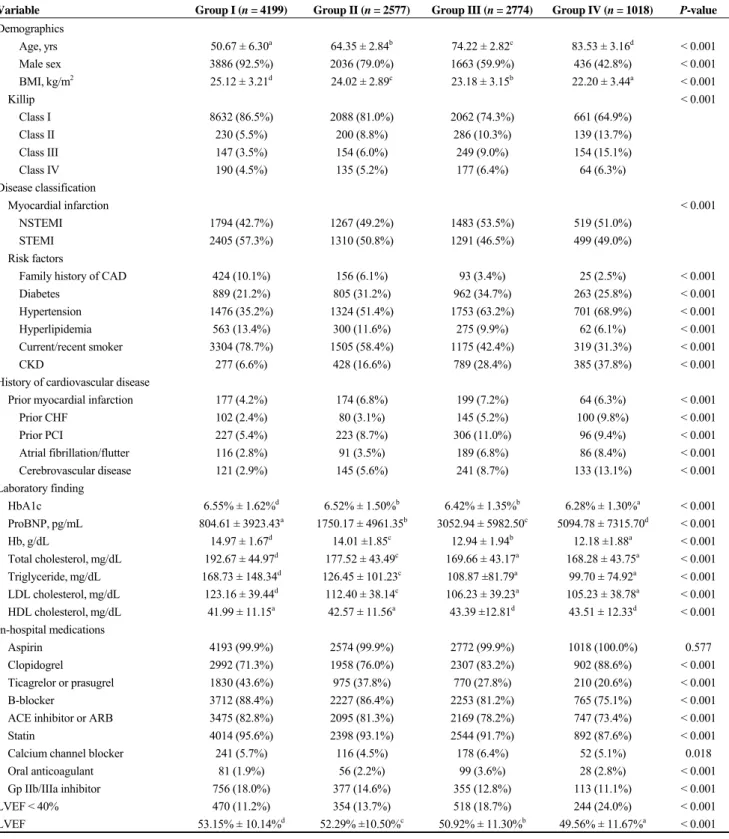
The population distributions for age, sex, BMI, Killip classification, risk factors, history of cardiovascular diseases, laboratory findings, the use of other medications except aspirin and left ventricular ejection fraction (LVEF) differed significantly among the four groups. Their mean ages of the groups were 50.6, 64.4, 74.2, and 83.5 years, respectively.
And the proportion of male was the highest in Group I and became lower as the age increased. The incidence of Killip III, IV tended to be relatively higher in the aged groups (Groups III and IV) than in the youthful groups (Groups I and II). Also, incidences of hypertension, CKD, prior CHF, atrial fibrillation/flutter, cerebrovascular disease, proBNP and LVEF < 40% became decremented as the age became younger. Interestingly, lipid profile of the groups were well controlled in the older group compared to the younger one.
In addition, while the use of clopoidogrel was higher in the Group IV than Group I, ticagrelor or prasugrel and GpIIb/IIIa inhibitor were used less in the Group IV than Group I. Also, the use of B-blocker, angiotensin converting enzyme (ACE) inhibitor or angiotensin II receptor blocker (ARB) and statin was used relatively less in the aged groups and evidence-based medical therapy is less likely given for this groups.
Meanwhile, in angiographic findings (Table 2), all of target vessels including left anterior descending (LAD), left circumflex (LCX), right coronary artery (RCA) and left main coronary artery (LMCA) occurred more frequently in the aged group than younger groups. There were no signifi-
Table 1. Baseline demographic, clinical and laboratory characteristics.
Variable Group I (n = 4199) Group II (n = 2577) Group III (n = 2774) Group IV (n = 1018) P-value
Demographics
Age, yrs 50.67 ± 6.30a 64.35 ± 2.84b 74.22 ± 2.82c 83.53 ± 3.16d < 0.001
Male sex 3886 (92.5%) 2036 (79.0%) 1663 (59.9%) 436 (42.8%) < 0.001
BMI, kg/m2 25.12 ± 3.21d 24.02 ± 2.89c 23.18 ± 3.15b 22.20 ± 3.44a < 0.001
Killip < 0.001
Class I 8632 (86.5%) 2088 (81.0%) 2062 (74.3%) 661 (64.9%)
Class II 230 (5.5%) 200 (8.8%) 286 (10.3%) 139 (13.7%)
Class III 147 (3.5%) 154 (6.0%) 249 (9.0%) 154 (15.1%)
Class IV 190 (4.5%) 135 (5.2%) 177 (6.4%) 64 (6.3%)
Disease classification
Myocardial infarction < 0.001
NSTEMI 1794 (42.7%) 1267 (49.2%) 1483 (53.5%) 519 (51.0%)
STEMI 2405 (57.3%) 1310 (50.8%) 1291 (46.5%) 499 (49.0%)
Risk factors
Family history of CAD 424 (10.1%) 156 (6.1%) 93 (3.4%) 25 (2.5%) < 0.001
Diabetes 889 (21.2%) 805 (31.2%) 962 (34.7%) 263 (25.8%) < 0.001
Hypertension 1476 (35.2%) 1324 (51.4%) 1753 (63.2%) 701 (68.9%) < 0.001
Hyperlipidemia 563 (13.4%) 300 (11.6%) 275 (9.9%) 62 (6.1%) < 0.001
Current/recent smoker 3304 (78.7%) 1505 (58.4%) 1175 (42.4%) 319 (31.3%) < 0.001
CKD 277 (6.6%) 428 (16.6%) 789 (28.4%) 385 (37.8%) < 0.001
History of cardiovascular disease
Prior myocardial infarction 177 (4.2%) 174 (6.8%) 199 (7.2%) 64 (6.3%) < 0.001
Prior CHF 102 (2.4%) 80 (3.1%) 145 (5.2%) 100 (9.8%) < 0.001
Prior PCI 227 (5.4%) 223 (8.7%) 306 (11.0%) 96 (9.4%) < 0.001
Atrial fibrillation/flutter 116 (2.8%) 91 (3.5%) 189 (6.8%) 86 (8.4%) < 0.001 Cerebrovascular disease 121 (2.9%) 145 (5.6%) 241 (8.7%) 133 (13.1%) < 0.001
Laboratory finding
HbA1c 6.55% ± 1.62%d 6.52% ± 1.50%b 6.42% ± 1.35%b 6.28% ± 1.30%a < 0.001 ProBNP, pg/mL 804.61 ± 3923.43a 1750.17 ± 4961.35b 3052.94 ± 5982.50c 5094.78 ± 7315.70d < 0.001 Hb, g/dL 14.97 ± 1.67d 14.01 ±1.85c 12.94 ± 1.94b 12.18 ±1.88a < 0.001 Total cholesterol, mg/dL 192.67 ± 44.97d 177.52 ± 43.49c 169.66 ± 43.17a 168.28 ± 43.75a < 0.001 Triglyceride, mg/dL 168.73 ± 148.34d 126.45 ± 101.23c 108.87 ±81.79a 99.70 ± 74.92a < 0.001 LDL cholesterol, mg/dL 123.16 ± 39.44d 112.40 ± 38.14c 106.23 ± 39.23a 105.23 ± 38.78a < 0.001 HDL cholesterol, mg/dL 41.99 ± 11.15a 42.57 ± 11.56a 43.39 ±12.81d 43.51 ± 12.33d < 0.001
In-hospital medications
Aspirin 4193 (99.9%) 2574 (99.9%) 2772 (99.9%) 1018 (100.0%) 0.577
Clopidogrel 2992 (71.3%) 1958 (76.0%) 2307 (83.2%) 902 (88.6%) < 0.001
Ticagrelor or prasugrel 1830 (43.6%) 975 (37.8%) 770 (27.8%) 210 (20.6%) < 0.001
B-blocker 3712 (88.4%) 2227 (86.4%) 2253 (81.2%) 765 (75.1%) < 0.001
ACE inhibitor or ARB 3475 (82.8%) 2095 (81.3%) 2169 (78.2%) 747 (73.4%) < 0.001
Statin 4014 (95.6%) 2398 (93.1%) 2544 (91.7%) 892 (87.6%) < 0.001
Calcium channel blocker 241 (5.7%) 116 (4.5%) 178 (6.4%) 52 (5.1%) 0.018
Oral anticoagulant 81 (1.9%) 56 (2.2%) 99 (3.6%) 28 (2.8%) < 0.001
Gp IIb/IIIa inhibitor 756 (18.0%) 377 (14.6%) 355 (12.8%) 113 (11.1%) < 0.001
LVEF < 40% 470 (11.2%) 354 (13.7%) 518 (18.7%) 244 (24.0%) < 0.001
LVEF 53.15% ± 10.14%d 52.29% ±10.50%c 50.92% ± 11.30%b 49.56% ± 11.67%a < 0.001 Data are presented as mean ± SD or n (%) where appropriate. Group was stratified according to age (Group I < 60 years, Group II 6070 years, Group III 7080 years, Group IV ≥ 80 years). Lesion based on American College of Cardiology/American Heart Association lesion classification. In ANOVA analysis, values labeled with the different superscripts in a row indicate significant differences between groups based on Scheffe's multiple comparison tests. ACE inhibitor: angiotensin converting enzyme inhibitor; ARB: angiotensin II receptor blocker; BMI: body mass index; CAD: coronary artery disease; CHF: congestive heart failure; CKD: chronic kidney disease; Hb: haemoglobin; HDL: high-density lipoprotein; LDL: low-density lipoprotein; LVEF: left ventricular ejection fraction; NSTEMI: non-ST segment eleva- tion myocardial infarction; PCI: percutaneous coronary intervention; proBNP: pro-brain natriuretic peptide; STEMI: ST segment elevation myocardial infarction; TC:
total cholesterol; TG: triglyceride.
Journal of Geriatric Cardiology | [email protected]; http://www.jgc301.com Table 2. Baseline angiographic characteristics.
Variable Group I
(n = 4497)
Group II (n = 2780)
Group III (n = 2993)
Group IV
(n = 1121) P-value
Target vessel
LAD 2832 (67.4%) 1821(70.7%) 2006 (72.3%) 770 (75.6%) < 0.001
LCX 1635 (38.9%) 1178 (45.7%) 1324 (47.7%) 494 (48.5%) < 0.001
RCA 2057 (49.0%) 1361 (52.8%) 1612 (58.1%) 618 (60.7%) < 0.001
LMCA 146 (3.5%) 119 (4.6%) 168 (6.1%) 64 ( 6.3%) < 0.001
Lesion classification
A 51 (1.2%) 38 (1.5%) 35 (1.3%) 10 (1.0%) 0.651
B1 502 (12.0%) 319 (12.4%) 315 (11.4%) 113 (11.1%) 0.588
B2 1557 (37.1%) 957 (37.1%) 1025 (37.0%) 421 (41.4%) 0.064
C 2088 (49.7%) 1263 (49.0%) 1399 (50.4%) 474 (46.6%) 0.188
Pre-PCI TIMI 0 or 1 2568 (61.2%) 1421 (55.1%) 1491 (53.7%) 540 (53.0%) < 0.001
Post-PCI TIMI 0 or 1 12 (0.3%) 3 (0.1%) 12 (0.4%) 4 (0.4%) 0.176
Post-PCI TIMI 3 4117 (98.0%) 2511 (97.4%) 2684 (96.8%) 963 (94.6%) < 0.001
Number of diseased vessel < 0.001
One-vessel disease 2388 (56.9%) 1230 (47.7%) 1192 (43.0%) 395 (38.8%) Two-vessel disease 1187 (28.3%) 836 (32.4%) 884 (31.9%) 346 (34.0%) Three-vessel disease 587 (14.0%) 466 (18.1%) 640 (23.1%) 249 (24.5%) Three-vessel disease with LM 37 (0.9%) 45 (1.7%) 58 (2.1%) 28 (2.8%)
Total number of stent 1.16 ± 0.39a 1.18 ± 0.42 1.21 ± 0.44b 1.21 ± 0.46b < 0.001 Stent size
Long, mm 28.68 ± 13.62 a 29.56 ± 13.93 30.43 ± 14.87b 30.05 ± 14.94b < 0.001 Diameter, mm 3.13 ± 0.56d 3.04 ± 0.53c 2.97 ± 0.53a 2.94 ± 0.50a < 0.001 Data are presented as mean ± SD or n (%) where appropriate. In ANOVA analysis, values labeled with the different superscripts in a row indicate significant differ- ences between groups based on Scheffe's multiple comparison tests. TIMI: thrombolysis in myocardial Infarction. LAD: left anterior descending artery; LCX: left circumflex artery; LMCA: left main coronary artery; PCI: percutaneous coronary intervention; RCA: right coronary artery; TIMI : thrombolysis in myocardial infarction.
cant differences in the lesion classification between the four groups. The aged group exhibited significantly more mul- tivessel disease and lower rates of post-PCI TIMI flow grade 3. In addition, the final stent size and total numbers of stent indicated more complex coronary lesion in the aged group.
3.2 Clinical outcomes of the overall population:
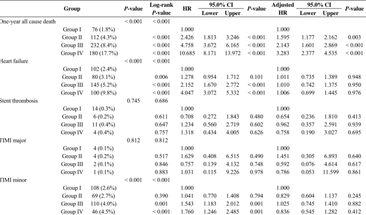
The median follow-up duration was one year. Among the patients with AMI, the cumulative rates of primary end- point including CD, MI, TVR and cerebrovascular events were significantly higher in the oldest age group (Group IV) than the youngest age group (Group I) at one year [172 (17.6%) vs. 145 (3.5%), P < 0.001, Table 3]. And the inci- dence of CD among all individuals at one year was signifi- cantly higher in the oldest age group (Group IV) than the youngest age group (Group I) at one year [138 (13.6%) vs.
62 (1.5%), P < 0.001, Table 3]. Multivariate Cox regression analysis revealed that age is a potent independent predictor for these events [primary end-points, aHR 1.975 (1.500
2.601), P < 0.001 at 12 months, Table 3]. Especially, these
primary cardiac events would be mainly driven by cardiac death in MACCE components [cardiac death, aHR 2.803 (1.9374.054), P < 0.001 at 12 months, Table 3] as well as cerebrovascular events [aHR 2.846 (1.2526.473), P = 0.013 at 12 months, Table 3]. Moreover, we revealed that the primary cardiac events in AMI could be independently affected in proportion to an increase in age (Group II, aHR
= 1.224, 95% CI: 0.9651.525, P = 0.096; Group III, aHR = 1.316, 95% CI: 1.0371.671, P = 0.024; Group IV, aHR = 1.975, 95% CI: 1.5002.601, P < 0.001) compared to Group I. In addition, only all cause death in secondary outcomes (Table 4) showed a significantly higher prevanlence in pro- portion to an increase in age (Group II, aHR = 1.595, 95%
CI: 1.1772.162, P = 0.003; Group III, aHR = 2.143, 95%
CI: 1.6012.869, P < 0.001; Group IV, aHR = 3.283, 95%
CI: 2.3774.535, P < 0.001) compared to Group I. Mean- while, the one-year MACCE in STEMI were 3.1% (Group I, n = 2405), 6.4% (Group II, n = 1310), 9.1% (Group III, n = 1291) and 18.8% (Group IV, n = 499) (Table 1S) vs. 4.0%
(Group I, n = 1794), 5.7% (Group II, n = 1267), 8.6%
(Group III, n = 1483) and 15.0% (Group IV, n = 519) in
Table 3. One-year primary clinical outcomes in MI patients stratified by age.
95.0% CI 95.0% CI
Group P-
value
Log-rank P-value HR
Lower Upper P-value Adjusted
HR Lower Upper P-value One-year primary end-point < 0.001 < 0.001
Group I 145 (3.5%) 1.000 1.000
Group II 156 (6.3%) < 0.001 1.780 1.420 2.232 < 0.001 1.224 0.965 1.525 0.096 Group III 245 (9.6%) < 0.001 2.665 2.170 3.273 < 0.001 1.316 1.037 1.671 0.024 Group IV 172 (17.6%) < 0.001 5.499 4.408 6.859 < 0.001 1.975 1.500 2.601 < 0.001
Cardiac death < 0.001 < 0.001
Group I 62 (1.5%) 1.000 1.000
Group II 78 (3.0%) < 0.001 2.068 1.481 2.886 < 0.001 1.335 0.941 1.895 0.106 Group III 148 (5.3%) < 0.001 3.698 2.749 4.975 < 0.001 1.575 1.122 2.210 0.009 Group IV 138 (13.6%) < 0.001 9.883 7.323 13.338 < 0.001 2.803 1.937 4.054 < 0.001
MI 0.003 < 0.001
Group I 44 (1.0%) 1.000 1.000
Group II 41 (1.6%) 0.020 1.546 1.010 2.366 0.045 0.996 0.635 1.563 0.985 Group III 55 (2.0%) < 0.001 1.985 1.335 2.951 < 0.001 0.956 0.598 1.529 0.852 Group IV 23 (2.3%) < 0.001 2.469 1.491 4.089 < 0.001 0.969 0.530 1.771 0.918 Target vessel revascularization 0.076 0.108
Group I 25 (0.6%) 1.000 1.000
Group II 25 (1.0%) 0.093 1.659 0.953 2.889 0.073 1.403 0.771 2.554 0.268 Group III 13 (0.5%) 0.787 0.828 0.424 1.618 0.581 0.602 0.274 1.320 0.205 Group IV 4 (0.4%) 0.877 0.773 0.269 2.220 0.632 0.536 0.163 1.762 0.304 Cerebrovascular events < 0.001 < 0.001
Group I 16 (0.4%) 1.000 1.000
Group II 25 (1.0%) 0.001 2.593 1.384 4.856 0.003 2.032 1.056 3.907 0.034 Group III 44 (1.6%) < 0.001 4.374 2.468 7.751 < 0.001 2.804 1.453 5.412 0.002 Group IV 16 (1.6%) < 0.001 4.753 2.377 9.506 < 0.001 2.846 1.252 6.473 0.013 Data are presented as n (%). HR: hazard ratio. Group was stratified according to age (Group I < 60 years, Group II 6070 years, Group III 7080 years, Group IV ≥ 80 years). MI: myocardial infarction.
Table 4. One-year secondary clinical outcomes in MI patients stratified by age.
95.0% CI 95.0% CI
Group P-value Log-rank
P-value HR
Lower Upper P-value Adjusted
HR Lower Upper P-value One-year all cause death < 0.001 < 0.001
Group I 76 (1.8%) 1.000 1.000
Group II 112 (4.3%) < 0.001 2.426 1.813 3.246 < 0.001 1.595 1.177 2.162 0.003 Group III 232 (8.4%) < 0.001 4.758 3.672 6.165 < 0.001 2.143 1.601 2.869 < 0.001 Group IV 180 (17.7%) < 0.001 10.685 8.171 13.972 < 0.001 3.283 2.377 4.535 < 0.001
Heart failure < 0.001 < 0.001
Group I 102 (2.4%) 1.000 1.000
Group II 80 (3.1%) 0.006 1.278 0.954 1.712 0.101 1.011 0.735 1.389 0.948 Group III 145 (5.2%) < 0.001 2.152 1.670 2.772 < 0.001 1.010 0.742 1.375 0.950 Group IV 100 (9.8%) < 0.001 4.047 3.072 5.332 < 0.001 1.006 0.699 1.445 0.976
Stent thrombosis 0.745 0.686
Group I 14 (0.3%) 1.000 1.000
Group II 6 (0.2%) 0.611 0.708 0.272 1.843 0.480 0.654 0.236 1.810 0.413 Group III 11 (0.4%) 0.647 1.234 0.560 2.719 0.602 0.962 0.357 2.591 0.939 Group IV 4 (0.4%) 0.757 1.318 0.434 4.005 0.626 0.758 0.190 3.027 0.695
TIMI major 0.812 0.812
Group I 4 (0.1%) 1.000 1.000
Group II 4 (0.2%) 0.517 1.629 0.408 6.515 0.490 1.451 0.305 6.893 0.640 Group III 2 (0.1%) 0.846 0.757 0.139 4.132 0.748 0.592 0.076 4.614 0.617 Group IV 1 (0.1%) 0.883 1.031 0.115 9.226 0.978 0.786 0.053 11.599 0.861
TIMI minor < 0.001 < 0.001
Group I 108 (2.6%) 1.000 1.000
Group II 69 (2.7%) 0.390 1.041 0.770 1.408 0.794 0.829 0.604 1.137 0.245 Group III 110 (4.0%) 0.001 1.543 1.183 2.012 0.001 1.025 0.745 1.410 0.882 Group IV 46 (4.5%) < 0.001 1.760 1.246 2.485 0.001 0.836 0.545 1.282 0.412 Data are presented as n (%). Group was stratified according to age (Group I < 60 years, Group II 6070 years, Group III 7080 years, Group IV ≥ 80 years). MI:
myocardial infarction; TIMI: thrombolysis in myocardial infarction.
Journal of Geriatric Cardiology | [email protected]; http://www.jgc301.com
NSTEMI, respectively (Table 2S). After adjustment for confounding parameters, patients with STEMI had the in- cremental risk of one-year MACCE (Group II, aHR = 1.451, 95% CI: 1.0432.018, P = 0.027; Group III, aHR = 1.441, 95% CI: 1.0192–0.036, P = 0.039; Group IV, aHR = 2.174, 95% CI: 1.4693.216, P < 0.001) compared to Group I (Ta- ble 1S). Cardiac death had a major role in this effect in pa- tients with STEMI (Group II, aHR = 1.567, 95% CI:
0.9852.493, P = 0.058; Group III, aHR = 1.808, 95% CI:
1.1402.869, P = 0.012; Group IV, aHR = 2.707, 95% CI:
1.6364.480, P < 0.001) compared to Group I (Table 1S).
However, there was only significant difference between the Group I and 4 in patients with NSTEMI (Group II, aHR = 1.034, 95% CI: 0.7341.457, P = 0.846; Group III, aHR = 1.197, 95% CI: 0.8591.667, P = 0.288; Group IV, aHR = 1.749, 95% CI: 1.1842.584, P = 0.005, Table 2S).
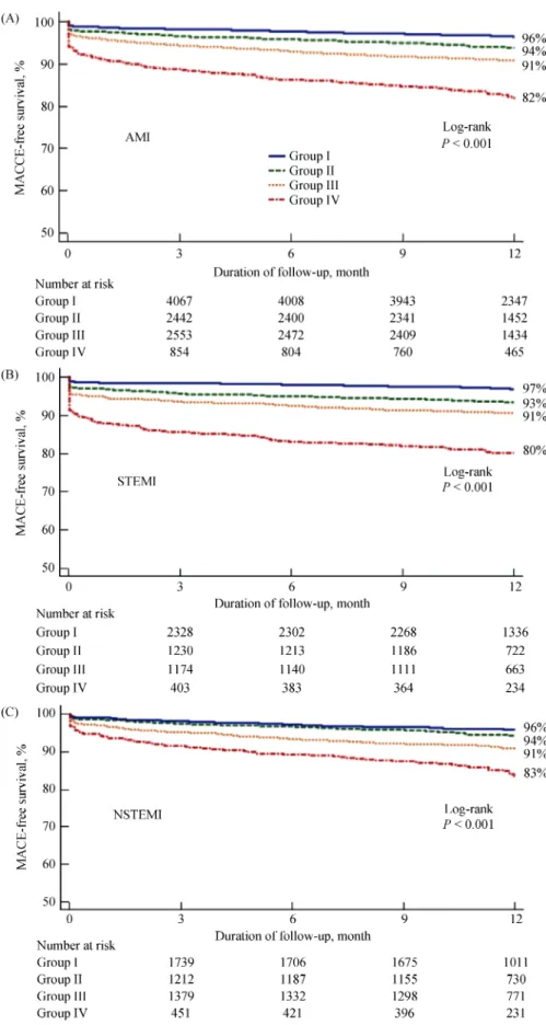
3.3 Kaplan-Meier and landmark analysis in the overall population
The Kaplan-Meier curve indicated a significantly higher incremental risk for primary end-point in the AMI patients undergoing primary PCI during one year (event-free sur- vival rate: 82% vs. 91% vs. 94% vs. 96%, P < 0.001, Table 3 and Figure 2). Also, Figure 2 showed Kaplan-Meier curves for the incidence of MACCE in both STEMI and NSTEMI patients during one year. They showed comparable results with a significantly higher incremental risk for primary outcomes (event-free survival rate: 80% vs. 91% vs. 93% vs.
97% in STEMI, P < 0.001 and 83% vs. 91% vs. 94% vs.
96% in NSTEMI, P < 0.001, Table 2S and Figure 2).
3.4 Predictors of the major adverse outcomes in AMI Univariable and multivariable logistic regression analy- ses were performed to identify independent predictors of MACCE in patients with AMI after PCI. In the multivari- able logistic regression model, killip classification 4, hyper- tension, CKD, atrial fibrillation, ACC/AHA type B2 were independent predictors of the MACCE (Table 5).
Expectantly, age was an independent predictor for the higher prevalence of the primary outcomes (adjusted OR = 1.018, P < 0.001). Meanwhile, not only LVEF and the mean stent diameter but also B-blocker, calcium channel blocker (CCB), ACE inhibitor/ARB and statin among medical therapies showed benign effects for the prevalence of MACCE. Interestingly, BMI also revealed counter-correla- tion with the occurrence of MACCE in AMI patients.
4 Discussion
This large, multicenter cohort analysis evaluated the
outcomes of STEMI and NSTEMI patients during one year according to an increase in age. This study included a rela- tively large number of patients with STEMI and NSTEMI who had undergone primary PCI. As life expectancy con- tinues to increase, interventional cardiologists can expect to encounter a significant increase in the number of patients with AMI who are ≥ 70 years old. In the era before reperfu- sion, elderly patients had one-month and one-year mortality rates of 30% and 75%, respectively.[8,9] Our study may help the clinician identify a high-risk subset of elderly patients with AMI, because most of clinical trials were based on a large proportion of relatively younger patients and the population of very elderly AMI patients constitute a very small portion. To our knowledge, this database reveals the largest published series of patients ≥ 70 years old undergo- ing primary percutaneous intervention for AMI.
Although thrombolytic therapy has been shown to im- prove survival in elderly patients when compared with pla- cebo,[10,11] multiple studies have shown lower mortality rates when elderly patients are treated with primary percutaneous transluminal coronary angioplasty.[1214] Mortality for pa- tients > 65 years was 5.7% in the angioplasty group versus 15.0% in the thrombolytic group. In our study, one-year mortality rate for AMI was 1.8% (< 60 years), 4.3% (6070 years), 8.4% (7080 years), 17.7% (≥ 80 years) (Table 4).
And the one-year rates of overall MACCE recorded in the present study were 3.5% (< 60 years), 6.3% (6070 years), 9.6% (7080 years), 17.6% (≥ 80 years). Although aged patients were more likely to have complex culprit lesions and multivessel disease, TIMI III flow was achieved in 96.8% (7080 years), 94.6% (≥ 80 years) (Table 2). Never- theless, the results found that mortality and MACCE rates even in the DES era were still higher in the older group.
This higher mortality and rates of adverse cardiac events in the elderly patients would be relevant to several comorbid- ities or cardiovascular risk factors (higher prevalence of hypertension, CKD, prior CHF, atrial fibrillation/flutter, cerebrovascular disease, etc, Table 1) prevalent in the eld- erly. Older AMI patients may often not receive the optimal medical treatment recommended by current guidelines be- cause of their conditions and comorbidities.[15] The record in the present study was roughly consistent with this result.
The use of B-blocker, ACE inhibitor or ARB and statin was relatively lower in the older patients after AMI (Table 1), which might be a possible reason for the higher incidence of adverse effects or suspected contraindications of medical therapy. Also, the present study indicates that this outcome were mainly driven by cardiac death (Table 3), surely al- though heart failure, cerebrovascular events and myocardial infarction as well as other comorbidities such as diabetes,
Figure 2. Kaplan-Meier curve for the 12-month probability of MACCE-free survival in patients with AMI (A), STEMI (B) and NSTEMI (C) undergoing primary PCI stratified by age. AMI: acute myocardial infarction; MACCE: major adverse cardiocerebrovascu- lar events; NSTEMI: non-ST segment elevation myocardial infarction; STEMI: ST segment elevation myocardial infarction.
Journal of Geriatric Cardiology | [email protected]; http://www.jgc301.com Table 5. Multivariate analysis of MACCE at one-year follow-up.
95% CI 95% CI
AMI Adjusted
OR Lower Upper P-value Adjusted
OR Lower Upper P-value
Killip 4 1.745 1.314 2.319 0.009 ACE inhibitor/ARB 0.563 0.462 0.685 0.002
Hypertension 1.403 1.164 1.690 < 0.001 Use of statin 0.242 0.193 0.303 < 0.001
CKD 1.325 1.073 1.636 0.009 ACC/AHA type B2 1.389 1.159 1.666 < 0.001
Atrial fibrillation 1.439 1.043 1.984 0.027 Age 1.018 1.009 1.028 < 0.001
B-blocker 0.532 0.432 0.654 < 0.001 BMI 0.964 0.938 0.991 0.010
CCB 0.533 0.351 0.809 0.003 LVEF 0.991 0.983 0.999 0.023
Mean stent diameter 0.784 0.666 0.921 0.003 ACE inhibitor: angiotensin converting enzyme inhibitor; ARB: angiotensin II receptor blocker; ACC/AHA: American College of Cardiology/American Heart Association; BMI: body mass index; CCB: calcium channel blocker; CKD: chronic kidney disease; LVEF: left ventricular ejection fraction.
renal failure, etc. might, in part, affect the mortality rates in older AMI patients. Our study suggests that elderly patients continue to have a higher risk of stroke and death after AMI.
However, with primary percutaneous intervention in DES era, the mortality rate of these high risk patients is lower than those observed in thrombotic trials.
Advanced age remains an independent predictor of major cardiac adverse events after acute AMI in our study (aOR = 1.018, 95% CI: 1.0091.028, P < 0.001, Table 5). There are overall obvious differences in cardiac risk factors between younger and older patients with AMI (Tables 1 & 2). Older patients have a higher incidence of hypertension, CKD, prior CHF, atrial fibrillation/flutter and cerebrovascular dis- ease. This is not surprising when considering the fact that these illnesses are closely correlated with advanced age.
Conversely, a proportion of male sex, obesity, family his- tory of CAD, hyperlipidemia, current/recent smoker seem to be strongly associated with the development of AMI in younger patients (Table 1). In the study analysis, older pa- tients have more advanced disease and more LV dysfunc- tion. Older patients tend to have a higher incidence of ag- gravated killip classification and LVEF (Table 1).
Besides age, this study identified the following indepen- dent predictors for adverse primary outcomes during 1 year.
Killip classification 4, hypertension, CKD, atrial fibrillation, ACC/AHA type B2 were the independent predictors for the prevalence of MACCE. The use of B-blocker, CCB, ACE inhibitor/ARB and statin as well as LVEF and the mean stent diameter were found to be relatively benign predictors for MACCE. Interestingly, an increase of BMI might be also favorably associated with the prevalence of MACCE.
Post-procedural coronary flow was not associated with one-year MACCE in this study. The overall high post-TIMI flow rate (96.7%) might have affected these results.
It is well known that elderly MI patients have a higher risk of all-cause death and major cardiac adverse events. DES has been reported to reduce the rate of restenosis and target
lesion revascularization compared with bare-metal stents (BMS).[16,17] Meanwhile, PCI with DES might be associated with the prevalence of stent thrombosis due to hypersensi- tivity reaction with extensive vasculitis,[18,19] delayed heal- ing process with endothelial dysfunction[20,21] and neo-athe- rosclerosis.[22] Most stent thrombosis in BMS era was early stent thrombosis, while stent thrombosis in 1st generation DES era was reported to happen regardless of stages even though late or very late stent thrombosis was more prob- lematic than early stent thrombosis. As the rate of late or very late stent thrombosis improved in 2nd generation DES era, concerns over the fatal matter in 1st generation DES was belittled.[23,24] In meta-analysis, early stent thrombosis in BMS was about 0.6%, while the rate of stent thrombosis in sirolimus-eluting stent and paclitaxel-eluting stent were reported as 0.5% and 0.5%, respectively.[25,26] In other stud- ies, early or late stent thrombosis using 2nd generation DES was at least comparable, not higher than BMS or 1st genera- tion DES.[27,28] The present study revealed that the rate of early or late stent thrombosis was very low (0.1% vs. 0.2%
vs. 0.1% vs. 0.1%, P = 0.745 according to an increase in age, Table 4) compared to previous studies, surprisingly even comparable regardless of age difference (Table 4). In addi- tion, the present report also indicated that there was no sig- nificant difference in the rate of TVR according to the age.
(0.6% vs. 1.0% vs. 0.5% vs. 0.4%, P = 0.076, Table 3).
The concerning matter is very late stent thrombosis hap- pening in aged population after 1 year. In our study, the duration was limited to one-year, accordingly we couldn’t evaluate the prevalence of very late stent thrombosis ac- cording to ARC definition. Elderly patients prescribed with dual anti-platelet agents presented with a high risk for bleed- ing.[29] However, the finding from this study did not show any significant difference in the occurrence of in-hospital major or minor bleeding between the four groups (Table 4).
The unexpected bleeding could be originated from the rela- tively lower use of ticagrelor or prasugrel (novel antiplatelet)
and the short term duration of follow-up. Large and pro- spective trials will be necessary to settle this issue more definitely and to assess long-term major or minor bleeding events and the optimal duration of anti-platelet treatment for aged population. According to our study, age over 70 years old is a potential risk factor to generate adverse cardiac outcomes. Especially, the advanced age population over 80 years has the most powerful influence on the outcomes even after the adjustment for several confounders (Table 3). This effect persists not only in STEMI but also in NSTEMI pa- tients (Tables 1S & 2S). Interestingly, the prevalence of MACCE and cardiac death in STEMI patients was signifi- cant in the patients over 70 years, while they became sig- nificant over 80 years old in NSTEMI patients. Age be- tween 70-80 years is found to be a potential factor affecting the prognosis of STEMI patients.
There are few studies which investigated the adverse outcomes after the DES implantation in aged patients with AMI. The results from the present study propose that oc- currence of TVR, stent thrombosis and TIMI major/minor bleeding are not affected by age. In particular, the oldest population over 80 years also showed consistent results.
4.1 Study limitations
There are some limitations in our study. First, it was a nonrandomized study and results might have been in- fluenced by selection bias and confounding factors. How- ever, this study was a prospective, large multicenter cohort study involving most confounders resulting in controlling the baseline differences to the greatest extent in a multi- variable Cox regression model. Second, these trials were conducted in a wide variety of hospital settings and the in- terventions were performed by operators with various de- grees of skill and experience. Third, our study only assessed one-year follow-up periods and the mortality and ischemic event rates were relatively low. Also, the proportion of the oldest aged population in this study was relatively low compared to other aged population. Fourth, our study was underpowered to evaluate the ischemic events of the older groups compared to the younger groups, even though we adjusted with statistical method. Lastly, this trial has an in- trinsic limitation itself due to several heterogenic compo- nents in the groups in terms of angiographic, procedural aspects and different routine laboratory tests performed separately by the different hospitals involved in this study.
4.2 Conclusions
The present study reveals that the elderly undergoing PCI in AMI patients still presents higher mortality and MACCE in the DES era. Several comorbidities and risk factors were
more prominent in the elderly and probably related with a higher prevalence of adverse cardiac events. Nevertheless, there were no significant differences in the occurrence of TVR, stent thrombosis and bleeding between the aged groups, which may be owing to the development of device, procedure technique and optimal medical treatments. We expect that the challenging coronary intervention in the elder patients with AMI would be promising and overcome in the near future.
Acknowledgements
This study was supported by the fund (2016-ER6304- 01) from Research of Korea Centers for Disease Control and Prevention and the Korea Health Technology R & D Project, Ministry of Health & Welfare (HI13C1527), Republic of Korea. There were no conflicts of interest to be declared.
References
1 Morice MC, Serruys PW, Sousa JE, et al. A randomized comparison of a sirolimus-eluting stent with a standard stent for coronary revascularization. N Engl J Med 2002; 346:
1773–1780.
2 Moses JW, Leon MB, Popma JJ, et al. Sirolimus-eluting stents versus standard stents in patients with stenosis in a na- tive coronary artery. N Engl J Med 2003; 349: 1315–1323.
3 Schofer J, Schluter M, Gershlick AH, et al. Sirolimus-eluting stents for treatment of patients with long atherosclerotic le- sions in small coronary arteries: double-blind, randomised controlled trial (E-SIRIUS). Lancet 2003; 362: 1093–1099.
4 De Gregorio J, Kobayashi Y, Albiero R, et al. Coronary artery stenting in the elderly: short-term outcome and long-term angiographic and clinical follow-up. J Am Coll Cardiol 1998;
32: 577–583.
5 DeGeare VS, Stone GW, Grines L, et al. Angiographic and clinical characteristics associated with increased in-hospital mortality in elderly patients with acute myocardial infarction undergoing percutaneous intervention (a pooled analysis of the primary angioplasty in myocardial infarction trials). Am J Cardiol 2000; 86: 30–34.
6 Lindsay J, Jr., Reddy VM, Pinnow EE, et al. Morbidity and mortality rates in elderly patients undergoing percutaneous coronary transluminal angioplasty. Am Heart J 1994; 128:
697–702.
7 Thygesen K, Alpert JS, Jaffe AS, et al. Third universal defini- tion of myocardial infarction. Eur Heart J 2012; 33: 2551–2567.
8 Yang XS, Willems JL, Pardaens J, et al. Acute myocardial infarction in the very elderly. A comparison with younger age groups. Acta Cardiol 1987; 42: 59–68.
9 Goldberg RJ, McCormick D, Gurwitz JH, et al. Age-related trends in short- and long-term survival after acute myocardial infarction: a 20-year population-based perspective (1975- 1995). Am J Cardiol 1998; 82: 1311–1317.
Journal of Geriatric Cardiology | [email protected]; http://www.jgc301.com 10 Gurwitz JH, Goldberg RJ, Gore JM. Coronary thrombolysis
for the elderly? Jama 1991; 265: 1720–1723.
11 Topol EJ, Califf RM. Thrombolytic therapy for elderly pa- tients. N Engl J Med 1992; 327: 45–47.
12 Grines CL, Browne KF, Marco J, et al. A comparison of immediate angioplasty with thrombolytic therapy for acute myocardial infarction. N Engl J Med 1993; 328: 673–679.
13 Stone GW, Grines CL, Browne KF, et al. Predictors of in- hospital and 6-month outcome after acute myocardial infarc- tion in the reperfusion era: the primary angioplasty in, myocardial infarction (PAMI) trail. J Am Coll Cardiol 1995;
25: 370–377.
14 Holmes DR, Jr., White HD, Pieper KS, et al. Effect of age on outcome with primary angioplasty versus thrombolysis. J Am Coll Cardiol 1999; 33: 412–419.
15 Wijns W, Kolh P, Danchin N, et al. Guidelines on myocardial revascularization. Eur Heart J 2010; 31: 2501–2555.
16 Stettler C, Wandel S, Allemann S, et al. Outcomes associated with drug-eluting and bare-metal stents: a collaborative net- work meta-analysis. Lancet 2007; 370: 937–948.
17 Marroquin OC, Selzer F, Mulukutla SR, et al. A comparison of bare-metal and drug-eluting stents for off-label indications.
N Engl J Med 2008; 358: 342–352.
18 Virmani R, Guagliumi G, Farb A, et al. Localized hypersen- sitivity and late coronary thrombosis secondary to a siroli- mus-eluting stent: should we be cautious? Circulation 2004;
109: 701–705.
19 Nebeker JR, Virmani R, Bennett CL, et al. Hypersensitivity cases associated with drug-eluting coronary stents: a review of available cases from the Research on Adverse Drug Events and Reports (RADAR) project. J Am Coll Cardiol 2006; 47:
175–181.
20 Joner M, Finn AV, Farb A, et al. Pathology of drug-eluting stents in humans: delayed healing and late thrombotic risk. J Am Coll Cardiol 2006; 48: 193–202.
21 Kotani J, Awata M, Nanto S, et al. Incomplete neointimal coverage of sirolimus-eluting stents: angioscopic findings. J Am Coll Cardiol 2006; 47: 2108–2111.
22 Park SJ, Kang SJ, Virmani R, et al. In-stent neoatherosclerosis:
a final common pathway of late stent failure. J Am Coll Cardiol 2012; 59: 2051–2057.
23 Silber S, Windecker S, Vranckx P, et al. Unrestricted rando- mised use of two new generation drug-eluting coronary stents:
2-year patient-related versus stent-related outcomes from the RESOLUTE All Comers trial. Lancet 2011; 377: 1241–1247.
24 Smits PC, Kedhi E, Royaards KJ, et al. 2-year follow-up of a randomized controlled trial of everolimus- and paclitaxel- eluting stents for coronary revascularization in daily practice.
COMPARE (Comparison of the everolimus eluting XIENCE-V stent with the paclitaxel eluting TAXUS LIBERTE stent in all-comers: a randomized open label trial). J Am Coll Cardiol 2011; 58: 11–18.
25 Bavry AA, Kumbhani DJ, Helton TJ, et al. Risk of thrombosis with the use of sirolimus-eluting stents for percutaneous coronary intervention (from registry and clinical trial data).
Am J Cardiol 2005; 95: 1469–1472.
26 Bavry AA, Kumbhani DJ, Helton TJ, et al. What is the risk of stent thrombosis associated with the use of paclitaxel-eluting stents for percutaneous coronary intervention?: a meta-analy- sis. J Am Coll Cardiol 2005; 45: 941–946.
27 Kedhi E, Joesoef KS, McFadden E, et al. Second-generation everolimus-eluting and paclitaxel-eluting stents in real-life practice (COMPARE): a randomised trial. Lancet 2010; 375:
201–209.
28 Serruys PW, Silber S, Garg S, et al. Comparison of zotaro- limus-eluting and everolimus-eluting coronary stents. N Engl J Med 2010; 363: 136–146.
29 Bhatt DL, Fox KA, Hacke W, et al. Clopidogrel and aspirin versus aspirin alone for the prevention of atherothrombotic events. N Engl J Med 2006; 354: 1706–1717.
95.0% CI 95.0% CI
Group P-
value
Log-rank P-value HR
Lower Upper
P-value Adjusted
HR Lower Upper P-value
1-year primary end-point < 0.001 < 0.001
Group 1 74 (3.1%) 1.000 1.000
Group 2 84 (6.4%) < 0.001 2.120 1.551 2.898 < 0.001 1.451 1.043 2.018 0.027 Group 3 117 (9.1%) < 0.001 3.064 2.290 4.099 < 0.001 1.441 1.019 2.036 0.039 Group 4 94 (18.8%) < 0.001 6.847 5.048 9.287 < 0.001 2.174 1.469 3.216 < 0.001
Cardiac death < 0.001 < 0.001
Group 1 35 (1.5%) 1.000 1.000
Group 2 46 (3.5%) < 0.001 2.431 1.566 3.774 < 0.001 1.567 0.985 2.493 0.058 Group 3 85 (6.6%) < 0.001 4.625 3.120 6.856 < 0.001 1.808 1.140 2.869 0.012 Group 4 77 (15.4%) < 0.001 11.294 7.573 16.843 < 0.001 2.707 1.636 4.480 < 0.001
Myocardial infarction 0.009 0.002
Group 1 17 (0.7%) 1.000 1.000
Group 2 21 (1.6%) 0.016 2.319 1.223 4.395 0.010 1.778 0.899 3.515 0.098 Group 3 16 (1.2%) 0.017 1.855 0.937 3.671 0.076 1.160 0.516 2.607 0.719 Group 4 11 (2.2%) < 0.001 3.645 1.707 7.782 < 0.001 1.892 0.725 4.940 0.193 Target vessel
revascularization 0.649 0.693
Group 1 12 (0.5%) 1.000 1.000
Group 2 9 (0.7%) 0.321 1.406 0.593 3.337 0.439 1.166 0.448 3.034 0.752 Group 3 6 (0.5%) 0.877 0.990 0.372 2.639 0.985 0.779 0.236 2.574 0.682 Group 4 2 (0.4%) 0.730 0.959 0.215 4.287 0.957 0.420 0.063 2.818 0.372
Cerebrovascular events 0.006 0.002
Group 1 9 (0.4%) 1.000 1.000
Group 2 12 (0.9%) 0.017 2.500 1.053 5.932 0.038 2.335 0.953 5.725 0.064 Group 3 15 (1.2%) < 0.001 3.305 1.446 7.552 0.005 2.181 0.836 5.695 0.111 Group 4 7 (1.4%) < 0.001 4.454 1.658 11.960 0.003 2.985 0.899 9.914 0.074 Data are presented as n (%). Group was stratified according to age (Group I , n = 2405, < 60 years; Group II , n = 1310, 6070 years; Group III, n = 1291, 7080 years; Group IV, n = 499, ≥ 80 years). STEMI: ST segment elevation myocardial infarction.