파킨슨병 모형 흰쥐의 줄무늬체에서 Apomorphine 투여 방법에 따른 도파민 D2 수용체의 발현 *
가톨릭대학교 의과대학 신경외과학교실
최승진·성재훈·손병철·박춘근·권성오·김문찬·이상원
= Abstract =
Expression of Dopamine D2 Receptor in Response to Apomorphine Treatment in the Striatum of the Rat with Experimentally Induced Parkinsonism
Seung Jin Choi, M.D., Jae Hoon Sung, M.D., Byung Chul Son, M.D., Choon Keun Park, M.D., Sung Oh Kwon, M.D.,
Moon Chan Kim, M.D., Sang Won Lee, M.D.
Department of Neurosurgery, College of Medicine, The Catholic University of Korea, Seoul, Korea
bjective: : :Parkinsonian rat models have generally been characterized by unilateral destruction of both the : nigrostriatal pathway and the mesolimbic pathway using the neurotoxin 6-hydroxydopamine. The induction of contraversive turning by apomorphine in these models is thought to reflect the stimulation of super- sensitive dopamine D2 receptor or receptor-mediated mechanisms in denervated neostriatum. The present study was undertaken to investigate the expression of dopamine D2 receptor in denervated striatum according to modalities of apomorphine(dopamine agonist) treatment after creating a hemiparkinsonian rat model in which there is 6-hydroxydopamine induced destruction of the unilateral dopaminergic nigrostriatal pathway.
Methods: : : :After making complete lesion in left side substantia nigra pars compacta(SNpc) by stereotactic injection of 6-hydroxydopamine into medial and lateral areas of SNpc, and confirming successful animal model by apomorphine induced contraversive turning behavior without recovery and complete destruction of ipsilateral SNpc with tyrosine hydroxylase immunostaining in 7th day after operation, 15 rats of parkinsonian model were studied with or without administration of apomorphine at varying doses and durations. According to the modalities of apomorphine treatment for 4 days, these rats were divided into 3 groups, as not-treated group, intermittently treated group and constantly treated group. For investigating the extent of the expression of dopamine D2 receptor in denervated striatum, immunohistochemical staining by dopamine D2 receptor antibody and Western blot were performed.
Results: : : :In the D2 receptor antibody immunohistochemical staining, the mean number of positive stained neurons was highest in not-treated group(20.5±1.14) of 3 groups. In constantly treated group, the mean number of positive stained neurons was less(3.9±1.79) than intermittently treated group(p<0.05). The Western blotting with the D2 receptor antibody revealed that expression of receptors was also highest in not-treated group and less in constantiy treated group than intermittently treated group.
Conclusion: : : :Dopamine D2 receptors in denervated striatum of parkinsonian rat models, which were not treated with apomorphine, revealed to be most highly expressed. And, according to doses and durations of apomorphine administration, desensitization of the receptor was more apt to develop with constant treatment than intermittent treatment. In clinical setting, the authors believe that, in long-term treated parkinsonian patients, desensitization of
OOOO
*본 논문은 가톨릭 중앙의료원 연구비의 일부로 이루어졌슴.
dopamine receptors due to chronic dopaminergic stimulation seems to be partially related to mechanisms of drug tolerance.
KEY WORDS:Parkinsonian rat model・6-hydroxydopamine・Apomorphine・Dopamine D2 receptor・Immunohist- ochemical stain・Western blot.
서 론
파킨슨병은 병리조직학적인 측면에서 흑색질에서 도파민 을 생성하는 신경세포의 소실에 의한, 흑색질과 줄무늬체간 신경경로의 퇴행성 변화에 의해 이차적인 줄무늬체에서의 도파민결핍에 의한 질환으로 정의되고 있다. 동물을 이용한 파킨슨병 모형에 있어서 지금까지는 흰쥐와 영장류를 대상 으로한 동물모델이 실험되고 있으며, 이중 1971년 Unge- rstedt가 처음 고안한 6-hydroxydopamine을 이용한 흰 쥐 파킨슨병 모델이 지금까지 가장 많이 이용되고 있다. 이 러한 동물모델에서 여러 도파민 작동제를 투여후 운동역학 적인 변화에 대한 연구가 많이 진행되고 있으며, 임상적으 로는 파킨슨병 치료를 위한 약리학적 선별검사나 새로운 약 물실험, 신경세포 이식과 같은 수술치료적인 접근을 연구하 기 위한 기본모델로 이용되고 있다.
한편 줄무늬체 도파민 수용체의 한 종류인 D2 수용체의 변화가 파킨슨병의 임상적인 경과와 관련성이 높을것으로 추정되고 있다. 사람 및 흰쥐에서 줄무늬체 도파민 수용체 의 대부분은 D2 수용체로 알려져 있고, 이러한 동물모델에 서 도파민세포의 소실시 줄무늬체의 D2 수용체가 증가됨이 보고되고 있다
6).
본 연구는 6-hydroxydopamine을 흑색질에 주입하여 편측 파킨슨병을 유발시킨 흰쥐모델을 만든후, 시냅스후 도 파민 작동제인 apomorphine을 일정기간 간헐적으로 주입 한 군과 지속적으로 주입한 군, 또한 주입하지 않은 군으로 나누어, 면역조직화학적 염색 및 Western blot을 시행하여 줄무늬체에서 도파민 D2 수용체의 발현양상을 알아 보고자 하였다.
재료 및 방법
1. 편측 파킨슨병 흰쥐모델 제작
1) 재료 및 방법
실험동물은 암수에 관계없이 체중 250~350g의 Sprague- Dawley계 흰쥐를 사용하였고, 50mg/kg의 ketamine과
2.5mg/kg의 xylazine을 혼합하여 복강내 주사로 마취하였 다. 뇌정위 수술기구인 David Kopf frame(David Kopf Instruments, CA, U.S.A.)에 치아고정기(toothbar)가 양 쪽 외이도선에서 3.3mm 아래에 위치하도록 실험쥐의 두 부를 고정시키고, 두피를 소독 후 절개하여 두개골을 노출 시켰다. 표적지점은 좌측 중뇌의 흑색질로 정하고 Paxinos 와 Watson
20)의 백서 뇌도보(rat brain atlas)에 따라서 표 적좌표를 정하였다. 가능한한 흑색질의 완전파괴 모델 (complete lesion)을 유도하기 위하여 흑색질을 내측과 외 측으로 나누었으며, 내측의 표적좌표는 전정(bregma)에서 후방으로 5.3mm, 중앙선에서 좌측으로 1.3mm, 깊이는 경막에서 7.1mm 아래로, 외측의 표적좌표는 전정에서 후 방으로 5.3mm, 중앙선에서 좌측으로 2.3mm, 깊이는 경 막에서 6.8mm 아래로 정하였다.
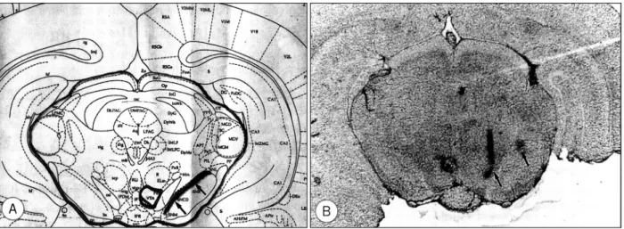
두군데 표적좌표위 두개골부위에 드릴을 이용하여 난원형 모양의 구멍(burr hole)을 만들어 뇌경막을 노출시킨뒤, 각 각의 표적지점을 확인하기 위하여 표적좌표에 Hamilton 주 사기를 이용하여 1μl의 gentian violet을 주입하였다. 주입 후 1.5ml의 KCl을 바로 심장에 주사한후, 뇌를 적출하여 gentian violet를 주입하였던 부위를 중심으로 50μm 두께 의 연속적인 동결절편을 만들어 광학현미경으로 관찰하였다.
Hamilton 주사기의 바늘이 통과한 부위(trajectory)와 ge- ntian violet이 주입된 부위를 Paxinos와 Watson의 백서 뇌도보와 비교하였으며, 흑색질의 표적지점이 정해진 좌표 에 만들어 졌슴을 확인하였다(Fig. 1). 편측 파킨슨병 모델 을 만들기 위하여 위와같이 실험쥐를 뇌정위 수술기구에 고 정후, 같은 방법으로 0.2mg/ml의 ascorbic acid를 함유한 3μg/μl 농도의 6-hydroxydopamine을 Hamilton 주사 기(26 G)를 이용하여 각각의 표적에 3μl씩 서서히 주입후, 2분간 기다렸다가 바늘을 제거하고 두피를 봉합하였다. 한 편 정상 대조군으로 2마리의 흰쥐에서도 같은 표적좌표에 같은 양의 생리적 식염수를 주입하였다.
2) Apomorphine 유발 회전운동 검사
6-hydroxydopamine을 주입후 1주후에 apomorphine
에 대한 회전반응검사를 시행하였다. 0.25mg/kg의 apomor-
phine을 피하주사하고 10분후에 전자식 회전측정기(Ro-
tometer, photobeam activity system, San Diego instru- ments, CA, U.S.A., 1998)를 이용하여 30분간 회전운동반 응을 관찰하였으며 1분간의 평균 회전수를 측정하였다. 이 들중 30분간 총회전수가 90회이상(1분간의 평균 회전수가 3회이상) 우측으로 회전운동을 보인 흰쥐를 완성된 편측 파 킨슨병 모델로 정하였다
22).
3) Tyrosine hydroxylase 면역조직화학적 염색
정상 대조군 중 1마리와 apomorphine 유발 회전운동 검 사로 확인된 파킨슨병 모델 흰쥐의 흑색질 도파민 생성세포 에 대한 tyrosine hydroxylase 면역조직화학 염색을 실시 하였다.
(1) 조직의 준비
Ketamine(50mg/kg)을 복강내 주사하여 마취시키고 흉 부를 절개한 후, 심장을 통하여 0.1M 인산완충용액(PBS, pH 7.4)과 4% paraformaldehyde로 관류시킨후 뇌를 적 출하여 약 24시간동안 4% paraformaldehyde in PBS에 후고정하였다. 중뇌부위를 Vibratome으로 30μm 두께의 연속절편으로 만들어 인산완충용액에 담가 10분간 3회 세 척하였다.
(2) 면역조직화학적 염색
1% Triton 100-X 용액에 30분간 2회 담그고 인산완충 용액에 10분간 3회 세척한 후 blocking 항체인 5% 정상 염소혈청에 1시간 담근후, 일차항체로 tyrosine hydroxy- lase에 대한 항체(11000으로 희석)를 4℃에서 48시간 배 양하였다. 0.05M의 Tris 완충액(pH 7.4, TB)으로 15분간 3회 세척한 다음 이차항체로 항토끼 항체를 넣어 30분간 배양하였다. 발색은 diaminobenzidine을 사용하여 5분간
시행하였다. 이후 10분간 인산완충용액에 담가 세척한 후 광학현미경으로 관찰하여 병변측 흑색질의 도파민 생성세포 의 퇴행변화 여부 및 정도를 확인하였다.
2. Apomorphine 투여
Apomorphine 유발 회전운동 검사로 30분간 90회이상 회전을 보여, 파킨슨병 모델로 정해진 실험쥐를 apomor- phine 투여방법에 따라 각각 5마리씩 3군으로 나누었다.
첫번째 군은 apomorphine을 투여하지 않았으며, 두번째 군과 세번째 군은 4일간 apomorphine을 투여하였다. 두번 째 군은 간헐적인 약물투여 군으로, 제1일에 0.2mg/kg의 용량으로 60분간격으로 8차례 투여후, 나머지 3일간은 0.4mg/kg의 용량으로 90분간격으로 하루에 8차례 투여하였 다. 세 번째군은 지속적인 약물투여군으로, 제1일에 0.2mg /kg의 용량을 20분간격으로 4시간 투여후, 나머지 3일간 역시 0.2mg/kg의 용량을 20분간격으로 하루에 6시간 투 여하였다.
3. 줄무늬체 도파민 D2 수용체의 면역조직화학적 염색
1) 조직의 준비
Ketamine(50mg/kg)을 복강내 주사하여 마취시키고 실 험쥐를 수술대에 고정시키고 흉부를 절개한 후, 심장을 통 하여 0.1M 인산완충용액으로 관류 세척시켰다. 뇌를 조심 스럽게 적출후 6-hydroxydopamine을 주입하였던 부위의 약 3mm 전방에서 약 5mm 두께로 관상으로 절편하여 면 역조직화학적 염색을 위하여 10% formalin에 24시간 고정 후 25μm 두께의 동결절편을 제작하였다. 한편 나머지 뇌 의 좌측 줄무늬체를 적출한 후 Western blot 시행을 위해 영하 70℃로 급속 냉각후 보관하였다.
Fig. 1. A:Photograph of Paxinos & Watson rat brain atlas(coronal section). Arrows indicate substantia nigra pars compacta.
B:Photomicrograph of rat brain(coronal section) showing trajectories toward substantia nigra and staining with gentian
violet in medial and lateral part of substantia nigra pars compacta(arrows)(10×).
2) 면역조직화학적 염색
세포막의 파괴를 위하여 citric acid(pH 6.0)에 슬라이드를 담가 121℃에서 15분간 가압 멸균후에 1시간동안 실온에서 건조시켰다. 45℃ 면역검정 완충액에 3번 세척하고 H
2O
2에 4분간 처리후 다시 면역검정 완충액에 3번 세척하였다. 1차 항체인 anti-rat dopamine D2 receptor polyclonal antib- ody(AB1792P, Chemicon, CA, USA)를 1:100으로 희석 하여 45℃에서 70분간 배양후 면역검정 완충액에 2분간 2번 세척하였다. 2차항체로 항토끼 항체를 45℃에서 8분간 배양 후 면역검정 완충액에 1분간 3번 세척하고 streptavidin 용 액에 8분간 배양하였다. 다시 면역검정 완충액에 3분간 3차 례 세척하고 발색제로 염색 및 대조염색으로 Mayer 헤마톡 실린 염색후, 광학현미경으로 병변측 줄무늬체의 배외측부위 에서 200배의 배율로 10구역을 관찰하였다.
4. Western blot
영하 70℃로 동결된 좌측 줄무늬체 조직을 homogeni-
zation 완충용액(20mM Tris-HCl(pH7.5), 1% Triton×
100, 150mM NaCl, 0.5% Sodium deoxycholate, 0.1%
SDS, 1mM EDTA, 5μg/ml leupeptin, 1 mM PMSF)에서 Polytron(PT 300, Brinmann Instruments, Inc., U.S.A.) 으로 갈은 후, 10,000G로 원심분리하여 상층액을 취하여 2X sample 완충용액(2% SDS, 10% glycerol, 5% merc- aptoethanol, 0.025% BPB, 60mM Tris, pH 6.8)을 가한 후 끓는 물에 5분간 가열한 후에 냉장고에 보관하여 사용 하였다. 단백질의 농도는 BCA protein Assay Reagent (Pierce, U.S.A.)를 사용하여 측정하였다. 각 실험조직에서 얻은 단백질 15μg 씩을 8% acrylamide gel에 전기영동시 킨 후에 전기영동된 단백질을 nitrocellulose 막(Schleicher
& Schuell, Germany)에 옮기고 비특이적 반응을 제거하 기 위하여 0.1% Tween-20 in 0.02 M Tris-buffered saline(TTBS, pH 7.6)에 녹인 5% 탈지분유용액으로 1시 간동안 처리하였다. 일차항체로 anti-rat dopamine D2 receptor polyclonal antibody를 TTBS에 1:100으로 희
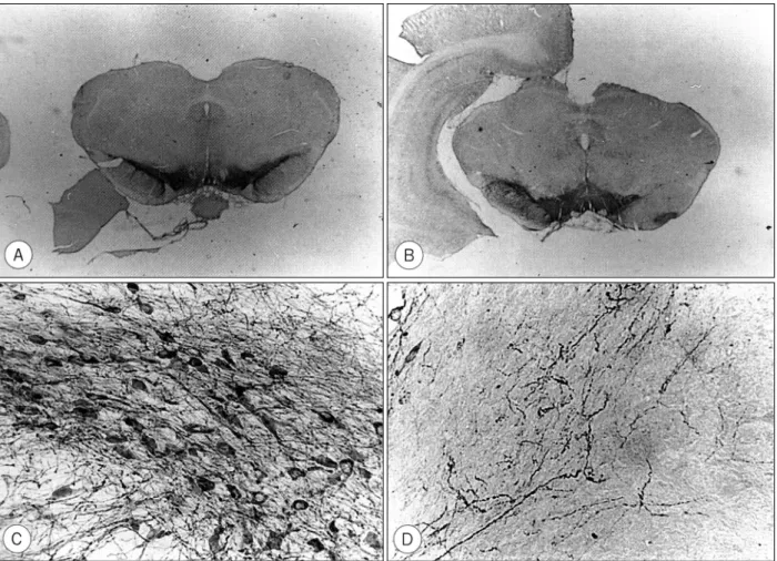
Fig. 2. A:Photomicrograph of tyrosine hydroxylase immunohistochemical stain of normal rat midbrain. Tyrosine hydroxylase
immunoreactive neurons are found in substantia nigra and ventral tegmental area on both sides(10×). B:Photomicr-
ograph of tyrosine hydroxylase immunohistochemical stain of 6-hydroxydopamine lesioned rat midbrain. Lesioned side
reveals marked degeneration of dopaminergic neurons in substantia nigra(10×). C : Intact side of substantia
nigra(200×) D:Lesioned side of substantia nigra(200×).
석하여 4℃에서 하루밤동안 반응시켰다. 다음날 TTBS로 씻은 후 다시 이차항체로 peroxidase가 표지된 항토끼 항 체를 TTBS에 1:1000으로 희석하여 2시간동안 반응시 킨 후 TTBS로 씻었다. ECL Western blotting detection reagents(Amersham, England)를 사용하여 실온에서 1 분간 처리 후 필름에 감광시키고 현상하였다.
결 과
1. 편측 파킨슨병 흰쥐모델 확인
1) Apomorphine 유발 회전운동 검사
회전측정기를 이용한 apomorphine 유발 회전운동에서, 편측 파킨슨병 모델로 정해진 실험군 15마리의 분당 평균 회전수는 각각 9.7회, 5.9회, 5.6회, 5.1회, 4.4회, 7회, 5.3 회, 4.2회, 9.4회, 8.4회, 6회, 5.6회, 6.9회, 4.6회, 4.7회로 이들의 분당 총평균 회전수는 6.2회(6.2±1.76)를 보였다.
2) Tyrosine hydroxylase 면역조직화학적 염색검사 정상 대조군에서 중뇌의 양측 흑색질과 복측 피개부가 강 한 양성 염색을 보인 반면, apomorphine 유발 회전운동을 보인 파킨슨병 모델에서는 병변측 흑색질에서 거의 전부 퇴 행성 파괴가 된 것을 관찰할 수 있었다(Fig. 2). 즉 표적지 점으로써, 흑색질의 경우 내측과 외측으로 나눈 표적좌표에 각각 9μg의 6-hydroxydopamine 주입으로 완전한 파괴 를 유도할 수 있었다. 또한 병변후 1주일후에 전 실험군에 서 apomorhine에 대한 회전반응을 보여, 도파민 생성세포 의 파괴정도가 약할때 생길 수 있는 회전운동의 회복이 발 생하지 않음을 알 수 있었다.
2. 도파민 D2 수용체의 면역조직화학적 염색
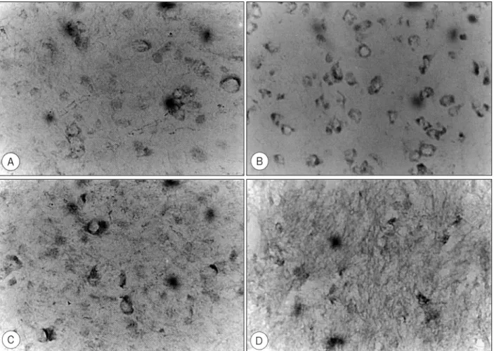
정상 대조군 2마리와 파킨슨병 모델에서 apomorphine을 투여하지 않은 5마리, 간헐적으로 투여한 5마리, 지속적으 로 투여한 5마리에서 도파민 D2 수용체 면역조직화학적 염 색 표본을 병변측 줄무늬체의 배외측부위에서 광학 현미경
Fig. 3. Photomicrograph of dopamine D2 receptor in immunohistochemical staining of dorsolateral area of corpus striatum
(200×). A:Case 1 of normal rat, showing rare positive stained cells. B:Case 5 of parkinsonian rat untreated with ap-
omorphine, showing many positive stained cells. C:Case 2 of intermittently treated group with apomorphine, showing
less positive stained cells compared to untreated case. D:Case 5 of constantly treated group with apomorphine,
showing few positive stained cells.
으로 200배의 배율로 10구역을 관찰하였다.
정상 대조군에서는 한 구역에서 면역반응 신경세포가 관찰되지 않거나 5개 이하를 보임에 비하여 apomorph- ine을 투여하지 않은 파킨슨병 모델에서는 한 구역당 6개 에서 41개까지 관찰되었다. Apomorphine을 간헐적으로 투여한 군과 지속적으로 투여한 군은 apomorphine을 투 여하지 않은 군보다 면역반응 신경세포의 수가 적고, 더구 나 지속적으로 투여한 군에서 가장 적게 나타났다(Fig. 3).
즉 apomorphine투여로 인하여 도파민 D2 수용체가 적어 지고 지속적으로 투여시 수용체의 출현이 더욱 낮아짐을 볼 수 있었으며, 이들 파킨슨씨 모델의 세 군에서 Duncan 의 다중비교로 각 군간에 유의한 차이가 있음을 알 수 있 었다(p<0.05)(Table 1).
3. 도파민 D2 수용체 단백질의 발현 양상
Western blot의 결과로 두 마리의 대조군인 정상 흰쥐와 각 군의 파킨슨병 모델의 도파민 D2 수용체 발현 양상을 비교하였다. 도파민 D2 수용체에 해당되는 80kDa의 단백 질이 발현된 띠가 형성되었으며, apomorphine을 투여하지 않은 파킨슨병 모델에서 가장 강하게 나타났고, 또한 도파 민 D2 수용체가 apomorphine을 지속적으로 투여한 군에 서 간헐적으로 투여한 군보다 약하게 발현됐슴을 알 수 있 었다(Fig. 4).
고 찰
신경독소인 6-hydroxydopamine(6-OHDA)은 화학적 구조가 catecholamine 신경전달물질과 매우 유사하며 말초 신경계에서 교감신경 말단부위의 선택적인 빠른 변성을 유 발시키고, 중추신경계의 monoamine 신경세포에 대한 효과 에 대해서는, 흑색질과 줄무늬체에 6-OHDA를 주입시 도 파민 신경말단 및 세포체내에서 신경전달물질의 고갈이 발 생됨이 알려져 있다. Ungerstedt
27)는 흰쥐의 편측 흑색질 에 역시 6-OHDA를 주입한 파킨슨병 모형에서, 도파민을 생성하는 세포체와 줄무늬체에서의 도파민 신경말단의 변성 을 확인하여, 6-OHDA를 주입한 부위의 catecholamine 세포체뿐만 아니라 축삭 및 신경말단부위에 변성이 유발됨 을 확인하였다. Maler 등
16)은 6-OHDA 주입후 2일 이내 에 흑색질에서 광범위한 신경세포의 괴사를 관찰할 수 있었 으며, 이러한 변성은 4일에서 6일째 최고조에 이른다
12).
이와같이 6-OHDA를 편측 흑색질에 주입하여 흑색질과 줄무늬체간 신경경로의 퇴행성 변화를 유발시킨 흰쥐 파킨 슨 모형에서, 행동역학적인 면을 관찰하면 병변측으로 회전 하는 비대칭적인 운동양상을 볼 수가 있다. Miklyaeva 등
18)은 이러한 운동양상이 병변 반대측 다리를 움직이는 능력의 상실에서 기인된 것이 아니라, 몸을 이동시키기 위하여 병 변 반대측 다리에 힘을 쓰고자하는 능력의 상실 즉 무동성 (akinetic)에 기인한 것으로 설명하였다. 이때 간접적 도파 민 작동제인 amphetamine을 투여시에는 병변부위의 방향
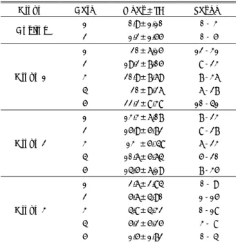
Table 1. The number of D2 receptor of immunohistochemi- cally stained neurons in lesioned striatum
Group Case Mean±SD Range 1 0.9±1.10 0- 3 Control
2 1.2±1.55 0- 5 1 20±6.15 12-31 2 19.2±7.05 8-23 3 20.9±7.69 7-36 4 20±9.26 6-27 Group 1
5 22.2±8.38 10-41 1 13.3±6.07 7-23 2 15.9±5.72 8-27 3 13 ±5. 48 6-23 4 10.6±5.64 5-20 Group 2
5 14.5±6.19 7-35 1 2.6±2.84 0- 9 2 5.6±4.90 1-15 3 4.8±4.32 0-18 4 5.2±5.25 3- 8 Group 3
5 1.5±1.72 0- 4 Group 1:Parkinsonian group without apomorphine treatment.
Group 2:Intermittently treated group with apomorphine.
Group 3:Constantly treated group with apomorphine.
Fig. 4. Western blot analysis of dopamine D2 receptor in rat
striatum. A:Two normal rats(Lane 1, 2) B:Five parki-
nsonian rats untreated with apomorphine(Lane 1-5)
C:Five rats of intermittently treated group with apo-
morphine(Lane 1-5) D:Five rats of constantly treated
group with apomorphine(Lane 1-5).
으로 회전운동을 보이고, 직접적 도파민 작동제인 apomo- rphine이나 levodopa등을 투여하면 병변부위의 반대방향으 로 회전운동을 보인다. amphetamine은 줄무늬체내의 도파 민 분비 증가, 재흡수 감소, 효소에 의한 도파민 분해의 감 소를 유도하여, 간접적으로 비병변측 줄무늬체내 도파민 수 용체의 활성을 증가시기 때문이며, apomorphine의 경우에 는 줄무늬체내에서 시냅스전과 시냅스후 도파민 수용체에 직접 작용하므로, 도파민 고갈로 인해 과민상태에 있는 병 변측 줄무늬체내 도파민 수용체의 활성을 증가시키기 때문 이다.
한편, 중뇌(mesencephalon) 도파민 생성세포의 파괴정 도에 따라서 비정상적인 회전운동의 양상이나 정도가 다르 게 나타나며
24), 도파민 생성세포의 파괴정도가 약하여 병변 측 줄무늬체내 도파민의 고갈정도가 심하지 않은 경우에는, 이러한 편측 회전운동이 일정기간후에 회복되는 것을 볼 수 가 있다. Steiner 등
26)은 병변측 줄무늬체내 도파민의 고갈 정도가 81~87%, Fornaguera 등
7)은 83% 이하인 경우에 병변직후에 나타난 비정상 회전운동이 며칠후 회복되는 것 을 보았으며, 이러한 운동기능의 회복기전은 일부 파괴되지 않은 도파민 생성세포에서의 도파민 방출 증가, 도파민의 재흡수 감소, 효소작용에 의한 분해의 감소등 보상기전에 의한 것으로 설명되며, 또한 비병변측 뇌에서 병변측 줄무 늬체와 시상으로 투사되는 신경경로의 작용증가로도 설명된 다. 따라서 본 연구자는 병변측 도파민 신경세포를 90%이 상 파괴하여, 병변제작후 발생된 비대칭적인 회전운동의 회 복을 막고 줄무늬체내 도파민의 고갈상태를 지속시키기 위 하여, 중뇌의 흑색질을 내측부와 외측부로 나누어 각각에 6-OHDA를 주입하였다. 본 연구의 초기 동물모델 제작시, 편측 흑색질의 표적좌표를 Marshall 등
17), Winn 등
29)의 단 일 표적좌표나 Perese 등
21)의 부분파괴 모델에서와 같은 내외측 표적좌표를 이용하였으나, 수술후 1~2주에 시행한 apomorphine 유발 회전운동 검사와 tyrosine hydroxy- lase 면역조직화학적 염색상 성공적인 모델은 20%이하를 보였다. 이에 본 연구자들은 이들의 경우와 달리 전정을 기 준점으로 하고 표적좌표를 보다 내측으로 설정한 결과, 수 술후 1주일에 tyrosine hydroxylase 면역조직화학적 염색 으로 확인된 바와 같이 병변측 흑색질의 완전한 파괴가 이 루어 졌으며, 90%이상의 흰쥐가 apomorphine 유발 회전 운동 검사에서 병변 반대측으로 회전운동을 보이고, 도파민 생성 신경세포의 파괴가 충분치 못할 때 나타날 수 있는 비 정상 운동의 회복이 없어, 본 연구자들의 표적좌표에 의한 편측 흑색질의 완전한 파괴로써 성공률이 매우 높은 파킨슨 병 모델이 될 수 있을것으로 생각한다.
도파민 수용체는 Kebabian 등
13)에 의하여 adenylate cyclase의 활성증가에 관계되는 D1 형과 이와 무관한 D2 형으로 구별되었으며, 그후 현재까지 도파민 수용체를 발 현시키는 5종류의 유전자(d1, d2, d3, d4, d5)가 밝혀졌다.
이들 도파민 수용체중에서 파킨슨병과 가장 관련이 있는 것 이 줄무늬체내의 D2수용체로, 흰쥐에서 D2수용체는 mes- ostriatal과 mesolimbic system 및 중뇌의 흑색질과 복측 피개부, 뇌하수체의 전엽과 중간엽에 주로 분포하며, 미상핵 과 피각부위에서는 배외측부위에 많이 분포한다
3). Unger- stedt의 흰쥐 파킨슨병 모형에서 apomorphine으로 유발되 는 회전운동은 병변부위 줄무늬체내 D2 수용체의 과민성 증가로 설명된다
25). 이러한 D2 수용체의 증가는 병변제작 후 약 1주일후에 발생되며
10), Savasta 등
23)은 자가방사기 록을 이용한 실험에서 D2 수용체가 줄무늬체의 외측부위에 서 30~40% 증가함을 보였고, 또한 병변측 D2 수용체의 mRNA 발현이 증가됨이 알려졌다
9).
반면에 위와같이 도파민 신경계 제거후 D1 수용체의 반 응을 보면, 변화가 없거나 도리어 감소를 보여
1)D1 수용체 와의 관계는 정립되지 않았으며, D1 수용체의 결합성이나 친화성은 회전운동과 큰 관련이 없다. 또한 D3나 D4 수용 체는 D2 수용체에 비하여 매우 적은 양이 존재하는 것으로 알려졌다
2). 이와같이 현재까지는 주로 D2 수용체가 파킨슨 병과 관련된 것으로 인식되고 있다.
한편 apomorphine의 지속적인 투여시 파킨슨병 흰쥐모 델에서 회전운동이 저하됨이 알려졌으며
11)28), 다른 도파민 작동제의 장기적인 혹은 지속적인 투여시에도 회전운동의 저하가 보고되었다
15). Gancher 등
8)은 apomorphine의 투 여량이 많고 투여시간이 잦을수록 회전운동이 저하됨을 보 았다. 따라서 이러한 회전운동의 저하는 본 연구에서와 같 이, 도파민 수용체에 대한 도파민 작동제의 지속적인 자극 으로 인한, 특히 자극의 정도가 많거나 잦을수록 도파민 수 용체의 발현이 낮아지는 탈과민화 반응에 의한 것으로 생각 된다.
임상적으로 levodopa를 장기적으로 투여받는 파킨슨병 환
자에서 levodopa의 효용성이 떨어지거나 운동양상의 불안전
성(motor fluctuation)을 볼 수가 있다. 이에대한 기전에 대
해서는 현재까지 논란이 되고 있으나, 일반적으로는 도파민
의 대사와 혹은 수용체 민감성의 장애
4), levodopa의 오랜기
간 복용으로 인한 시냅스전 도파민 저장능력의 저하
14), 도파
민 저장의 감소와 도파민 대사회전의 증가
40), 혈중 levod-
opa농도의 불안전성을 완충하는 도파민의 능력저하
30)등으
로 생각되고 있으며 또한 시냅스후 도파민 수용체의 장애도
언급되었다.
본 연구에서와 같이 지속적인 자극에 의한 도파민 수용체 의 탈과민화가 장기적으로 levodopa를 복용하는 파킨슨병 환자에서 약의 효용성이 떨어지거나 운동양상의 불안전성 (motor fluctuation)을 유발하는 한 원인으로 생각되며, 약 에 대한 효과를 지속시키기 위해서는 일정한 휴약기간을 갖 는 것이 바람직할 것으로 생각된다.
결 론
결론적으로 6-OHDA를 중뇌의 편측 흑색질에 주입하여 파킨슨병을 유발시킨 흰쥐모형에서, 도파민 작동제인 apo- morphine의 투여 여부와 투여시 용량 및 투여시간 간격에 따라 병변측 줄무늬체내 도파민 D2 수용체의 발현정도에 차이를 보였으며, apomorphine을 투여하지 않은 군에서 수 용체의 발현이 가장 높았고, 간헐적으로 투여한 군보다 지 속적으로 투여한 군에서 가장 낮은 발현정도를 보였다. 이 는 지속적인 도파민 작동제의 자극에 의한 도파민 수용체의 탈과민화로 생각되며, 특히 수용체에 대한 자극의 정도가 많거나 잦을수록 탈과민화 반응이 심해질 것으로 여겨진다.
이러한 탈과민화 반응은 오랜기간 도파민 작동제를 복용하 는 파킨슨병 환자에서 약에 대한 감수성이 떨어지는 일부 원인이 될 수 있을것으로 생각된다.
•
논문접수일:1999년 12월 27일•
심사완료일:2000년 2월 11일•
책임저자:이 상 원442-060 경기도 수원시 팔달구 지동 93번지 가톨릭대학교 의과대학 신경외과학교실
전화:031) 249-7190, 전송:031) 245-5208 E-mail:[email protected]
References
1) Ariano MA:Striatal dopamine receptor distribution following
chemical lesions of the nigrostriatal pathway. Brain Res 443
:204-214, 1988
2) Ariano MA, Sibley DR:Dopamine receptor distribution in
the rat CNS
:elucidation using anti-peptide antisera directed against DIA and D3 subtypes. Brain Res 649
:95-110, 1994
3) Brock JW, Farooqui S, Ross K, Prasad C:Localization ofdopamine D2 receptor protein in rat brain using polyclonal antibody. Brain Res 578
:244-250, 1992
4) Calne DB, Stoessl AJ:Transmitter receptor alterations in
Parkinson’s disease. Adv Neurol 45
:45-49, 1986
5) Caronti B, Calderaro C, Passarelli F, Palladini G, Pontieri FE:
Dopamine receptor mRNAs in the rat lymphocytes. Lif Sci 62
:1919-1925, 1998
6) Creese I, Burt DR, Snyder SH:Dopamine receptor binding
enhancement accompanies lesion-induced behavioral super- sensitivity. Science 197
:596-598, 1977
7) Fornaguera J, Carey RJ, Huston JP, Schwarting RK:Behavi-
oral asymmetries and recovery in rats with different degrees of unilateral striatal dopamine depletion. Brain Res 664
:178- 188, 1994
8) Gancher S, Crabbe J, Garland A, Lea E, Woodward W:Dose-
and duration-dependent tolerance to rotational effects of apo- morphine in a rat model of parkinsonism. J Pharmacol Exp Ther 272
:275-281, 1995
9) Gerfen CR, Engber TM, Mahan LC, Susel Z, Chase TN, Mo- nsma FJ, et al:D1 and D2 dopamine receptor-regulated gene
expression of striatonigral and striatopallidal neurons. Science 250
:1429-1432, 1990
10) Guerin B, Silice C, Mouchet P, Feuerstein C, Demenge P:
Changes of the striatal 3H-spiperone binding 3-6 weeks after nigro-striatal denervation and after two years. Life Sci 37
:953-961, 1985
11) Hargraves R, Freed WJ:Chronic intrastriatal dopamine inf-
usions in rats with unilateral lesions of the substantia nigra.
Life Sci 40
:959-966, 1986
12) Jeon BS, Jackson-Lewis V, Burke RE:6-Hydroxydopamine
lesion of the rat substantia nigra
:time-course and morphol- ogy of cell death. Neurodegeneration 4
:131-137, 1995
13) Kebabian JW, Calne DG:Multiple receptors for dopamine.Nature 277
:93-96, 1979
14) Leenders KL, Salmon EP, Tyrrell P, Perani D, Brooks DJ, Sa- ger H, et al:The nigrostriatal dopaminergic system assess-ed
in vivo by positron emission tomography in healthy volunteer subjects and patients with Parkinson’s disease. Arch Neurol 47
:1290-1298, 1990
15) Lieberman AN, Goldstein M, Leibowitz M, Gopinathan G, Neophytides A, Hiesiger E, et al:Long-term treatment with
pergolide
:Decreased efficacy with time. Neurology 34
:223- 226, 1984
16) Maler L, Fibiger HC, McGeer PL:Demonstration of the nigro-
striatal projection by silver staining after nigral injections of 6-hydroxydopamine. Exp Neurol 40
:505-515, 1973
17) Marshall TJ, Gotthelf T:Sensory inattention in rats with 6-hydroxydopamine-induced degeneration of ascending dopam- inergic neurons
:Apomorphine-induced reversal of deficits.
Exp Neurol 65
:398-411, 1979
18) Miklyaeva EI, Martens DJ, Whishaw IQ:Impairments and
compensatory adjustments in spontaneous movement after un- ilateral dopamine depletion in rats. Brain Res 681
:23-40, 1995
19) Nutt JG, Woodward WR, Hammerstad JP, Carter JH, Anderson JL, et al:The “
on-off
”phenomenon in Parkinson's disease.
N Eng J Med 310
:483-488, 1984
20) Paxinos G, Watson C:The rat brain in stereotaxic coordinates,
4nd ed. UK
:Academic Press, 1998
21) Perese DA, Ulman J, Viola J, et al:A 6-hydroxydopamine-in-
duced selective parkinsonian rat model. Brain Res 494
:285- 293, 1989
22) Pycock CJ:Turning behaviour in animals. Neuroscience 5:
461-514, 1980
23) Savasta M, Dubois A, Feuerstein C, Manier M, Scatton B:
Denervation supersensitivity of striatal D2 dopamine receptors is restricted to the ventro- and dorso-lateral regions of the str- iatum. Neurosci Lett 74
:180-186, 1987
24) Schwarting RK, Bonatz AE, Carey RJ, Huston JP:Rela-tion-
ships between indices of behavioral asymmetries and neuroc- hemical changes following mesencephalic 6-hydroxydopa-mine injections. Brain Res 554
:46-55, 1991
25) Staunton DA, Wolfe BB, Groves PM, Molinoff PB:Dopamine
receptor changes following destruction of the nigros-triatal pathway
:Lack of relationship to rotational behavior. Brain Res 211
:315-327, 1981
26) Steiner H, Bonatz AE, Huston JP, Schwarting R:Lateral-
ized wall-facing versus turning as measures of behavioral as-
ymmetries and recovery of function after injection of 6-hydro- xydopamine into the substantia nigra. Exp Neurol 99
:556- 566, 1988
27) Ungerstedt U:6-Hydroxy-dopamine induced degeneration of
central monoamine neurons. European J Pharmacol 5
:107- 110, 1968
28) Winkler JD, Weiss B:Effect of continuous exposure to selec-
tive D1 and D2 dopaminergic agonists on rotational behavior in supersensitive mice. J Pharmacol Exp Ther 249
:507-516, 1989
29) Winn SR, Wahlberg L, Tresco PA, Aebischer P:An encapsu- lated dopamine releasing polymer alleviates experimen-tal
parkinsonism in rats. Exp Neurol 105
:244-250, 1989
30) Woodward WR, Olanow CW, Beckner RM, Hauser RA, Gau-ger LL, Cedarbaum JM, et al:The effect of levodopa infusion