Received February 3, 2011; Revised February 17, 2011;
Accepted March 3, 2011
Corresponding author: Woo-Geon Jeong Tel: +82 (55) 640-3101 e-mail: [email protected] 1225-3480/24375
Effect of Polycyclic Aromatic Hydrocarbon (PAH) on Shell Repair in the Pacific oyster,
Crassostrea gigas
Sang-Man Cho
1, You-Me Lee
2and Woo-Geon Jeong
31
Department of Aquaculture and Aquatic Science, Kunsan National University 1170 Daehak-Ro, Kusna, Jeongbuk 573-701, Republic of Korea
2
Geyongnam Fisheries Institute, 240 Seohodong, Tongyeong,Gyeongnam 560-090, Republic of Korea
3
Department of Marine biology and Aquaculture & Insitute of Marine industry, Gyeongsang National University, 445 Inpyeong, Tongyeong, Gyeongnam 650-160, Republic of Korea.
ABSTRACT
In order to understand effect of polycyclic aromatic hydrocarbon (PAH) on shell repair of the Pacific oyster, Crassostrea gigas, shell regeneration experiments were carried out using oysters drilled a hole on the right valve.
The change of pH and hemocytic characteristics in both extrapallial fluid and hemolymph were observed during the shell repair. The thickness of mantle tissue was apparently decreased, while necrosis in epithelium and periostracal gland was increased in response to PAH exposure. Our finding suggested that PAH could adversely influence on shell repair.
Key words: Pacific oyster, Crassostrea gigas, Shell repair, Hemocyte, Polycyclic aromatic hydrocarbon (PAH).
Introduction
Shells can give a shelter for marine bivalves to protect themselves from predators and unfavorable environmental condition. Many authors have been studied on shell forming proteins and found that bivalve shell is composed by a significant proportion of soluble protein which has been regarded as the template for biomineralizaiton (Weiner and Hood, 1975; Lee et al., 2010). Several reports have been suggested that involvement of hemocyte in shell regeneration process (Mount et al., 2004; Cho, 2006;
Kadar et al., 2009) but many aspects of the biomineralization process still remain unclear (Simkiss and Wilbur 1989).
Hemocyte has been considered as a crucial physiological controller in oyster and also involve in a
various function on immune system, nutrition transport as well as shell repair (Cheng, 1996). Great differences in shell growth exist depending on locality and environmental differences, which suggest that environmental stress can influence on shell growth.
Therefore, shell growth and repair may closely associate with hemocyte characteristics.
Many authors have been reported the decreased or
suppressed hemocytic function and activities when
oyster exposed to polycyclic aromatic hydrocarbons
(PAH), one of most common contaminant in coastal
water (Sami et al., 1992; Capuzzo, 1996; Jeong and
Cho, 2005). Furthermore, retarded growth derived
from significant energy lose is also an important
change when oyster exposed to PAH contamination
(Capuzzo, 1996; Jeong and Cho, 2007; Kim et al.,
2007). Production of reactive oxygen species (ROS),
subsequent DNA damage, enzyme activation and
possible cell death (Regoli et al., 2002; Boutet et al.,
2004) can be also possible adverse impact when oyster
exposed to chemicals. These changes can adversely
influence on hemocytic function which may cause a
malformation of oyster shell.
Abnormal shell formation of eastern oyster has been issued in France which was characterized with anomalous conchiolin layer and basophilic round calcium structure in gill and mantle connective tissue despite the lack of an abious pathogen (Renault et al., 2002). Juvenile oyster disease is also a shell associated disease in eastern oyster (Christopher and Barber, 1999; Ford and Borrero, 2001). Some reports suggest that shell diseases are closely related to environmental stress (Soko owski et al., 2004) and pathogenic stress (Novoa et al., 1998; Lopez-Cortes et al., 1999).
Depending on our pervious finding on role of hemocyte on shell repair (Cho, 2006), we investigated possible impact of PAH on on shell repair, with an emphasis on the alteration of hemocytic and histological features of mantle tissue after exposure to PAH.
Materials and Methods
1. Experimental animal
After cleaning epibiota and fouling matter, oysters (80-100 mm in shell height) were then kept in a flow-through tank for 24 hours. For the in vivo shell regeneration experiment, a hole (I.D. 5.0 mm) was carefully drilled by a flexible hand grind on the right valve and allocated to three groups: seawater only (CON), acetone only (ACE) and acetone + PAH (PAH;
US EPA 16 species cocktail) in duplicate. The shell repair experiment was carried out for 28 days, and the oysters were fed mixed microalgae: Chaetoceros simplex, C. gracillis and Ishochrisis glabana twice a day on 09:00 and 18:00.
2. Hemocyte characteristics
Hemolymph (0.5 ml per animal) and extrapallial fluid (EPF) werecollected from the pericardial cavity and hole on shell serface using a 1 ml syringe. The HE and EPF applied to pH measurement and then total hemocyte count using Improved Neubauer hemocytometer. Cell viability was measured by dye exclusion using the red dye, eosin Y (Birmelin et al., 1998). The hemocyte composition in both hemolymph and extrapallial fluid was microscopically observed by
differential interference contrast (DIC) mode with confirmation of Fura-2AM calcium staining. For the peroxidase activity, hemocyte smears were incubated in 0.5 diaminobenzidine (DAB) mg l
-1, 0.2 M Tris-HCL buffer, pH 7.6, puls 0.02% hydrogen peroxidase for 35min at 20℃(details are in Xing et al., 2002). For calcium determination, hemocyte smears were allowed to calcium stain using the vonKossa method (Sheehan and Hrapchak, 1980).
3. Histological observation of mantle tissue
For histological observation, sampled oysters were fixed in 10% formalin and embedded following a routine paraffin method. Harris-HE staining was applied to 5 μm tick tissue sections. Changes in mantle lobe, periostracal gland and excreted conchiolin were observed by microscope (BX-50; Olympus, Tokyo, Japan).
4. Statistical analysis
For the hemocytic characteristics, the homogeneities of each measurement was tested by ANOVA (P <
0.05) and with the Dunnett test as a post hoc test (control versus experiment) using SigmaStat 3.11 (Systat Software, Inc., Point Richmond, CA, USA).
Results and Discussion
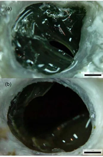
In previous report (Cho, 2006), we reported two different response of shell repair: EMR, extrapallial fluid mediated repair and MMR, mantle lobe mediated repair. When exposed to PAHs, oyster was not showed any EMR by pumping extrapallial fluid, while immediate MMR response was observed to avoid invasion of seawater and toxic compoundthrough the holl. The shell repair was significantly delayed and mostly remained uncovered even at the end of the experiment in PAH-exposed oyster (Fig 1).
The prismatic shell of oyster was formed by
aggregated hemocytes (Cho, 2006) and its endogenous
calcium ion could be a source for nucleation of
calcium crystallization but we could not observe any
cell aggregation from the the memebrane-like shell
layerfrom PAH-exposed oysters. This might be
attributed to differences between EMR and MMR
response.
Fig. 1. Photograph of regenerated shell at 28
thday.
Compared to control (a), shell repair was apparently retarded in PAH-exposed oyster even at the end of the experiment. Scale bar = 1 mm.
Fig. 2. SEM of calcification on the surface of newly regenerated shell sheet. S:sperm, M: microorganism, G:
calcium granule.
SEM observation of the layers revealed a honeycomb-like structure with various organic matrixes on the surface such as microorganism and sperm (Fig. 2). At magnified views, each prismatic cell had an irregular shaped prism in the center of the cell and surrounded by organic sheaths. At 4th day, each of the prismatic cells possessed several granular structures on the surface of the shell (Fig. 3) while apparently grown at size in comparision to that at 2
ndday. The size of the structure ranged 100 nm to 500 nm depending on the elapsed time for experiment.
Carriker (1996) and Taylor et al. (1969) represented similar observation on bivalve shell formation study.
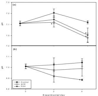
In both the extrapallial fluid and the hemolymph,
significant pH alteration had occurred during the
E x p e r im e n t a l d a y
0 2 4
pH
6 . 8 6 . 9 7 . 0 7 . 1 7 . 2
C o n t r o l A C E P A H
pH
7 . 0 7 . 1 7 . 2 7 . 3
* *
*
( b )
*
( a )
Fig. 4. Alteration of pH in extrapallial fluid (a) and hemolymph (b). *P < 0.05.
Fig. 3. SEM of prismatic cell at 2nd (a) and 4th (b) experimental day
experimet. In extrapallial fluid, pH decreased significantly on 4
thday in both the ACE and PAH group but only in the ACE of hemolymph on 4
thday (Fig. 4). Decrease of pH is natural when oyster exposed to shell generation environment because of chemical reaction between calcium and bicarbonate ions (Ca
2++ HCO
3-CaCO
3+ H
+, Wheeler, 1975;
Rousseau et al., 2003). Crenshow (1980) reported that active calcification had been occurred in extrapallial space within the range of 7.0-7.2 in pH. Thus, hypersuppression of pH in chemically exposed group could result in suppression of shell repair. This sysmtoms were alos observed in histological observation of artney which showed a significant increase of basophilic cells and hemocytic inflammation in the artery in comparison to control (Fig. 5).
The mean of total hemocyte count ranged 87.42 to 120.71 × 10
4cells ml
-1in hemolymph and 60.17 to 115.88 × 10
4cells ml
-1in extrapallial fluid without significant difference between groups (P > 0.05).
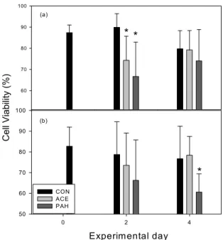
Viability of hemocytes significantly decreased only in the PAH group at the 2
ndday for hemolymph (P <
0.05) and on the 4
thday for EPF (Fig. 6). This finding might be related to the increased hemocytic inflammation in the artery caused by PAH exposure.
In ordinary oyster without shell repair condition, refractive (REF) granulocytes ranged from 5 to 15%
of total hemocyte in oyster (Mount et al., 2004). REF grnulocytes increased significantly during shell repair in the EPF (P < 0.05) in accordance with Mount et al. (2004). The slight decreases of REF granulocytes in EPF at PAH group might reflect internal oxidative stress caused by PAH exposure, which might result in the hemocytic inflammation in the artney. However, close correlation was not observed between REF granulocytes content in hemoclymph between groups.
Peroxidase activity in hemolymph increased
significantly in PAH groups (P < 0.05, Fig. 7), which
was consistenet with Pan et al. (2006). Increased
peroxidase activity of EPF in ACE group might,
while, be related to shell regeneration because
Fig. 5. Histological observation around the circumpallial artery in the control (a) and PAH (b) groups. Highly dense inflammation of the hemocyte (IF) in the artery and increased basophilic cells (B) and necrosis (N) observed in the PAH group. Scale bar = 20 μ.
Cell Viabili ty (% )
60 70 80 90 100
* *
Experimental day
0 2 4
50 60 70 80 90 100
CON ACE PAH
*
(a)
(b)
Fig. 6. Changes in positive percentage of viability in hemocyte from hemolymph (a) and extrapallial fluid (b).
Positive cell for peroxidase (%)
5 10 15 20 25 30 35
Experimental day
0 2 4
0 5 10 15 20 25 30 35
CON ACE PAH
* *
*
*
*
*
(a)
(b)