Cloning and Characterization of BTG-1 Gene from Pacific Oyster (Crassostrea gigas)
In Young Chung
1, Jeong Hwan Oh
2and Young Hwan Song
1*
1Department of Microbiology, Pukyong National University, Busan 46041, Korea
2Institute of Fisheries Science, College of Fisheries Science, Pukyong National University, Busan 46041, Korea Received February 8, 2017 /Revised February 5, 2017 /Accepted February 27, 2017
BTG 1 (B-cell translocation gene 1) gene was first identified as a translocation gene in a case of B-cell chronic lympocytic leukemia. BTG1 is a member of the BTG/TOB family with sharing a conserved N-terminal region, which shows anti-proliferation properties and is able to stimulate cell differ- entiation. In this study, we identified and characterized the pacific oyster Crassostrea gigas BTG1 (cg-BTG1) gene from the gill cDNA library by an Expressed Sequence Tag (EST) analysis and its nu- cleotide sequence was determined. The cg-BTG1 gene encodes a predicted protein of 182 amino acids with 57% 56% identities to its zebrafish and human counterparts, and is an intron-less gene, which was confirmed by PCR analysis of genomic DNA. Maximal homologies were shown in conserved Box A and B. The deduced amino acid sequence shares high identity with other BTG1 genes of human, rat, mouse and zebrafish. The phylogenic analysis and sequence comparison of cg-BTG1 with other BTG1 were found to be closely related to the BTG1 gene structure. In addition, the predicted promoter region and the different transcription-factor binding site like an activator protein-1 (AP-1) response el- ement involved in negative regulation and serum response element (SRE) were able to be identified by the genomic DNA walking experiment. The quantitative real-time PCR analysis showed that the mRNA of cg-BTG1 gene was expressed in gill, heart, digestive gland, intestine, stomach and mantle.
The cg-BTG1 gene was expressed mainly in heart and mantle.
Key words : Abundant in neuroepithelium area, B-cell translocation gene, Crassostrea gigas, PMRT1,
protein arginine methyltransferase 1
*Corresponding author
*Tel : +82-51-629-5614, Fax : +82-51-629-5619
*E-mail : [email protected]
This is an Open-Access article distributed under the terms of the Creative Commons Attribution Non-Commercial License (http://creativecommons.org/licenses/by-nc/3.0) which permits unrestricted non-commercial use, distribution, and reproduction in any medium, provided the original work is properly cited.
Journal of Life Science 2017 Vol. 27. No. 4. 398~407 DOI : https://doi.org/10.5352/JLS.2017.27.4.398
Introduction
The cell cycle is a very important biological process con- trolled a set of proto-oncogenes, which promote cell division and genes that negatively regulate cell proliferation, which are 12 different sets of tumor suppressor genes. It is well known that proliferation-related genes such as retino- blastoma protein (Rb) and p53 are key regulators of the cell cycle progression or cell apoptosis [14]. However, some physiologically important anti-proliferative genes also have a role in cell cycle regulation, which controls cell growth, differentiation and apoptosis [3, 5, 9, 13, 14, 18, 21, 26, 30, 31]. In vertebrate, BTG family of anti-proliferative gene products consists of at least nine distinct members: BTG1,
BTG2/TIS21/PC3 (orthologs of human, mouse and rat re-
spectively), BTG3/ANA, TOB, TOB2, B9.10, PC3K, PC3B and
B9.15. This family has two short conserved domains (BoxA and Box B).
The BTG1 was identified near the breakpoint of chromo- somal translocation found in a B-cell chronic lymphocytic leukemia [21] and share high homology with the previously identified Tis21 [7] and Pc3 [2] genes. It is expressed by sev- eral types of non-endothelial cells and is highly regulated during cell growth and proliferation [15, 21, 28]. Expression of BTG1 was maximal in the G
0/G
1phases of the cell cycle and down-regulated when cells progressed throughout G
1[16, 28], and also increased in response to DNA damage [6, 28].
The deduced amino acid sequence of Btg1 displays a high
degree of conservation in two regions of Box A and B [8],
which determine the localization of Btg1 in cytoplasm and
nucleus. Cytoplasmic retention sequence located in the first
43 amino acids residues of Btg1 and in a region encompass-
ing the A Box [19]. The well-conserved Box B, a Nuclear
Localization Sequence (NLS), whose activity is enhanced by
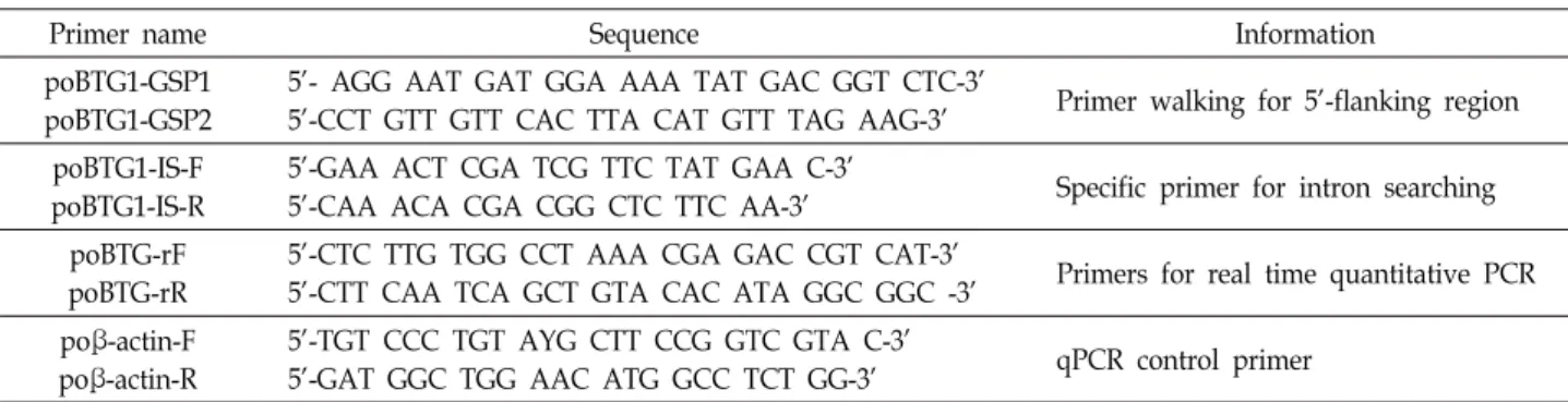
Table 1. Oligonucleotide primers used for pacific oyster BTG1 amplification and expression studies
Primer name Sequence Information
poBTG1-GSP1 poBTG1-GSP2
5’- AGG AAT GAT GGA AAA TAT GAC GGT CTC-3’
5’-CCT GTT GTT CAC TTA CAT GTT TAG AAG-3’ Primer walking for 5’-flanking region poBTG1-IS-F
poBTG1-IS-R
5’-GAA ACT CGA TCG TTC TAT GAA C-3’
5’-CAA ACA CGA CGG CTC TTC AA-3’ Specific primer for intron searching poBTG-rF
poBTG-rR
5’-CTC TTG TGG CCT AAA CGA GAC CGT CAT-3’
5’-CTT CAA TCA GCT GTA CAC ATA GGC GGC -3’ Primers for real time quantitative PCR poβ-actin-F
poβ-actin-R
5’-TGT CCC TGT AYG CTT CCG GTC GTA C-3’
5’-GAT GGC TGG AAC ATG GCC TCT GG-3’ qPCR control primer (All oligonucleotide primers were designed using Primer3 [22] and synthesized from Bioneer, Korea)
the C-terminal domain of Btg1 showing a nuclear retention
activity, mediate the interaction of BTG1 with the nuclear protein CAF1 [20]. In addition, the Btg1 amino-terminal do- main bears an LXXLL motif acting as a weak Nuclear Localization Sequence [18].
The Box C (GXXXVLYXX) sequence is present only in Btg1 and Btg2 [10, 14], which interacts with the methyl- transfease class I, Protein-arginine N-Methyltransferase 1 (PRMT 1). Both BTG1 and BTG2 may modulate signal trans- duction pathways leading to growth arrest and/or differ- entiation by activating PRMT1.
Recent data report that BTG1 may play an important role in the process of angiogenesis [11]. And act as a novel im- portant co-activator involved in the regulation of myoblast differentiation [4]. It does not only stimulate the activity of myogenic factors, but also of nuclear receptors already known as positive myogenic regulators. Transcriptional co- factors Btg1 and Btg2 act as important regulators of B-cell differentiation, providing an important link to their role as tumor suppressors in B-cell malignancies [25, 29].
To date, more than 20 members of this family have been found from several species by other many laboratories.
Though a biological common feature of these gene family was their ability to inhibit cell proliferation, their molecular role is not definite clearly. In pacific oyster, BTG1 could prompt the apoptosis in oyster hemocytes and vertebrate cancer cells, and possessed an analogous function of angio- genesis in adductor muscle of oyster [12].
The pacific oyster is one of the important aquaculture ani- mals and is a physiologically well studied bivalve species.
Because of the filter-feeder characteristics of pacific oyster, bacterial and non-bacterial pathogens become concentrated within them will threaten the human health [27]. The goal of our research is to define the set of genes which can de- termine the degree of pacific oyster’s health quality from
chemical contaminants, bacterial and viral pathogens. In this study, we have cloned and characterized cg-BTG1 gene from the pacific oyster and analyze its tissue specific expression.
This is the report on cloning of Btg1 gene of invertebrates.
Materials and Methods
Animal and preparation of tissue
The Pacific oyster (Crassostrea gigas), weighed between 150 g and 200 g, was provided from the Department of Food Safety in National Institute of Fisheries Science, Republic of Korea (http://www.nifs.go.kr/). The following tissues were obtained by dissection: gill, heart, digestive gland, intestine, stomach and mantle. The tissues were kept on liquid nitro- gen until preparation of total RNA.
cDNA library construction
Poly(A)
+RNA was isolated from pacific oyster gill using the Micro-FastTrack
TM2.0 (Invitrogen, USA) according to the manufacturer’s instruction, and the cDNA library was con- structed from this isolated mRNA using a ZAP-cDNA syn- thesis kit (Stratagene, USA) following the manufacturer’s instructions. The size-fractionated cDNA fragments were li- gated into the EcoR I and Xho I sites of the Uni-ZAP XR vector. Ligated vectors were packaged using Gigapack II Gold extract (Stratagene, USA).
Cloning of the pacific oyster BTG1 gene, nucleotide sequencing and phylogenetic analysis
Initially, full-length cDNA of pacific oyster BTG1 was ob-
tained by the random sequencing of gill cDNA library and
EST analysis. The cDNA library was sequenced on both
strands with a capillary DNA sequencer (ABI PRISM
®3700
DNA Analyzer, Applied Biosystems, Foster, CA, USA) at
Genotech (Deajeon, Republic of Korea). Nucleotide and pre-
dicted protein sequences were analyzed using BLAST pro- grams in not-redundant databases of the National Center for Biotechnology Information (NCBI BLAST, http://www.ncbi.
nlm.nih.gov/BLAST). For phylogenetic analysis, the de- duced amino acids sequences between three conserved re- gions (Box A, Box B and Box C) of pacific oyster BTG1 gene and other BTG1 genes obtained from GenBankwere aligned using CLUSTAL omega. Multiple sequence alignment was utilized to generate the phylogenetic tree using neigh- bor-joining methods with 1,000 bootstrap replicates. A neighbor-joining tree was generated based on this genetic distance matrix by using MEGA4 to identify possible phylo- genetic clade. The predicted transcription factor binding re- gion was searched using the MatInspector at Genomatix http://www. genomatix.de).
Genomic DNA extraction from pacific oyster and primer walking for 5’ flanking region
The genomic DNA of the pacific oyster gill was prepared by the method of Blin and Stafford [1].
To identify the existence of possible introns in BTG1 gene, two separate PCR reactions were performed by using pacific oyster genomic DNA and BTG1 cDNA clone as for PCR tem- plates with same set of PCR primers (poBTG1-IS-F and poBTG1-IS-R). PCR was conducted under the following cy- cle parameters: initial denaturation for 5 min at 94℃, fol- lowed by 35 cycles of a 30 sec denaturation step at 94℃, a 30 sec annealing step at 55℃ and 1 min extension step at 72℃, with a final extension of 5 min at 72℃. The PCR product was analyzed on a 1.5% agarose gel stained with EtBr along with DNA 100 bp size marker (Bioneer, Republic of Korea).
Primer walking PCR primer sets was constructed using a sequence of pacific oyster BTG1 from degenerate PCR. For primer walking PCR, two specific primers of BTG1 were de- signed as follows; poBTG1-GSP1 and poBTG1-GSP2.
Primer walking was conducted using GenomeWalker
TMKit (Clontech, USA) following the manufacturer's protocol.
Primer walking PCR reaction mixture was comprised of 1 ul of each adaptor ligated DNA, 1 pmol of AP1 and GSP1, each 10 mM dNTP mix, 10X Tth PCR reaction buffer, 50X Advantage Genomic Polymerase mix and deionized H
2O in a final volume of 50 ul.
PCR was conducted under the following conditions, two-step cycle parameters: initial denaturation for 5 min at 94
oC, followed by 7 cycles of a 25 sec denaturation step at
94
oC, a 3 min annealing and extension step at 72
oC, and 32 cycles of 25 sec denaturation step at 94
oC, a 3 min annealing and extension step at 67
oC, with a final extension of 7 min at 67
oC.
After amplification, the PCR product was cloned into pGEM
®-T easy vector system (Promega, USA) and se- quenced (Genotech, Republic of Korea).
RNA isolation and Expression profile by quantitative real-time PCR
Real-time quantitative PCR was used to evaluate the tis- sue distribution of pacific oyster BTG1 mRNA expression.
The following tissues were obtained by dissection: gill, heart, digestive gland, intestine, stomach and mantle. The tissues were kept on liquid nitrogen until preparation of total RNA.
Total RNA was prepared using the RNeasy RNA purifica- tion kit (Qiagen, USA). One microgram of RNA was used for cDNA synthesis using Superscript
TMII RNase H- reverse transcriptase (Invitrogen, USA) in a reaction mixture con- taining Random Hexamer, pd(N)6 (Bioneer, Republic of Korea). Reactions were run at 42
oC for 60 min and then in- activated at 75℃ for 10 min.
Each quantitative PCR reaction for pacific oyster BTG1 and β-actin gene was done in a 20 ul reaction mixture con- taining 80 ng of cDNA, a set of forward/reverse primers (1 pmol each), QuantiTect
TMSYBR
®Green PCR Kit (Qiagen, USA). The fluorescence was detected by DNA Engine Opticon 2 Real-Time PCR Detection System (Bio-Rad, USA).
The set of primers used for pacific oyster BTG1 is poBTG-rF and poBTG-rR and for pacific oyster β-actin is poβ-actin-F and poβ-actin-R. The PCR protocol was as follows: 95℃ for 15 min, 45 cycles of 95℃ for 15 sec, 65℃ for 20 sec, 72℃
for 25 sec and 72
oC for 5 min. Standard curves were obtained by amplifying known amounts of PCR fragments of pacific oyster BTG1 (35 ng ~ 0.0035 pg) and pacific oyster β-actin (7.04 ng ~ 0.074 pg) under the same PCR conditions. The real-time qPCR was analyzed on the Opticon Monitor 3 soft- ware (Bio-rad, USA) and PRISM4, statistical graph software (Graphpad, USA).
Results
Cloning, sequencing and structure of pacific oyster BTG1
The cg-BTG1 was identified from gill cDNA library dur-
ing EST analysis. As shown in Fig. 1. A, the full-length
A
B
Fig. 1. A. cDNA and deduced amino acid sequence of pacific oyster (Crassostrea gigas) B-cell translocation gene 1 (BTG1). Start and stop codons are shown in bold. The Box A, B and C are boxed. Polyadenylation signal is underlined. B. The remarkable structural feature of the BTG1 protein residues in the existence of three highly conserved domains (Box A, B and C) in deduced amino acid sequence of pacific oyster.
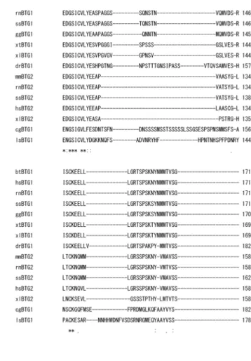
Fig. 2. The multiple alignment of the deduced amino acid sequence of the pacific oyster BTG1 with other BTG families (Table 2). An asterisk indicates the identical amino acids.
cDNA of cg-BTG1 yielded a 1419-bp sequence that included an initiation codon ATG, and in-frame stop codon, and a poly (A) tail. The cDNA of cg-BTG1 comprised a 171-bp 5’-untranslated region (5’-UTR) and 549-bp coding region, followed by a 699-bp 3’-untranslated region (3’-UTR) con- taining on potential poly-adenylation signal, AATAA (nt 1318-1322).
The nucleotide of cg-BTG1 was predicted to encode 182 amino acids from the first methionine according to universal codon usage. A translational termination codon (nt 718-720, TGA) was observed in-frame following codon 182.
The deduced amino acid sequence of cg-BTG1 demon- strated that cg-BTG1 as like other BTG1 genes, contains a LXXLL-related sequence (LQNLL, aa 34-38), a Box A (aa 43-63), a Box B (aa 88-107) and Box C (aa 109-119), which were all previously defined as conserved elements in BTG1 family (Fig. 1). The multiple serine residues were found in C-terminus region (from aa 121 to aa 155) of cg-Btg1. It is known that the serine residue (aa 159) in C-terminus of hu-
man BTG1 acts as the phosphorylation site, which enhance to drive the BTG1 protein in the cytoplasm. The nucleotide sequence of cg-BTG1 ORF was submitted to NCBI Genbank with accession number of FJ009577.
To determine the existence of possible introns in the ge- nome of cg-BTG1 gene, two separate PCR experiments were performed with pacific oyster genomic DNA and full length of cg-BTG1 cDNA clone by using primer set, poBTG1-IS-F and poBTG1-IS-R, which locate in the juxtaposition of BTG1 ORF region. PCR product obtained from pacific oyster ge- nomic DNA showed exactly same nucleotide sequence with cg-BTG1 cDNA sequence. It is revealed that pacific oyster BTG1 is an intron-less gene.
Analysis of the predicted protein sequence of pacific oyster BTG1
The educed amino acid sequences of cg-BTG1was ana-
lyzed for multiple alignments (Fig. 2) and phylogenetic anal-
ysis (Fig. 3).
Table 2. The amino acid sequence identity and similarity between BTG1 of pacific oyster (Crassostrea gigas) and other BTG families Sequence for
comparison Species Identity (%) Similarity (%) GenBank
Accession No.
hsBTG1 rnBTG1 btBTG1 ggBTG1 ssBTG1 lsBTG1 xlBTG1 xtBTG1 drBTG1 hsBTG2 mmBTG2
rnBTG2 ssBTG2 xlBTG2
Homo sapiens Rattusnorvegicus Bostaurus Gallus gallus Susscrofa
Lycosas ingoriensis Xenopus laevis Xenopustropicalis Danio rerio Homo sapiens Mus musculus Rattusnorvegicus Susscrofa Xenopus laevis
47 47 47 46 47 44 48 52 47 53 44 44 44 53
65 65 65 66 65 64 66 75 70 75 66 66 66 76
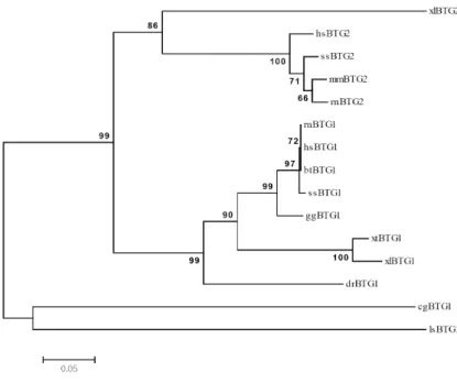
NP_001722 NP_058954 NP_776424 NP_990681 NP_001093406 ABX75488 NP_001079380 NP_988926 NP_956314 NP_006754 NP_031596 NP_058955 ABW83760 NP_001088812 Fig. 3. Phylogenetic relationships of pacific oyster
BTG1 (cgBTG1) with other BTG families (Table 2). In this neighbor-joining phylo- gram, all individuals are represented and the branches are based on the number of inferred substitutions as indicated by the bar.
The accession numbers of the amino acid sequences ap- plied to the multiple alignments of cg-BTG1 (pacific oyster) were hsBTG1 (human), hsBTG2 (human), rnBTG1 (rat), rnBTG2 (rat), mmBTG2 (mouse), btBTG1 (cattle), ggBTG1 (chicken), ssBTG1 (pig), ssBTG2 (pig), lsBTG1 (spider), xlBTG1 (frog), xlBTG2 (frog), xtBTG1 (frog) and drBTG1 (zebrafish). The pacific oyster BTG1 gene showed 44%~53%
amino acid sequence identity to BTG family of all available sequence information. As shown in Table 2, it shows about 53% of sequence identity and about 76% similarity to hsBTG2 (Homo sapiens), xlBTG2 (Xenopus laevis), and xtBTG1 (Xenopus tropiclais). This data appear to be some obscure to classified BTG1 and BTG2 gene. Although both BTG1 and
BTG2 show very high sequence homologies and have three
functional domains of same Box A, Box B and Box C region,
it is different that BTG1 have longer amino acid residues
than BTG2 in C-terminus region. Human Btg1 has 171 amino
acids residues while human Btg2 has 158 amino acids,
whereas cg-Btg1 has 182 amino acids residues, which is lon-
ger than human Btg1. From the phylogenetic relationship
of BTG family as shown in Fig. 3, it reveals that cg-BTG1
is closely related to lsBTG1 (Lycosas ingoriensis) as a good
example of invertebrate and shows more clear molecular
classification of complex and similar BTG family. From these
data, we designated our cDNA clone of cg-BTG1 gene as
a member of BTG1 family not in BTG2.
Fig. 4. Genome-walked 5’ flanking region sequence of pacific oyster BTG1. These contain the predicted binding site of transcription factor, Tata-binding protein factor (TBPF), cAMP response element binding protein (CREB), CCAAT binding factor (CAAT) and Serum response element binding factor (SRFF).
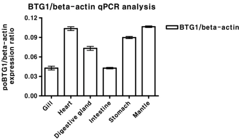
BTG1/beta-actin qPCR analysis
Gill Heart
Digesti ve gland
Intestine Stoma
ch Mantle 0.00
0.03 0.06 0.09 0.12
BTG1/beta-actin
poBTG1/beta-actin expression ratio
Fig. 5. Quantitative real-time PCR analysis of BTG1 in pacific oyster. BTG1 was measured in 6 dif- ferent tissue, gill, heart, digestive gland, intes- tine, stomach and mantle. All data were taken from a representative assay and were normal- ized relative to β-actin, representing the mean (n=3) of three identical replicates ± standard deviation.
Analysis of the predicted transcription factor binding site
Primer walking was conducted using GenomeWalker
TMKit (Clontech, USA) following the manufacturer's protocol.
Genome-walked 5’ flanking region sequence of cg-BTG1 is 750-bp upstream from the initiation codon. As shown in Fig.
4, the 5’-flaning region contains the predicted binding site of transcription factor, TATA-binding protein factor (TBPF, nt568-572), cAMP response element binding protein (CREB, nt 661-668), CCAAT binding factor (CAAT, nt 681-685) and
Serum response element binding factor (SRBF, nt 589-599).
Expression profile study by quantitative real-time PCR
In order to determine the tissue specific mRNA ex- pression of pacific oyster BTG1 genes, quantitative real-time PCR analysis was carried out using total RNA isolated from the gill, heart, digestive gland, intestine, stomach and mantle of pacific oyster. As shown in Fig. 5, mRNA transcripts of BTG1 were higher in heart, stomach, and mantle than gill, digestive gland, and intestine. This data suggests that cg-BTG1 mRNA is widely expressed in most tissues of the pacific oyster.
Discussion
In this study, the cDNA clone of cg-BTG1 was obtained from the gill cDNA library and the full-length sequence of the cg-BTG1 cDNA was determined, and the cg-BTG1 mRNA expression was analyzed by real-time quantitative PCR.
In vertebrate, BTG (or BTG/TOB) family consists of at least six distinct members and show high similarity to each other. Box A and Box B, the functional domains in BTG1, exist in BTG2, BTG3, BTG4, TOB and TOB 2 and involve in the cellular localization of the protein such as the cytoplas- mic retention of Box A and the nuclear localization of Box B [17, 18, 20]. Box C, which binds with PRMT1 and is the third functional domain of BTG1 and BTG2, exists only in BTG1 and BTG2 [10, 14]. The predicted amino acids se- quence of cg-BTG1 also shows strong similarity to other BTG1 and BTG2. The multiple sequence alignment of all available BTG1 and BTG2 proteins shows strong homology.
Although it is not easy to classify the cg-BTG1 in this study
as BTG1 not as BTG family, the following reasons can sup- port our designation of cg-BTG2. First, cg-BTG1 contains Box C (aa 109 -119), which is a typical domain in only BTG1 and BTG2. Second, the polypeptide of BTG1 is longer than BTG2 as shown in Fig. 2. Human, bovine, and pig BTG2 have 158 amino acids whereas BTG1 have 171 amino acids (for human, bovine, and pig), 170 amino acids (for chicken), 169 amino acids (for frog), 182 amino acids (for zebrafish), and 178 amino acids (for spider). Finally, the phylogenetic tree reveals that cg-BTG1 is closely related to spider BTG1 (Fig. 4.).
Interestingly, unlike the human and mouse, the genomic organization of the BTG1 in pacific oyster have not con- tained intron. Moreover, like the human and mouse se- quences, the pacific oyster BTG1 cDNA contained a long AT-rich region in 3’ un-translated region which may be im- portant in post-transcriptional regulation of the genes [21].
Some copies of ATTTA motif related RNA stability were found.
Also, transcripts for pacific oyster BTG1 in 6 different tis- sues of pacific oyster detected by real-time PCR, BTG1 was measured in all tissues, gill, heart, digestive gland, intestine, stomach and mantle. Most remarkably, expression of BTG1 genes mostly high detected in heart and mantle, this two tissues consists of many muscle, especially. Many other re- ports have already shown that BTG1 strongly inhibited myo- blasts proliferation and stimulated myoblasts differentiation [17, 18] which suggested that BTG/TOB family may play a key role in muscle development. One or several members of BTG/TOB family may be related to muscle growth.
BTG/TOB family in muscle development seems to be evolu- tionary conserved, as suggested by experiments in Xenopus [23], and by the expression pattern of the family in zebrafish [24]. In particular, the expression of BTG1 and BTG2 in the heart primordia suggests a novel role for BTG genes in car- diomyogenesis, and it is noteworthy that Btg1 expression precedes that of Btg2, which, in turn, is very restricted and transiently expressed.
Further studies will focus on the existence of BTG2 gene in oyster and the regulation of cg-BTG1 and cg-BTG2 by p53 activation which induced by hypoxia, genotoxic stress, and metabolic changes.
Acknowledgement
This work was supported by a Research Grant of Pukyong
National University (2015Year).
References
1. Blin, N. and Stafford, D. W. 1976. A general method for isolation of high molecular weight DNA from eukaryotes.
Nucleic Acids Res. 3, 2303-2308.
2. Bradbury, A., Possenti, R., Shooter, E. M. and Tirone, F.
1991. Molecular cloning of PC3, a putatively secreted pro- tein whose mRNA is induced by nerve growth factor and depolarization. Proc. Natl. Acad. Sci. USA 88, 3353-3357.
3. Buanne, P., Corrente, G., Micheli, L., Palena, A., Lavia, P., Spadafora, C., Lakshmana, M. K., Rinaldi, A., Banfi, S., Quarto, M., Bulfone, A. and Tirone, F. 2000. Cloning of PC3B, a novel member of the PC3/BTG/TOB family of growth inhibitory genes, highly expressed in the olfactory epithelium. Genomics 68, 253-263.
4. Busson, M., Carazo, A., Seyer, P., Grandemange, S., Casas, F., Pessemesse, L., Rouault, J. P., Wrutniak-Cabello, C. and Cabello, G. 2005. Coactivation of nuclear receptors and myo- genic factors induces the major BTG1 influence on muscle differentiation. Oncogene 24,1698-1710.
5. Corjay, M. H., Kearney, M. A., Munzer, D. A., Diamond, S. M. and Stoltenborg, J. K. 1998. Antiproliferative gene BTG1 is highly expressed in apoptotic cells in macro- phage-rich areas of advanced lesions in Watanabe heritable hyperlipidemic rabbit and human. Lab. Invest. 78, 847-858.
6. Cortes, U., Moyret-Lalle, C., Falette, N., Duriez, C., Ghissassi, F. E., Barnas, C., Morel, A. P., Hainaut, P., Magaud, J. P.
and Puisieux, A. 2000. BTG gene expression in the p53-de- pendent and -independent cellular response to DNA damage. Mol. Carcinog. 27, 57-64.
7. Fletcher, B. S., Lim, R. W., Varnum, B. C., Kujubu, D. A., Koski, R. A. and Herschman, H. R. 1991. Structure and ex- pression of TIS21, a primary response gene induced by growth factors and tumor promoters. J. Biol. Chem. 266, 14511-14518.
8. Guehenneux, F., Duret, L., Callanan, M. B., Bouhas, R., Hayette, S., Berthet, C., Samarut, C., Rimokh, R., Birot, A.
M., Wang, Q., Magaud, J. P. and Rouault, J. P. 1997. Cloning of the mouse BTG3 gene and definition of a new gene family (the BTG family) involved in the negative control of the cell cycle. Leukemia 1, 370-375.
9. Iacopetti, P., Michelini, M., Stuckmann, I., Oback, B., aku-Saraste, E. and Huttner, W. B. 1999. Expression of the antiproliferative gene TIS21 at the onset of neurogenesis identifies single neuroepithelial cells that switch from pro- liferative to neuron-generating division. Proc. Natl. Acad. Sci.
USA 96, 4639-4644.
10. Ikematsu, N., Yoshida, Y., Kawamura-Tsuzuku, J., Ohsugi, M., Onda, M., Hirai, M., Fujimoto, J. and Yamamoto, T.
1999. Tob2, a novel anti-proliferative Tob/BTG1 family member, associates with a component of the CCR4 tran- scriptional regulatory complex capable of binding cyclin-de- pendent kinases. Oncogene 18, 7432-7441.
11. Iwai, K., Hirata, K., Ishida, T., Takeuchi, S., Hirase, T., Rikitake, Y., Kojima, Y., Inoue, N., Kawashima, S. and Yokoyama, M. 2004. An anti-proliferative gene BTG1 regu- lates angiogenesis in vitro. Biochem. Biophys. Res. Commun.
316, 628-635.
12. Liu, R., Cheng, Q., Wang, X., Chen, H., Wang, W., Zhang, H., Wang, L. and Song, L. 2017. The B-cell translocation gene 1 (CgBTG1) identified in oyster Crassostrea gigas exhibit multiple functions in immune response. Fish Shellfish Immunol. 61, 68-78.
13. Marchal, S., Cassar-Malek, I., Magaud, J. P., Rouault, J. P., Wrutniak, C. and Cabello, G. 1995. Stimulation of avian my- oblast differentiation by triiodothyronine: possible involve- ment of the cAMP pathway. Exp. Cell. Res. 220, 1-10.
14. Matsuda, S., Rouault, J., Magaud, J. and Berthet, C. 2001.
In search of a function for the TIS21/PC3/BTG1 /TOB family. FEBS Lett. 497, 67-72.
15. Raburn, D. J., Hamil, K. G., Tsuruta, J. K., O'Brien, D. A.
and Hall, S. H. 1995. Stage-specific expression of B cell trans- location gene 1 in rat testis. Endocrinology 136, 5769-5777.
16. Rimokh, R., Rouault, J. P., Wahbi, K., Gadoux, M., Lafage, M., Archimbaud, E., Charrin, C., Gentilhomme, O., Germain, D. and Samarut, J. 1991. A chromosome 12 coding region is juxtaposed to the MYC protooncogene locus in a t(8;12) (q24,q22) translocation in a case of B-cell chronic lympho- cytic leukemia. Genes Chromosomes Cancer 3, 24-36.
17. Rodier, A., Marchal-Victorion, S., Rochard, P., Casas, F., Cassar-Malek, I., Rouault, J. P., Magaud, J. P., Mason, D.
Y., Wrutniak, C. and Cabello, G. 1999. BTG1: a triiodothyr- onine target involved in the myogenic influence of the hormone. Exp. Cell. Res. 249, 337-348.
18. Rodier, A., Rochard, P., Berthet, C., Rouault, J. P., Casas, F., Daury, L., Busson, M., Magaud, J. P., Wrutniak-Cabello, C. and Cabello, G. 2001. Identification of functional domains involved in BTG1 cell localization. Oncogene 20, 2691-2703.
19. Rouault, J. P., Prevot, D., Berthet, C., Birot, A. M., Billaud, M., Magaud, J. P. and Corbo, L. 1998. Interaction of BTG1 and p53-regulated BTG2 gene products with mCaf1, the murine homolog of a component of the yeast CCR4 tran- scriptional regulatory complex. J. Biol. Chem. 273, 22563-22569.
20. Rouault, J. P., Rimokh, R., Tessa, C., Paranhos, G., Ffrench, M., Duret, L., Garoccio, M., Germain, D., Samarut, J. and Magaud, J. P. 1992. BTG1, a member of a new family of antiproliferative genes. EMBO J. 11, 1663-1670.
21. Rouault, J. P., Samarut, C., Duret, L., Tessa, C., Samarut, J. and Magaud, J. P. 1993. Sequence analysis reveals that
the BTG1 anti-proliferative gene is conserved throughout evolution in its coding and 3' non-coding regions. Gene 129, 303-306.
22. Rozen, S. and Skaletsky, H. 2000. Primer3 on the WWW for general users and for biologist programmers. Methods Mol. Biol. 132, 365-386.
23. Saka, Y., Tada, M. and Smith, J. C. 2000. A screen for targets of the Xenopus T-box gene Xbra. Mech. Dev. 93, 27-39.
24. Sakaguchi, T., Kuroiwa, A. and Takeda, H. 2001. Expression of zebrafish btg-b, an anti-proliferative cofactor, during ear- ly embryogenesis. Mech. Dev. 104, 113-115.
25. Scheijen, B., Boer, J. M., Marke, R., Tijchon, E., van Ingen, S. D., Waanders, E., van, E. L., van der Meer, L. T., Pieters, R., Escherich, G., Horstmann, M. A., Sonneveld, E., Venn, N., Sutton, R., la-Pozza, L., Kuiper, R. P., Hoogerbrugge, P. M., den Boer, M. L. and van Leeuwen, F. N. 2016. Tumor suppressors BTG1 and IKZF1 cooperate during mouse leu- kemia development and increase relapse risk in B-cell pre- cursor acute lymphoblastic leukemia patients. Haematologica 102, 541-551.
26. Seo, M. S., Lee, M. S. and Lim, I. K. 1999. Expression of rat BTG(3) gene, Rbtg3, is regulated by redox changes. Gene 240, 165-173.
27. Starks, A. M., Schoeb, T. R., Tamplin, M. L., Parveen, S., Doyle, T. J., Bomeisl, P. E., Escudero, G. M. and Gulig, P.
A. 2000. Pathogenesis of infection by clinical and environ- mental strains of Vibrio vulnificus in iron-dextran-treated mice. Infect. Immun. 68, 5785-5793.
28. Suk, K., Sipes, D. G. and Erickson, K. L. 1997. Enhancement of B-cell translocation gene-1 expression by prostaglandin E2 in macrophages and the relationship to proliferation.
Immunology 91, 121-129.
29. Tijchon, E., van, E. L., Yuniati, L., van Ingen, S. D., Havinga, J., Rouault, J. P., Hoogerbrugge, P. M., van Leeuwen, F. N.
and Scheijen, B. 2016. Tumor suppressors BTG1 and BTG2 regulate early mouse B-cell development. Haematologica 101, e272-e276.
30. Tirone, F. 2001. The gene PC3 (TIS21/BTG2), prototype member of the PC3/BTG/TOB family: regulator in control of cell growth, differentiation, and DNA repair? J. Cell Physiol. 187, 155-165.
31. Yoshida, Y., Matsuda, S., Ikematsu, N., Kawamura-Tsuzuku, J., Inazawa, J., Umemori, H. and Yamamoto, T. 1998. ANA, a novel member of Tob/BTG1 family, is expressed in the ventricular zone of the developing central nervous system.
Oncogene 16, 2687-2693.
초록:참굴( Crassostrea gigas )의 BTG1 유전자의 특성
정인영
1․오정환
2․송영환
1*
(1국립부경대학교 미생물학과, 2국립부경대학교 수산과학연구소)