제주 구멍갈파래 가수분해물에 의한 노화된 섬유아세포 증식 및 콜라겐 합성증진 효과
고 현 주
†⋅김 경 범⋅이 동 환⋅이 근 수⋅표 형 배
한불화장품(주) 기술연구소
(2013년 3월 12일 접수, 2013년 4월 4일 수정, 2013년 5월 16일 채택)
The Effect of Hydrolyzed Jeju Ulva pertusa on the Proliferation and Type I Collagen Synthesis in Replicative Senescent Fibroblasts.
Hyun Ju Ko
†, Gyoung Bum Kim, Dong Hwan Lee, Geun Soo Lee, and Hyeong Bae Pyo
R&D Center, Hanbul Cosmetics Co. Ltd, 547-62, Daesung-ro, Samsung-myun, Umsung-kun, Chungbuk 369-834, Korea (Received March 12, 2013; Revised April 4, 2013; Accepted May 16, 2013)
요 약: 피부 섬유아세포는 인간 피부의 주요 콜라겐 생산 세포이다. 노화가 진행되면, 섬유아세포에서의 콜라겐
생산이 감소되고, matrix metalloproteinase-1 (MMP-1)에 의해 시작되는 콜라겐 조각화가 증가된다. 즉 섬 유아세포의 콜라겐 항상성의 불균형으로 인해 피부 collagenous, 세포외기질(ECM)의 구조와 기능이 변형되어, 피부노화가 촉진되는 것이다. Cysteine rich protein 61 (CCN1)는 CCN family의 일부이며, 인간피부의 섬유 아세포에서 콜라겐 항상성을 조절하는 단백질이다. 노화된 인간 피부 섬유아세포에서의 CCN1 과 발현은 실질 적으로 유형 Ⅰ procollagen 생성을 감소시킴과 동시에 MMP-1의 발현을 증가시켜 섬유의 콜라겐 저하를 일으 킨다. 그리고 노화된 섬유아세포는 노화 전 섬유아세포에 비해 증식률이 감소한다. 본 연구에서 만들어 사용한 복제 노화 피부 섬유아세포는 유형 Ⅰ procollagen의 생성량이 감소하였고, MMP-1의 발현 수준이 증가하는 특징을 나타냈다. 또한 CCN1 단백질의 발현이 증가되고, 증식률이 감소하는 특징을 나타냈다.
가수분해 구멍갈파래 추출물은 노화 전 섬유아세포에서 새로운 콜라겐의 합성을 촉진하고 자외선에 의해 증가 된 MP-1의 발현을 감소시켜 광노화를 개선하는 물질로 알려져 있다. 본 연구에서는 이러한 활성을 나타내는 가수분해 구멍갈파래 추출물을 사용하여, 복제 노화 피부 섬유아세포에서 가수분해 구멍갈파래 추출물에 의한 CN1 단백질의 발현 억제 여부를 조사하였으며, 이들 추출물은 배양된 복제 노화 피부 섬유아세포에서 유형 Ⅰ procollagen의 생성을 증가시켰으며, MMP-1 발현을 억제시키는 것을 확인하였다. 또한, 콜라겐 항상성을 조 절하는 단백질인 CN1 발현을 크게 감소시켰으며, 노화세포의 증식률을 증가시켰다. 이 결과는 복제 노화 섬유아 세포가 in vitro 자연 노화모델로 화장품 원료 활성 연구에 사용될 수 있음을 말한다. 그리고 가수분해 구멍갈파 래 추출물은 광노화 뿐 아니라 자연노화를 개선하는 피부미용제로 주름개선 기능성 화장품에 사용가능 하다는 것을 의미한다.
Abstract: Skin dermal fibroblast is the major collagen-producing cell type in human skin. As aging process continues in human skin, collagen production is reduced and fragmentation is increased, which is initiated by matrix metal- loproteinase-1 (MMP-1). This imbalance of collagen homeostasis impairs the structure and function of dermal collage- nous extracellular matrix (ECM), thereby promoting skin aging. Cysteine-rich protein 61 (CCN1), a member of the CCN family, negatively regulates collagen homeostasis in primary human skin dermal fibroblast cells. It is known in aging fibroblast cells that elevated CCN1 expression substantially reduces type Ⅰ procollagen and concurrently increases MMP-1, which initiates fibrillar collagen degradation. And proliferation rate of aging fibroblast cells is reduced compared 1)
† 주 저자 (e-mail: [email protected])
to the pre-aging fibroblast cells. In this study, we confirmed that the replicative senescence dermal fibroblast cells in- creased the expression levels of MMP-1 and decreased the production of type I procollagen. Our results also showed that the replicative senescence dermal fibroblast cells increased in the expression of CCN1 and decreased in the pro- liferation rate. Hydrolyzed Ulva pertusa extracts are the materials to improve photo-aging by reducing the expression of MMP-1 that was increased by ultraviolet and by promoting the synthesis of new collagen from fibroblast cells. In this study, we also investigated the hydrolyzed U. pertusa extract to see whether it inhibits CCN1 protein expression in the senescence fibroblasts. Results showed that the hydrolyzed U. pertusa extract inhibited the expression of MMP-1 and increased the production of type I procollagen in the aging skin fibroblast cells cultured. In addition, the proteins that regulate collagen homeostasis CCN1 expression were greatly reduced. The hydrolyzed U. pertusa extract increased the proliferation rate of the aging fibroblast cells. These results suggest that replicative senescent fibroblast cells may be used in the study of cosmetic ingredients as a model of the natural aging. In conclusion, the hydrolyzed U. pertusa extract can be used in anti-wrinkle functional cosmetic material to improve the natural aging skin care as well as pho- to-aging.
Keywords: cysteine-rich protein 61 (CCN1), homeostasis, hydrolyzed Ulva pertusa, replivative senescent dermal fibroblast, natural aging
1. 서 론
피부의 노화를 일으키는 원인은 다양하며 이에 대 한 이론도 다양하게 제시되고 있으나 그 요인에 따라 크게 자연노화와 광노화로 나눈다. 광노화의 경우 유 전적으로 이미 결정된 자연 노화에 비해 적절한 피부 관리 습관에 따라 충분히 예방가능 하다는 측면에서 의의가 있다고 할 것이다. 그러나 피부의 노화는 자연 노화와 광노화가 동시에 작용한다. 피부가 노화가 됨 에 따라 여러 가지 대사 활성이 저하되며 세포 활성도 떨어진다. 콜라겐 합성과 콜라겐의 전사 이후에 일어 나는 과정도 자연노화로 감소한다[1].
노화에 대한 연구는 굉장히 복잡하고 다양한 인자 들이 관여 되어 있다. 최근 연구에서 피부노화를 조절 하는데 중요한 유전자 요인에 대한 연구가 활발히 진 행되고 있으며, 피부노화에 관련된 유전자 변화는 젊 은 피부와 내인성 노화, 광노화 피부에서 비교 연구되 고 있다. 이러한 연구들을 통하여 피부노화는 지방생 합성, keratin filaments의 형성, 콜라겐 항상성 조절 등 에 관련된 유전자 발현의 감소 및 증가와 관련이 있다 는 것이 밝혀지고 있다[2]. 피부의 결합조직은 주로 콜 라겐과 엘라스틴 등에 의해 이루어진다. 콜라겐과 엘 라스틴은 피부에 탄성과 힘을 주며 이들이 노화로 약 해지면 피부는 쉽게 손상되고 늙는다. 피부 진피층의 주요한 구성 성분인 콜라겐은 진피 건조중량의 70 ~ 80%를 차지한다. Type Ⅰ, Ⅲ 콜라겐은 사람 피부 진
피층의 주요한 성분이며, type Ⅰ은 총 콜라겐의 80%
를, type Ⅲ는 15%를 차지한다. 피부의 주름은 콜라겐 의 합성과 분해의 불균형에 기인한다. 보통 피부에서 는 type Ⅰ콜라겐의 합성과 그 분해 효소인 matrix met- alloproteinase-1 (MMP-1) 이 균형을 이루고 있다 (collagen homemostasis). 진피 섬유아세포는 결합조직 에서 가장 중요한 세포로 세포외기질(extracellular ma- trix) 안에 들어 있으며 콜라겐 등 조직성분 및 분해 물 질을 합성한다.
최근의 연구결과들을 보면 노화된 인간 피부에서 cysteine rich protein 61 (CCN1) 이 콜라겐의 합성과 분 해의 균형을 조절하는 중요한 매개단백질 역할을 하 는 것을 알 수 있다. CCN1은 세포외기질(extracellular matrix)과 관련된 신호단백질로 섬유아세포에 의해 분 비된다. 그리고, CCN1은 세포배양에서 세포의 다양한 세포 접착, 세포이동(migration), 세포외기질의 상호작 용, 그리고 세포외기질(extracellular matrix)을 구성하 는 단백질의 합성을 조절하는 것으로 보고되었다 [3-5]. Thihao Quan등은 CCN1이 과 발현된 복제 노화 피부 섬유아세포에 CCN1의 발현을 억제하는 siRNA 를 주입한 후, 콜라겐 합성량과 MMP-1 발현량을 평가 하여 콜라겐의 항상성이 CCN1의 발현에 의해 조절됨 을 증명하였다[6].
이러한 연구 결과는 CCN1 단백질 발현과 콜라겐 항상성 사이에 관계가 있음을 입증한 것이다. 따라서 복제 노화 섬유아세포는 in vitro 피부자연노화 모델로
연구에 사용할 수 있음을 말한다. 본 연구에서는 복제 노화 섬유아세포를 확립하여 피부자연노화 모델로 사 용 하였다. 확립된 복제 노화 섬유아세포에서는 CCN1 단백질이 과 발현 되었으며, 이에 따라 Procollagen의 합성은 감소하였고, MMP-1의 발현은 증가하였다.
피부에서 MMPs의 발현을 억제하고 콜라겐의 합성 을 촉진하여 피부 광노화를 개선하거나 억제하는 것 으로 알려진 비타민 A는 자연노화 피부에서도 동일한 효과를 지니며 이는 노화가 많이 진행된 경우 더욱 효 과적이었다는 연구 결과가 있다[7].
본 연구에서는 제주도 연안 자생 해조류인 구멍갈 파래(Ulva pertusa)의 가수분해물을 이용하였다. 갈파 래과(Ulvaceae) 해조류인 구멍갈파래는 제주 연안에서 주로 4월에서 8월까지 대량 발생하여 번무하고 또한 장소에 따라서는 연중 번무하고 있는 종으로 제주해 역에서 주로 우점하며 대량으로 번식하여 개체수가 풍부한 해조류이다. 따라서 개체수가 풍부한 구멍갈 파래로부터 피부에 유용한 화장품 신소재를 개발할 경우, 대량 채집으로 인한 환경에 피해를 주지 않으면 서도 현재 해외에서 수입하여 사용하고 있는 해조류 기원의 고가 화장품 원료에 대한 수입대체제로도 가 치가 크다고 할 수 있다. 우리는 구멍갈파래 가수분해 물(이하 가수분해 구멍갈파래 추출물)을 이용하여, 이 활성성분이 자연 노화모델에서도 효과가 있는지 확인 해 보고자 하였다. 따라서 노화된 섬유아세포에서 가 수분해 구멍갈파래 추출물이 CCN1 발현에 어떤 영향 을 미치는가와 그로 인해 콜라겐의 합성과 분해에 미 치는 영향에 대해서 조사하여, 비타민 A와 같이 자연 노화를 억제할 수 있는지에 대해 알아보았다.
2. 실험재료 및 방법
2.1. 실험재료
본 실험에서 사용한 각종 시약은 Sigma Aldrich (Sigma, USA) 제품을 사용하였다. 가수분해 구멍갈파 래 추출물은 건조하여 마쇄한 구멍갈파래에 10배량의 정제수를 가해 환류 추출기에서 4 h 동안 추출한 후, 여과하고 제조된 열수추출물을 냉각한 후, 추출물량 과 동일한 양의 에틸알코올을 가하고 교반, 4 ℃에서 하룻밤 동안 정치한 후 형성된 침전물을 여과하여 회 수하고 이를 정제수에 재분산하고 동결 건조하여 구
멍갈파래 고분자 분획을 제조한다. 제조된 고분자를 휘발성 유기산을 함유한 산 용액에 가해 분산한 후, 100 ℃에서 2 내지 6 h 가열하여 산 분해 시료를 제조 하였다. 제조된 산 분해물은 여과한 후, 감압농축을 통해 잔여 유기산을 제거하였다. 이때 감압농축물의 pH가 3.0 이상이 될 때까지 정제수를 더해 감압 농축 하는 공정을 수회 반복한 후, 동결 건조하여 각각의 산 분해물을 제조하여 사용하였다.
2.2. 세포배양 및 복제노화 섬유아세포
신생아의 포피조직에서 분리한 human dermal fibro- blast (HDFn)는 Cascade (Biologics Inc, USA)로부터 구 입하였다. 구입한 HDFn을 DMEM/F12 (3 : 1) 배지에 10% FBS (fetal bovine serum), 1% penicillin-streptomycin 을 첨가하여 37 ℃, 5% CO2 조건 하에 배양하고 tryp- sinization으로 계대 배양한 뒤 β-galactosidase activity 를 평가하여 50% 이상 양성이 나오는 세대 (p24 ~) 세 포를 실험에 이용하였다. Pre-senescent control 섬유아 세포는 p6 ~ p10세대의 세포를 실험에 이용하였다.
2.3. β-galactosidase 염색을 이용한 노화 지표의 확인 복제 노화 세포임을 확인하기 위해 노화의 지표 단 백질로 이용되고 있는 β-galactosidase (Senescence-as- sociated β-galactosidase, SA-β-Gal) 활성을 다음과 같 이 senescent cell staining kit (sigma, USA)를 사용하여 염색/ 평가하였다.
35 mm culture dishes에 섬유아세포를 1×10⁵ cells를 seeding 한 후 24 h 동안 안정화 한 뒤에 PBS로 wash 한 후, 0.2% glutaraldehyde와 2% formaldehyde로 7분간 고정시킨 후 40 mM citric acid/phosphate (pH 6.0), 5 mM K3FeCN6, 5 mM K4FeCN6, 150 mM NaCl과 2 mM MgCl2에 1 mg/mL 5-bromo-4-chloro-3-indolyl β-D-gal- actopyranoside이 되도록 녹인 용액을 CO2가 없는 37
℃에서 24 h 둔 후, 파란색으로 염색된 세포들을 광학 현미경으로 관찰하여 세포 수를 카운터 한 후 사진 촬 영을 하였다.
2.4. 가수분해 구멍갈파래 추출물의 노화세포활성 확인 계대를 통하여 확립한 노화 섬유아세포를 DMEM/
F12 (3 : 1) 배지에 10% fetal bovine serum (FBS), 1%
penicillin-streptomycin을 첨가하여 37 ℃, 5% CO2 조건
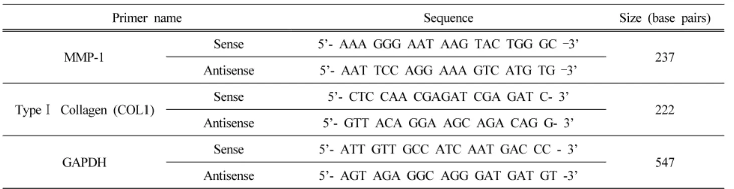
Primer name Sequence Size (base pairs)
MMP-1
Sense 5’- AAA GGG AAT AAG TAC TGG GC –3’
237 Antisense 5’- AAT TCC AGG AAA GTC ATG TG –3’
Type Ⅰ Collagen (COL1) Sense 5’- CTC CAA CGAGAT CGA GAT C- 3’
222 Antisense 5’- GTT ACA GGA AGC AGA CAG G- 3’
GAPDH
Sense 5’- ATT GTT GCC ATC AAT GAC CC - 3’
547 Antisense 5’- AGT AGA GGC AGG GAT GAT GT -3’
Table 1. Sequences of Primers and Fragment Sizes of the Investigated Genes in RT-PCR Analysis
하에 배양하여 사용하였다. 24 well plate에 1 × 10⁵ cell/well로 세포를 분주하고 안정화시킨 후, 100% 세 포 생존을 나타내는 농도로 시료 처리 후 약 72 h 동 안 37 ℃ CO2배양기에서 배양한 후, 배지를 제거하고 CCK-8 method를 이용, microplate reader (Sunrise, Tecan, Austria) 450 nm에서 흡광도를 측정하였다.
2.5. Western blot을 이용한 CCN1 단백질 발현평가 세포를 회수한 후 RIPA buffer (0.1% SDS, 10 mM Sodium Fluoride, 1% NP-40, 1 mM DTT, 500 µM Sodium Orthovanadate, 10 µg/mL Aprotinin, 10 µg/mL leupeptin, 1 mM PMSF in PBS)를 넣고 cell을 깬 후 원 심분리하고, 상층액 속에 포함된 전체 단백질을 Brad- ford assay로 정량 하였다. 이를 취해 sample buffer를 넣고 100 ℃에서 5분간 가열하였다. Caster에 gel을 붓 고 12% SDS- polyacrylamide gel 을 만들었고, 준비한 sample을 loading (50 µg)하였다. 전기영동 장치에 run- ning buffer를 채우고 단백질 전기영동을 실시하였다.
Primary antibody로 Anti-cystein rich protein 61 (Santacruz, USA)을 1 : 200 희석시켜 24 시간 4 ℃에서 반응하고 세척하였다. Secondary antibody로 anti-mouse IgG를 1 시간 반응시킨 후 washing하고 Amplified Opti-4CN Detection kit (Bio-Rad Laboratories, Inc, USA)를 이용 하여 검출하였다.
2.6. Enzyme-linked immunosorbent assay (ELISA) Human dermal fibroblast (p8 ~ 10)와 replicative sen- escent dermal fibroblast (p24 ~)를 10% fetal bovine se- rum이 들어 있는 DMEM에 37 ℃, 5% CO2배양기에서 배양하고 배양된 세포는 trypsin-EDTA을 이용하여 세 포를 회수하였다. 회수된 세포는 48 well에 5 × 10⁴
cell/well로 분주하고 24 h 동안 안정화 시킨다. 시료 처리 시에는 serum free상태로 48 h 배양하여 이용하 였다. Procollagen type I C-peptide enzyme immunoassay (EIA) kit (Takara, Japan)를 사용하여 콜라겐 합성량을 정량 하였다.
MMP-1과 CCN1은 세포배양액을 96 well immuno- plate에 coating하여 ELISA 법을 이용하여 정량 하였다.
2.7. 유전자 발현량 측정(RT-PCR)
Total RNA추출은 RNeasy mini kit (Qiagen Hilden, Germany)을 이용하였고, cDNA합성은 1 µg의 total RNA를 oligo (dT) 15 primer, dNTP (0.5 µM), 1 unit RNase inhibitor 그리고 4 unit Omniscript reverse tran- scriptase (Qiagen Hilden, Germany)로 37 ℃에서 60 min, 93 ℃에서 5 min heating 시킴으로서 반응을 중지 시켰다. PCR은 cDNA로부터 MMP-1, GAPDH를 증폭 하기 위하여 1 µL cDNA, 1 µM의 5'과 3'primer, per- fectshot EX Taq premix (Takara, Japan)를 섞고 distilled water로 전체를 25 µL로 맞춘 다음 PCR을 실시하였 다. PCR증폭은 94 ℃ 0.5 min, 52 ℃ 0.5 min, 72 ℃ 1 min, 28 cycles로 반응시켰다. PCR에 의하여 생성된 산물을 1.5% agarose gel에서 전기영동 후 MMP-1, GAPDH 유전자의 발현을 Molecular Imager (Gel doc XR System, Bio-Rad Laboratories, Inc, USA)로 확인 하 였으며, 각 band의 density는 image J program (NIH Image software, Maryland, USA)을 이용하였다. 각 유 전자의 서열은 Table 1에 나타내었다.
(A) (B)
(C) (D)
Figure 1. Characteristics of replicative senescent dermal fibroblast cells. Senescent cells were prepared from primary normal human skin dermal fibroblasts. The cells were cultured by multiple serial passages until they decreased proliferation.
A : Senescence-associated-β-galactosidase staining was performed using kit (Sigma, USA) according to manufacturer’s instruction.
Images are representative of three experiments. B : Intracellular CCN1 protein levels were determined by western analysis and the intensities were quantified and normalized using β-actin as loading control. Insets show representative western blot. Data are expressed as mean ± SE, N=3, * p < 0.05. Secreted CCN1 protein levels from culture media were determined by ELISA.
O.D value measured and normalized by cell numbers. Data are expressed as mean ± SE, N = 3, * p < 0.05. C : MMP-1 mRNA levels were quantified by RT-PCR and were normalized to GAPDH. Data is expressed as mean ± SE, N = 3, * p <
0.05. Secreted MMP-1 protein levels from culture media were determined by ELISA. Data are expressed as mean ± SE, N = 3,
* p < 0.05. D : Type I procollagen mRNA levels were quantified by RT-PCR and were normalized to GAPDH. Data are
expressed as mean ± SE, N = 3, * p < 0.05. Secreted Type I procollagen protein levels from culture media were determined by
ELISA. Data are expressed as mean ± SE, N = 3, * p < 0.05.
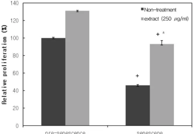
Non-treatment extract (250 µg/ml)
Figure 2. Cell proliferation activity of hydrolyzed Ulva per- tusa extract on senescent dermal fibroblast cells. The hydro- lyzed Ulva pertusa extract treated senescent dermal fibro- blast cells and pre-senescent cells were maintained for 72h.
Viable cells measurement indicated a significant prolifera- tion activity of extract on human dermal fibroblast cells.
Data are expressed as mean ± SE, N = 3, (+ p < 0.05 vs.
pre-senescent; * p < 0.05 vs. Non-treated)
3. 결과 및 고찰
3.1. 복제 노화 섬유아세포의 확립
복제노화 섬유아세포는 일주일에 두 번씩 계대하여 3개월 뒤(P24 ~)에 β-Gal activity를 측정하였다. 그 결과 50% 이상의 세포들이 양성을 보였다(Figure 1A).
이를 복제 노화 섬유아세포로 사용하여 그 특징을 살 펴보았다.
CCN1은 in vivo 노화된 섬유아세포에서 과 발현된 다. 과 발현된 CCN1은 콜라겐의 합성과 분해의 균형 을 파괴한다. 이 연구에서 확립된 복제노화 섬유아세 포의 세포 내 CCN1 단백질의 발현 양을 웨스턴 실험 방법을 통해 분석하였더니, 노화 전 세포에 비해 1.5 배 증가하였다. 또한 ELISA 법을 통해 세포외로 분비 된 CCN1의 양을 비교 분석하였더니 노화 전 세포에 비해 2배 상승하는 등 확립된 노화 섬유아세포에서 CCN1 단백질이 과 발현됨을 확인하였다(Figure 1B).
이로 인해 콜라겐 항상성이 깨어졌는지를 확인하기 위하여 MMP-1 및 Collagen의 단백질 및 mRNA 수준 을 분석하였다. 세포외로 분비된 MMP-1 단백질의 경 우 노화 전 세포에 비해 100% 이상 증가한 것이 확인 되었다. 또한 MMP-1의 mRNA 경우 노화 전 세포에 비해 2배 이상 증가 하였다(Figure 1C).
Type Ⅰ 콜라겐은 피부의 세포외기질의 주요 구성 성분이다. 분비된 콜라겐의 경우 노화 전 세포에 비해 35% 전 후로 줄어든 것을 확인하였다. 또한 mRNA 경 우 노화 전 세포에 비해 40% 정도 줄어든 것을 확인 하였다(Figure 1D). 이것은 확립된 노화 섬유아세포에 서 CCN1의 과 발현에 의해 MMP-1의 발현은 증가되 고 콜라겐의 합성양은 감소된 것이라고 생각된다.
3.2. 가수분해 구멍갈파래 추출물의 노화된 피부세포 증 식효과
내인성 피부노화의 조직학적 소견은 표피 및 진피 의 두께가 얇아지고, 혈관이 감소되며, 진피 내의 섬 유아세포의 수가 감소한다[8]. 따라서 가수분해 구멍 갈파래 추출물에 의한 노화된 섬유아세포의 증식효과 가 있는지를 확인한 결과 Figure 2와 같은 결과를 얻 었다. Figure 2에서 대조군으로서 노화 전 섬유아세포 와 노화된 섬유아세포의 증식률의 차이를 알아보기 위해, 가수분해 구멍갈파래 추출물을 처리하지 않고
세포 분열정도를 측정하였다. 노화 전 세포를 100%로 하였을 때, 노화된 세포는 46%로 세포 증식이 유의적 으로 감소하였다(p < 0.05). 이들 세포에 가수분해 구 멍갈파래를 처리하였을 때, 비 처리군과 비교하여 노 화 전 세포에서는 131%, 노화세포에서는 147%로 유 의적 증가를 보였다(p < 0.05).
3.3. 가수분해 구멍갈파래 추출물에 의한 노화된 세포의 CCN1 단백질 발현 감소
노화된 섬유아세포에서는 CCN1 단백질의 과 발현 으로 인해 MMP-1이 증가 하여 콜라겐 분해가 시작되 고 콜라겐 합성양은 감소하여 세포외기질(ECM)이 붕 괴된다. Epigallocatechin-3-gallate (EGCG)는 녹차 및 동백 잎의 주요 성분이다. Ping 등에 의해 밝혀진 바에 의하면, EGCG는 골수 유래 osteoblasts와 U2OS cells에 서 염증성 Cytokine에 의해 발현 유도된 CCN1을 억제 한다[9]. 이러한 결과를 참고하여 우리는 EGCG를 노 화 섬유아세포에 처리하여 실험한 결과, CCN1의 발 현을 유의적으로 감소시키는 것을 확인하였다. 그래 서 이 실험에 양성컨트롤로 사용하였다. 노화된 섬유 아세포에 가수분해 구멍갈파래 추출물을 처리하였을 경우, 노화된 섬유아세포에서 세포질 내 CCN1 단백 질의 발현에 있어 변화가 있는지 확인한 결과, 비 처
(A) (B)
Figure 3. Hydrolyzed Ulva pertusa extract treatment reduces CCN1 expression in senescent dermal fibroblast cells. Senescent dermal fibroblast cells were treated with hydrolyzed Ulva pertusa extract for 48 h. EGCG used as a positive control.
A : Intracellular CCN1 protein (western blot). B : Secreted CCN1 protein (ELISA). Western analysis were normalized using β -actin as loading control. Inset shows representative western blots. Secreted CCN1 protein in culture medium was determine by ELISA. Data are expressed as mean ± SE, N = 3, * p < 0.05.
(A) (B)
Figure 4. Hydrolyzed Ulva pertusa extract increases type I procollagen and reduces MMP-1 in senescent dermal fibroblast cells.
Senescent dermal fibroblast cells were treated with hydrolyzed Ulva pertusa extract for 48 h.
A : Secreted MMP-1 protein (ELISA) and MMP-1 mRNA. B : Secreted type I procollagen protein (ELISA) and type I
procollagen mRNA. mRNA levels were quantified by RT-PCR and were normalized to GAPDH as an internal control for
quantitative analysis. Secreted type I procollagen and MMP-1 in culture medium was determine by ELISA. O.D value measured
and normalized by cell numbers. Data are expressed as mean ± SE, N = 3, (* p < 0.05 vs. Non-treated)
리군과 비교하여 구멍갈파래 처리군이 70%로 유의적 감소를 보였다(p < 0.05). 또한 세포외로 분비된 CCN1 단백질을 확인한 결과 비 처리군에 비하여 처리군이 53%로 유의적 감소를 보였다(p < 0.05).
3.4. 노화된 세포에서 가수분해 구멍갈파래 추출물의 MMP-1 발현 감소 및 Type Ⅰ procollagen 합성증 가 효과
노화세포는 CCN1 단백질의 과 발현으로 인해 MMP-1 의 발현은 증가되고 콜라겐의 합성양은 감소되어있 다. CCN1 단백질의 발현 수준의 감소는 MMP-1의 발 현은 줄이고, 콜라겐의 합성양을 증가시켜 콜라겐 항 상성을 키워 세포외기질을 유지 하는 것으로 알려져 있다. 이에 CCN1 단백질의 발현 수준을 감소시킨 가 수분해 구멍갈파래 추출물을 노화 섬유아세포에 처리 하여 MMP-1 발현 수준과 콜라겐 합성양에 어떤 영향 을 미치는지 알아보고자 하였다. 그 결과는 Figure 4 (A)에 나타내었다. 노화 전 세포의 MMP-1의 발현 수 준은 노화 섬유아세포에 비해 낮은 수준이었다. 이 섬 유아세포들에 가수분해 구멍갈파래 추출물을 처리하 였을 때, 노화 전 세포에서는 MMP-1의 단백질 및 mRNA를 수준에 변화가 거의 없었으나, 노화세포에 서는 MMP-1의 단백질 및 mRNA 수준이 비 처리군에 비하여 각각 48%, 50%로 유의적으로 감소되었다(p <
0.05). 또한 가수분해 구멍갈파래 추출물이 노화 섬유 아세포의 콜라겐 합성에 미치는 영향을 알아보았다.
가수분해 구멍갈파래 추출물에 의한 노화된 섬유아세 포의 콜라겐 합성 정도는 Figure 4 (B)와 같다. 노화 전 및 노화 섬유아세포에 가수분해 구멍갈파래를 250 µg/mL로 처리한 군에서 노화 전 세포에서는 24% 정 도 증가되는 경향이 보였으나 노화세포에서는 103%
의 유의적 증가를 보였다(p < 0,05). mRNA의 수준에 서도 노화 전 세포에서는 거의 차이가 없었으나 노화 세포에서는 1.6배의 유의적 증가를 보였다(p < 0.05).
이는 노화된 섬유아세포에서 가수분해 구멍갈파래 추 출물의 효과를 잘 설명해주고 있다.
4. 결 론
인간은 시간의 흐름에 따라 피부 진피층에 존재하 는 섬유아세포의 기능저하로 인해 피부의 탄력이 소
실되고, 주름이 형성된다. 이에 우리는 기능이 저하된 노화 섬유아세포에 가수분해 구멍갈파래 추출물을 처 리하여, 섬유아세포의 저하된 기능이 개선되는지 알 아보고자 하였다. 이에, 복제 노화 섬유아세포를 확립 하여 노화 섬유아세포로 사용하였다. 또한, 세포질 외 기질의 주요 성분인 콜라겐의 항상성을 조절하는 단 백질인 CCN1의 발현 변화 여부를 노화 섬유아세포의 기능 개선의 타켓으로 삼아, 가수분해 구멍갈파래 추 출물을 노화 섬유아세포에 처리하였을 때, CCN1의 발현 수준의 변화가 있는지 확인하였다. 확립된 노화 섬유아세포에서는 CCN1의 발현량이 노화 전 섬유아 세포에 비해 증가하였다. 또한, 콜라겐 분해 효소인 MMP-1의 경우 노화 전 섬유아세포에 비해 증가하였 고, Procollagen의 경우 노화 전 섬유아세포에 비해 생 성이 감소하였다.
가수분해 구멍갈파래 추출물을 확립된 노화 섬유아 세포에 처리한 결과, 노화 섬유아세포의 기능 개선의 타켓으로 정한 CCN1 단백질의 발현을 억제하였고, 그로 인해 콜라겐 합성이 촉진되었다. 또한 노화 섬유 아세포를 증식시켜 노화 섬유아세포의 기능 개선 효 과가가 우수하였다. 이러한 결과는, 가수분해 구멍갈 파래 추출물이 자연노화 및 광노화에 의한 섬유아세 포의 콜라겐 항상성 파괴 및 기능저하를 효율적으로 방어하여 노화를 지연시키는 항노화 물질임을 증명하 는 것이다. 따라서, 가수분해 구멍갈파래 추출물은 주 름 개선 기능성 화장품 분야에서 이용 가능성을 높일 수 있을 것이다.
감사의 글
본 연구는 지식경제부에서 시행한 “지역산업 기술 개발 사업(과제번호:A000200480)”의 지원에 의하여 이루어진 것으로 이에 감사드립니다.
Reference
1. B. A. Gilchrest and Yaar M. Ageing, photoageing of the skin: observations and the cellular and molecular level, Br. J. Dermatol., 127, 25 (1992).
2. M. A. Farage, K. W. Miller, E. Berardesca, and H.
I. Maibach, Clinical implications of aging skin: cuta-
neous disorders in the elderly, Review. Am J. Clin Dermatol., 10, 73 (2009).
3. C. C. Chen, N. Chen, and L. F. Lau, The angiogenic factors Cyr61 and connective tissue growth factor in- duce adhesive signaling in primary human skin fibro- blasts, J. Biol Chem., 276, 10443 (2001).
4. M. L. Kireeva, F. E. Mo, G. P. Yang, and L. F. Lau, Cyr61, a product of a growth factor-inducible imme- diate-early gene, promotes cell proliferation, migra- tion, and adhesion, Mol Cell Biol., 16, 1326 (1996).
5. F. E. Mo, A. G. Muntean, C. C. Chen, D. B. Stolz, S. C. Watkins, and L. F. Lau, CYR61 (CCN1) is es- sential for placental development and vascular in- tegrity, Mol Cell Biol., 22, 8709 (2002).
6. T. Quan, Z. Qin, John J. Voorhees, and Gary J.
Fisher., Cysteine-Rich Protein 61 (CCN1) Mediates Replicative Senescence-Associated Aberrant Collagen Homeostasis in Human Skin Fibroblasts, J. Cellular Biochemistry, 113, 3011 (2012).
7. D. R. Brigstock, The CCN family: A new stimulus package, J. Endocrinol., 178, 169 (2003).
8. M. D. West, The cellular and molecular biology of skin aging, Arch Dermatology, 130, 87(1994).
9. P. H. Wu1, S. K. Lin, B. S. Lee, S. H. Kok, J. H. Wang, K. L. Hou, H. Yang, J. S. Wang, and C. Y. Hong, Epigallocatechin-3-gallate diminishes cytokine-stimu- lated Cyr61 expression in human osteoblastic cells: a therapeutic potential for arthritis, Rheumatology, 51, 1953 (2012).
10. T. Quan, T. He, Y. Shao, L. Lin, S. Kang, J. J.
Voorhees, and G. J. Fisher, Elevated cysteine-rich 61 mediates aberrant collagen homeostasis in chronolog- ically aged and photoaged human skin, Am J.
Pathol., 169, 482 (2006).
11. T. Quan, Z. Qin, Y. Xu, T. He, S. Kang, J. J.
Voorhees, and G. J. Fisher, Ultraviolet irradiation in- duces CYR61/CCN1, a mediator of collagen homeo- stasis, through activation of transcription factor AP-1 in human skin fibroblasts, J. Invest Dermatol., 130, 1697 (2010).
12. T. Quan, Z. Qin, P. Robichaud, J. J. Voorhees, and
G. J. Fisher, CCN1 contributes to skin connective tissue aging by inducing age-associated secretory phenotype in human skin dermal fibroblasts, J. Cell Commun Signal, 5, 201 (2011).
13. T. Quan, Z. Qin, Y. Shao, Y. Xu, J. J. Voorhees, and G. J. Fisher, Retinoids suppress cysteine-rich protein 61 (CCN1), a negative regulator of collagen homeostasis, in skin equivalent cultures and aged hu- man skin in vivo, Exp Dermatol., 20, 572 (2011).
14. J. Varani, M. K. Dame, L. Rittie, S. E. Fligiel, S.
Kang, G. J. Fisher, and J. J. Voorhees, Decreased collagen production in chronologically aged skin:
Roles of age-dependent alteration in fibroblast func- tion and defective mechanical stimulation, Am J.
Pathol., 168, 1861 (2006).
15. L. F. Lau and S. C. Lam, The CCN family of an- giogenic regulators: The integrin connection, Exp.
Cell Res., 248, 44 (1999).
16. G. J. Fisher, S. Kang, J. Varani, Z. Bata-Csorgo, Y.
Wan, S. Datta, and J. J. Voorhees, Mechanisms of photoaging and chronological skin aging, Arch Dermatol., 138, 1462 (2002).
17. G. J. Fisher, Z. Q. Wang, S. C. Datta, J. Varani, S.
Kang, and J. J. Voorhees, Pathophysiology of pre- mature skin aging induced by ultraviolet light, N.
Engl J. Med., 337, 1419 (1997).
18. S. E. Fligiel, J. Varani, S. C. Datta, S. Kang, G. J.
Fisher, and J. J. Voorhees, Collagen degradation in aged/photodamaged skin in vivo and after exposure to matrix metalloproteinase-1, in vitro, J. Invest Dermatol., 120, 842 (2003).
19. D. R. Holt, S. J. Kirk, M. C. Regan, M. Hurson, W.
J. Lindblad, and A. Barbul, Effect of age on wound healing in healthy human beings, Surgery., 112, 293 (1992).
20. N. J. Raine-Fenning, M. P. Brincat, and Y. Muscat- Baron, Skin aging and menopause: Implications for treatment, Am J. Clin Dermatol., 4, 371 (2003).
21. M. R. Khorramizadeh, E. E. Tredget, C. Telasky, Q.
Shen, and A. Ghahary, Aging differentially modu- lates the expression of collagen and collagenase in
dermal fibroblasts, Mol Cell Biochem., 194, 99 (1999).
22. T. Quan, T. He, S. Kang, J. J. Voorhees, and G. J.
Fisher, Solar ultraviolet irradiation reduces collagen
in photoaged human skin by blocking transforming growth factor-beta type II receptor/Smad signalin, Am J. Pathol., 165, 741 (2004).