Inhibitory Effects of Chrysanthemum boreale Makino on 3T3-L1 Preadipocyte Differentiation and Down-regulation of Adipogenesis and Lipogenesis
Dae Il Hwang1†, In-Ho Choi1,2†, Do Yoon Kim1,3, Soo Min Park1, Ha Bin Kim1, YaLi Li1 and Hwan Myung Lee1*
1Division of Cosmetic and Biotechnology, College of Life and Health Science, Hoseo University, 20, Hoseo-ro 79beon-gil, Baebang-eup, Asan-city, Chungcheongnam-do 31499, Korea
2Department of Integrated Biosciences, College of Biomedical and Health Science, Konkuk University, 268, Chungwon-daero, Chungju-city, Chungcheongbuk-do 27478, Korea
3College of Chemistry and Chemical Engineering, Hunan Institute of Science and Technology, Yueyang 414006, Hunan, China Received October 29, 2018 /Revised March 1, 2019 /Accepted March 3, 2019
Obesity is associated with an increased risk of many diseases including type 2 diabetes mellitus, hy- pertension, and hyperlipidemia. The flowers of Chrysanthemum boreale have been used as traditional medicines for the treatment of diseases such as obesity and type 2 diabetes mellitus. This study aimed to evaluate the effect of C. boreale Makino flower essential oil (CFEO) on adipocyte differentiation us- ing preadipocyte cell line 3T3-L1. CFEO at concentrations between 0.1 and 5 μg/ml did not affect 3T3- L1 cell viability. A CFEO concentration of between 0.1 and 1 μg/ml significantly inhibited lipid accu- mulation during MDI-induced differentiation in 3T3-L1 cells in a dose-dependent manner, reaching a maximal level at 1 μg/ml (28.94±2.01%; approximately 30% of control treated with MDI alone).
Western blot analysis revealed that CFEO concentrations between 0.1 and 1 μg/ ml suppressed the acti- vations of three adipogenic transcription factors in the MDI-stimulated 3T3-L1 cells: peroxisome pro- liferator-activated receptor γ; CCATT/enhancer binding protein α; and sterol regulatory element bind- ing protein-1. Moreover, the expressions of lipogenic enzymes, acetyl-CoA carboxylase, and fatty acid synthase were also inhibited by treatment with CFEO between 0.1 and 1 μg/ml. CFEO may therefore be a promising functional material for obesity prevention.
Key words : 3T3-L1 cells, adipogenesis, Chrysanthemum boreale Makino, essential oil, obesity
†Authors contributed equally.
*Corresponding author
*Tel : +82-41-540-9551, Fax : +82-41-540-9538
*E-mail : [email protected]
This is an Open-Access article distributed under the terms of the Creative Commons Attribution Non-Commercial License (http://creativecommons.org/licenses/by-nc/3.0) which permits unrestricted non-commercial use, distribution, and reproduction in any medium, provided the original work is properly cited.
Journal of Life Science 2019 Vol. 29. No. 3. 332~336 DOI : https://doi.org/10.5352/JLS.2019.29.3.332
서 론
현대사회에서 비만은 심각한 건강 문제로 대두되고 있다.
비만이란, 에너지 섭취가 소비를 초과하는 에너지 불균형에 의해, 지방 세포수 및 세포내 지질의 크기를 증가 시킴으로서 인체내 지방이 과도하게 축적되어 나타나는 현상이다[4, 6, 9].
비만은 대사 증후군으로 간주되며 고지혈증, 고혈압, 제2형 당뇨병 및 특정 유형의 암을 포함한 많은 질병과 관련이 되어 있다[1]. 지방 세포의 분화는 세포내 지방구 형성과 지질 생합 성 과정에서 필수적인 과정이며, 융합(confluence), 호르몬 유 도(hormonal induction), 클론 확장(clonal expansion), 성장 정지(growth arrest) 및 말단 분화(terminal differentiation)가
단계적으로 진행된다[3]. 지방세포의 분화의 과정은 perox- isome proliferator-activated receptor γ (PPARγ), sterol regu- latory element binding protein (SREBP-1) 및 CCATT/en- hancer binding protein α (C/EBPα) 등의 전사 인자에 의해 조절된다[8]. 각각의 전사 인자는 fatty acid synthase (FAS) and acetyl-CoA carboxylase (ACC)를 비롯한 다양한 지질 대 사와 관련된 효소의 발현을 조절하게된다[4]. 현재 비만 치료 를 위해 사용되는 다수의 약물은 혈압상승, 변비, 심장질환 등의 심각한 부작용으로 인해 약물의 안전성의 확보가 필요한 실정이다. 약용 식물 추출물과 식물성 화학 물질(phytochem- icals)은 오랜기간동안 인체 적용을 통해 안전성이 확보되어 있으며, 비만 치료를 위한 높은 잠재력을 지니고 있어서 식이 보충제로도 많이 사용되고 있다[5].
산국화(Chrysanthemum boreale Makino)는 국화과(Compo- sitae)에 속하며 아시아에서 널리 분포하는 다년생 식물로서 노란색 꽃을 피우며 산과 들에서 쉽게 찾아볼 수 있는 식물이 다[7, 8]. 국화과 식물은 전통적으로 열을 제거하고 시력을 향 상시키는데 사용된 약재이다[10]. 산국화 에센셜오일(Chry- santhemum boreale Makino essential oil)은 항박테리아 및 항바 이러스 작용 등의 다양한 생물학적 활성을 가지는 2차 대사산
물로 보고되고 있다[9, 12]. Kim 등의 연구에 의하면 산국화 에센셜오일은 camphor, α-thujone, cis-chrysanthenol, 1,8-cin- eole, α-pinene, β-caryophyllene 등의 주요 화합물을 포함한 87가지의 성분으로 구성되어 있다고 확인 되었다[7]. 그러나 산국꽃 에센셜오일의 지방세포 분화 유도 및 지방 생합성과 관련된 항비만 효과에 대해서는 아직까지 연구 된 적이 없다.
따라서 본 연구에서는 산국꽃 에센셜오일의 전지방세포주 (preadipocyte, 3T3-L1)에 대한 지방세포 분화 및 지방생합성 억제 효능을 확인하고, 이를 활용한 항비만 소재로서의 활용 가능성을 확인 하고자 하였다.
재료 및 방법
시약
본 실험에 사용된 dulbecco's modified eagle medium (DMEM), fetal bovine serum (FBS), fetal calf serum, penicillin/strepto- mycin (P/S), phosphate buffered saline (PBS), trypsin-ethyl- enediamine tetraacetic acid는 Hyclone (Logan, UT, USA) 또 는 Welgene (Daegu, Korea)에서 구입하여 사용하였다. Bovine serum albumin, insulin, dexamethasone, 3- isobutyl-methyl xanthine (IBMX), oil red O reagent는 Sigma (St. Louis, MO, USA), EZ-CyTox kit 은 DAEIL LAB Service (Seoul, Korea) 구입하여 사용하였다. 단백질 발현에 사용된 anti-PPARγ, an- ti-C/EBPα, anti-FAS와 anti-ACC는 Cell signaling (MA, USA) 에서, anti-SREBP-1는 Santacruz (CA, USA)에서 anti-β-actin 은 Sigma (MO, USA)에서 각각 구입하여 사용하였다.
산국꽃 에센셜오일의 제조
본 실험에 사용한 산국화는 호서대학교 아산캠퍼스 화장품 과학과 실습지에서 재배하여 채취하였으며, 국립수목원 양종 철 박사에 의해 동정되었다. 산국화는 표본으로 제작하여 호 서대학교 화장품과학과 에센셜오일소재과학연구실에 보관하 고있다(voucher no. CFEO-0001). 산국꽃은 10월에 꽃이 피기 직전의 꽃망울을 20 kg을 채취하였고, 수증기 증류법을 활용 하여, 10 l의 증류액과 에센셜오일 혼합액을 확보하였다. 혼합 액으로부터 분별깔대기(separating funnel)를 이용하여 에센 셜오일 50 ml을 분리하고 빛이 차단된 4℃ 저온고에 보관하며 사용하였다.
세포 배양 및 지방세포 분화
Mouse embryo 3T3-L1 전지방 세포주는 American type culture collection (ATCC)에서 구입하여 10% bovine calf se- rum, 1% P/S이 첨가된 DMEM 배지에 2.5×10⁵ cells/ml로 분 주한 후 confluent 상태가 될 때까지 37℃ CO2 배양기에서 배양 하였다. 세포가 confluent한 상태(D-0, 분화 시작 시점)가 되면 10% FBS, 1% P/S, 5 μg/ml insulin, 1 μM dexamethasone,
0.5 mM IBMX (MDI)가 포함된 배지로 교환하여 2일 동안 배 양하였다. 2일 후(D-2)에 10% FBS, 1% P/S, 5 μg/ml insulin이 포함된 배지로 바꿔주고, D-5일째부터 insulin을 첨가하지 않 은 배지로 D-8이 되는 시점까지 배양하여 지방세포로 분화를 유도하였다. 세포독성은 WST (water soluble tetrazolium salt) assay를 통해 확인하였다. 3T3-L1 세포를 96well plate (5x10³ cells/well)에 분주한 다음, 12시간 동안 배양하였다. 그 후, 산국꽃 에센셜오일 농도별로 처리한 후 37℃, 5% CO2 배양기 에서 48시간 배양하였다. 세포독성을 측정하기 위하여 well 당 10 μl의 EZ-CyTox kit를 처리하여 37℃, 5% CO2 배양기에 서 30 min 동안 반응시킨 후 , multi-well plate reader (Syner- gy 2, Bio-Tek Instruments, Winooski, VT, USA)를 이용하여 450nm에서 흡광도를 측정하였다.
Oil red O를 활용한 세포내 지방 염색
분화된 지방세포의 지방방울(lipid droplet)을 염색하기 위 해 지방질에 특이적으로 반응하는 oil red O시약을 사용하였 다. 분화된 지방세포를 PBS로 3회 세척하고 4% formalin 첨가 하여 30분간 세포를 고정하였다. 고정한 세포를 증류수로 3회 세척 후 마지막으로 60% isopropanol로 세척하고 well을 완전 히 건조시켰다. 건조된 세포는 oil red O 시약으로 1시간 염색 하고, 증류수로 4회 세척한 후 현미경으로 분화된 세포의 사진 을 촬영하였다. 촬영이 끝난 세포는 증류수를 제거한 후 완전 히 건조시키고 100% isopropanol로 10분간 처리하여 oil red O를 용출시켰다. Oil red O는 multi-well plate reader (Syner- gy 2, Bio-Tek Instruments, Winooski, VT, USA)를 이용하여 520 nm에서 흡광도를 측정하였다.
Western Blot
세포를 ice cold-PBS로 2-3회 세척한 후 protease inhibitors 와 phosphatase inhibitors (GenDEPOT, Barker, TX, USA)가 포함된 RIPA buffer (Cell signaling, Beverly, MA, USA)를 사 용하여 균질화하고 17,000x g, 20분간 원심분리하여 상층액을 분리하였다. 10~30 μg의 단백질을 8~10%의 sodium dodesyl sulfate-polyacrylamide gel electrophoresis (SDS- PAGE)로 분리하고, polyvinylidene fluoride (PVDF) membrane으로 이 동시켰다. PVDF membrane은 blocking buffer (3% skim milk/PBS)로 2시간 동안 blocking하고 1차 antibody (PPARγ, C/EBPα, SREBP-1, ACC, FAS)를 blocking buffer에 희석하여 4℃에서 overnight (O/N)하였다. PBS-T (0.05% Tween 20)로 washing 후 2차 antibody를 blocking buffer에 희석하여 실온 에서 반응시켰다. PBS-T로 세척하고 ECL 용액(Welgene Inc, Daegu, Korea)에 반응시켜 단백질의 발현정도를 분석하였다.
Statistical analysis
실험결과는 mean ± S.E.M으로 표기하였으며, Graphpad
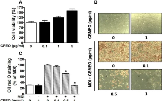
Fig. 1. Effects of Chrysanthemum boreale Makino flowers essential oil (CFEO) on the viability and accumulation level of lipid droplets in 3T3-L1 cells. (A) Effect of CFEO on the viability in 3T3-L1 cells. Cytotoxic effects of CFEO (0.1-5 μg/ml) on 3T3-L1 cells in the quiescent state were analyzed using the WST assay (n=5). Response in the quiescent state is expressed as 100%. *P<0.005.
(B and C) Effect of CFEO on accumulation level of lipid droplets in 3T3-L1 cells. 3T3-L1 cells were differentiated by treatment with 3-isobutyl-1-methylxanthine (0.5 mM), dexamethasone (1 μM) and insulin (5 μg/ml) (MDI) as described in the Methods section. 3T3-L1 cells incubated in either the presence or absence of CFEO (0.1-1 μg/ml) for 8 days under MDI treatment condition and were subjected to oil red-O staining. (B) Representative photomicrographs of lipid-laden 3T3-L1 cells. Red colors indicate oil red-O positive cells. Magnification: ×100. (C) Statistical data obtained from panel C (n=4). The response of cells treated with MDI alone we considered as 100%. *P<0.005 versus the group treated with MDI alone.
Prism software (version 5.00 for Windows, San Diego, CA, USA) 프로그램을 이용한 student’s t-test 방법을 통해 유의성 여부를 분석하였고, 그 결과 p<0.05일 경우에 한하여 유의성을 인정하였다.
결과 및 고찰
산국꽃 에센셜오일의 지방세포 분화 및 지방 생합성 억제 효능
지방세포는 에너지 항상성 유지 및 지질대사에 중요한 역할 을 하고, 지방전구세포인 3T3-L1은 다양한 호르몬과 전사인자 들에 의해 지방세포로 분화되면서 세포 내 지방을 생성 및 축적한다[11]. 산국꽃 에센셜오일은 0.1~5 μg/ml에서 3T3-L1 cell에 대해 유의한 독성이 확인되지 않았다(Fig. 1A). 세포생 존율에 영향을 미치지 않는 산국꽃 에센셜오일 0.1, 0.5 및 1 μg/ml을 3T3-L1세포에 처리하여 세포내 지방 생합성에 미치 는 영향을 확인하기 위해 oil red O로 염색을 통해 관찰하였다.
지방세포로 분화되지 않은 MDI 무처리군 세포에서는 자연발 생 및 산국꽃 에센셜오일에 의한 세포내 지방 생합성이 확인 되지 않았으나, MDI에 의해 지방세포 분화가 유도된 세포군 에서는 세포내 지방구 형성 및 생합성에 의한 축적이 관찰
되었다(Fig. 1B). 반면, MDI에 의해 지방세포로 분화가 유도된 세포내 지방구 및 지방생합성은 산국꽃 에센셜오일 0.5 및 1 μg/ml 처리군에서 농도의존적으로 유의하게 억제됨을 확인 하였다(Fig. 1B, Fig. 1C). 특히, 산국꽃 에센셜오일 1 μg/ml 처리군에서 71.06±4.45%의 억제율을 나타내어 새로운 항비만 소재로서의 가능성을 확인할 수 있었다(Fig. 1C).
산국꽃 에센셜오일이 지방세포 분화 및 지방생합성 관련 단 백질 발현에 미치는 영향
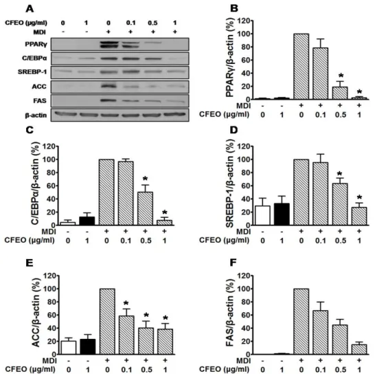
산국꽃 에센셜오일에 의한 지방세포 분화조절 인자인 PPARγ, C/EBPα 및 SREBP-1와 지방생합성 관여하는 ACC 및 FAS의 발현 정도는 western blot 기법을 통해 확인하였다.
PPARγ 및 C/EBPα는 지방세포의 분화에 의한 지방조직형성 에 관여하며, SREBP-1은 지방세포 분화 조절 뿐만 아니라, 지 방생합성을 조절하여 체지방량을 조절에 결정적인 역할을 하 는 것으로 알려져 있다[2]. 지방세포의 분화에 의한 지방조직 의 생성과 관련된 PPARγ, C/EBPα 및 SREBP-1의 발현은 MDI 처리군에 비하여 산국꽃 에센셜오일 0.5 μg/ml 처리군에서 19.11±8.60%, 50.49±10.75%, 63.73±8.18%, 1 μg/ml 처리군에 서 2.65±1.83%, 7.78±4.51%, 27.30±6.53% 각각 발현의 감소를 나타내었다(Fig. 2A, Fig. 2B, Fig. 2C, Fig. 2D).
Fig. 2. Effects of Chrysanthemum boreale Makino flowers essential oil (CFEO) on the expressions of adipogenesis and lipogenesis proteins in 3T3-L1 adipocytes. (A) 3T3-L1 cells were differentiated by treatment with MDI and maintained as described in the Methods section. 3T3-L1 cells were incubated in the presence or absence of CFEO (0.1-1 μg/ml) for 8 days from MDI treatment to cell harvest for Western blot analysis. The adipocyte lysates were immunoblotted with indicated antibodies.
(B–F) Statistical graphs showing the expression levels of each protein were obtained from panel A. The responses in cells treated with MDI alone was considered as 100%(n=4) *P<0.005 versus the group treated with MDI alone.
중성지방 합성에 관여하는 ACC는 0.1 μg/ml (58.63±11.04
%), 0.5 μg/ml (40.15±10.52%) 및 1 μg/ml (38.66±8.34%)의 발현 억제효과가 확인되었으며(Fig. 2E), 지방산 합성효소 조 절인자인 FAS는 산국꽃 에센셜오일 0.5 μg/ml (44.94±8.59%) 과 1 μg/ml (15.06±3.85%) 처리군에서 각각 유의한 발현 조절 효능이 관찰되었다(Fig. 2F).
본 연구에서 PPARγ, C/EBPα, SREBP-1, ACC 및 FAS의 발현이 MDI처리군에 비해 산국꽃 에센셜오일 처리군에서 농 도의존적으로 발현이 억제됨을 확인하였다. 이와 같은 사실을 토대로 산국꽃 에센셜오일이 전지방세포의 지방세포 분화뿐 만 아니라, 지방생합성의 조절에 관여함으로서 비만 억제 소 재로서 활용 가능성을 확인할 수 있었다(Fig. 2).
그러나 산국꽃 에센셜오일의 구체적인 비만 치료 메커니즘 을 밝히기 위해 지방 세포에서 항비만 활성을 나타내는 산국
꽃 에센셜오일의 주요 성분의 분리 및 동정이 향후 연구의 중요한 목표가 될 것이다.
본 연구는 산국꽃 에센셜오일을 이용하여 비만관련 in-vivo 실험 및 산국꽃 에센셜오일의 주요 성분에 관한 추가 실험이 필요할지라도, 비만 예방 또는 치료를 위한 잠재적인 예방 및 치료제 개발에 유용한 정보를 제공 할 수 있다.
감사의 글
본 연구는 2017년도 산업통상자원부 한국산업단지공단 산 업집적지경쟁력강화사업(이전기술사업화, RCC17015)과 산 림청(한국임업진흥원) 산림과학기술 연구개발사업(FTIS 2016 016B10-1919-AB02)의 지원에 의하여 이루어진 것입니다.
초록:산국(
Chrysanthemum boreale
Makino) 꽃 유래 에센셜오일(Essential oil)이 지방세포 분화 및 지방생성에 미치는 영향황대일1†․최인호1,2†․김도윤1,3․박수민1․김하빈1․이야려1․이환명1*
(1호서대학교 화장품생명공학부, 2건국대학교 바이오융합과학부, 3중국 호남이공대학교 화학화공 단과대학)
비만은 2 형 당뇨병, 고혈압 및 고지혈증을 포함한 다양한 질병과 관련이 있으며, 산국꽃은 전통적으로 비만과 2형 당뇨병 치료제로 사용되어왔다. 본 연구는 전지방세포(preadipocyte, 3T3-L1)를 사용하여 산국꽃 에센셜오일 이 지방세포 분화 및 지방 생합성에 미치는 영향을 확인하였다. 산국꽃 에센셜오일은 0.1-5 μg/ml의 농도에서 3T3-L1 세포에 대해 독성을 나타내지 않았다. 산국꽃 에센셜오일은 지방세포 분화 유도물질(MDI)을 처리한 3T3-L1세포에서 농도 의존적으로 지방분화를 억제하였으며, 1 μg/ml (28.94±2.01%)농도에서 최대 효과를 나타내 었다. 산국꽃 에센셜오일은 MDI에 의해 분화가 유도된 3T3-L1 세포에서 지방생성전사인자인 peroxisome pro- liferator-activated receptor γ (PPARγ), CCATT/enhancer binding protein α (C/EBPα) 그리고 sterol regulatory element binding protein (SREBP-1)의 단백질 발현을 억제하였다. 중성지방 생성 및 지방산합성효소 조절인자인 acetyl-CoA carboxylase (ACC)와 fatty acid synthase (FAS)의 발현 또한 산국꽃 에센셜오일에 의해 억제되었다.
따라서 본 연구를 통해 산국꽃 에센셜오일은 천연 항비만 기능성 소재로써의 사용이 가능할 것으로 사료 된다.
References
1. Ahn, J., Lee, H., Kim, S. and Ha, T. 2010. Curcumin-induced suppression of adipogenic differentiation is accompanied by activation of Wnt/beta-catenin signaling. Am. J. Physiol. Cell Physiol. 298, 1510-1516.
2. Darlington, G. J., Ross, S. E. and MacDougald, O. A. 1998.
The role of C/EBP genes in adipocyte differentiation. J. Biol.
Chem. 273, 30057-30060.
3. Gregoire, F. M., Smas, C. M. and Sul, H. S. 1998. Under- standing adipocyte differentiation. Physiol. Rev. 78, 783-809.
4. Ho, J. N., Choi, J. W., Lim, W. C., Lim, M. K., Lee, I. Y.
and Cho, H. Y. 2013. Kefir inhibits 3T3-L1 adipocyte differ- entiation through down-regulation of adipogenic transcrip- tion factor expression. J. Sci. Food. Agric. 93, 485-490.
5. Kazemipoor, M., Radzi, C. W., Hajifaraji, M., Haerian, B.
S., Mosaddegh, M. H. and Cordell, G. A. 2013. Antiobesity effect of caraway extract on overweight and obese women:
a randomized, triple-blind, placebo-controlled clinical trial.
Evid-based. Complement. Altern. Med. 2013, 928582.
6. Kim, D. M., Choi, H. R., Park, A., Shin, S. M., Bae, K. H., Lee, S. C., Kim, I. C. and Kim, W. K. 2013. Retinoic acid inhibits adipogenesis via activation of Wnt signaling path- way in 3T3-L1 preadipocytes. Biochem. Biophys. Res. Com- mun. 434, 455-459.
7. Kim, K. J., Kim, Y. H., Yu, H. H., Jeong, S. I., Cha, J. D., Kil, B. S. and You, Y. O. 2003. Antibacterial activity and chemical composition of essential oil of Chrysanthemum boreale.
Planta Med. 69, 274-277.
8. Kim, Y., Sung, J., Sung, M., Choi, Y., Jeong, H. S. and Lee, L. 2010. Involvement of heme oxygenase-1 in the anti-in- flammatory activity of Chrysanthemum boreale makino extract on the expression of inducible nitric oxide synthase in
RAW264.7 macrophage. J. Ethnopharmacol. 131, 550-554.
9. Kwon, T. H., Wu, Y. X., Kim, J. S., Woo, J. H., Park, K.
T., Kwon, O. J., Seo, H. J., Kim, T. and Park, N. H. 2015.
6,6‘-Bieckol inhibits adipocyte differentiation through down- regulation of adipogenesis and lipogenesis in 3T3-L1 cells.
J. Sci. Food. Agric. 95, 1830-1837.
10. Perry, L. M. 1980. Medicinal plants of East and Southeast Asia: attributed properties and uses, pp. 296, The MIT Press, Cambridge, MA, USA.
11. Rosen, E. D., Hsu, C. H., Wang, X., Sakai, X., Freeman, M.
W., Gonzalez, F. J. and Spiegelman, B. M. 2001. C/EBPα in- duces adipogenesis through PPAR: a unified pathway. Genes Dev. 16, 22-26.
12. Willuhn, G. 1998. Arnica flowers: Pharmacology, toxicology, and analysis of the sesquiterpene lactones-their main active substances, pp. 118, American Chemical Society Press, Washington D. C., USA.