비타민나무 잎 에탄올추출물의 AMPK 활성화를 통한 3T3-L1 지방전구세포의 adipogenesis 억제효과
정현주
1
, 박주희1
, 김명조2
*1
삼성생약(주) 바이오생명공학연구소,
2강원대학교 식물자원응용과학과
Ethanol Extract of Hippophae Rhamnoides L. Leaves Inhibits Adipogenesis through AMP-activated protein kinase (AMPK)
Activation in 3T3-L1 Preadipocytes
Hyeon Ju Jeong
1
, Ju Hee Park1
and Myong-Jo Kim2
*1
Biotechnology institute, Samsung Herb Medicine Agricutural Co., Chuncheon 24341, Korea
2
Department of Applied Plant Sciences, Kangwon National University, Chuncheon 24341, Korea
Abstract - In the present study, we investigated the effect of 70% EtOH extract from Hippophae Rhamnoides L. leaves (HRL) on the anti-obesity effect in 3T3-L1 cells. The effects of HRL on lipid accumulation in 3T3-L1 cells were examined using Oil Red O staining. In addition, we examined the gene expression levels by using RT-PCR and western blot. The results of this analysis showed that 100 ㎍/㎖ HRL significantly increased the inhibition of lipid accumulation by 82.25%;
significantly decreased the mRNA expression of sterol regulatory element binding protein-1c (SREBP-1c), peroxisome proliferator-activated receptor γ (PPARγ), CCAAT/enhancer binding proteins α (C/EBPα), and fatty acid synthase (FAS) in 3T3-L1 cells as well as the stimulated protein expression of AMP-activated protein kinase (AMPK); and suppressed the expression level of PPARγ. These results suggest that HRL can prevent adipogenesis through activation of AMPKα and inhibition of adipogenesis transcription factors.
Key words - Hippophae Rhamnoides L. leaves, HRL, Adipogenesis, 3T3-L1, AMPK
*교신저자: [email protected] Tel. +82-33-250-6413
서 언
최근 서구화된 식습관과 생활환경의 변화로 전 세계적으로 비만이 심각한 질환으로 대두되고 있다(No, 2012). 비만은 신체 에너지의 섭취와 소비의 불균형으로 생기는 질환으로 고혈압, 고지혈증, 당뇨 및 심혈관질환과 같은 성인병 발생 가능성을 증 가시키는 요소로 밝혀지고 있다(Spiegelman and Flier, 2001).
그로 인해 현재 다양한 비만 관련 치료제가 출시되고 있지만, 이 들은 지방변, 두통, 오심, 우울증, 불안 및 인지 관련 증상 등의 부작용이 보고되고 있다(Ballinger and Peikin, 2002; Lee, 2013). 따라서 이러한 부작용이 적으면서 우수한 효과를 기대할
수 있는 천연물을 이용한 항비만 식이 개발이 요구되는 실정이 다(Kim et al., 2010; Jeon et al., 2014; Choi et al., 2013; Shon et al., 2013).
지방세포 내의 지방구(lipid droplet)는 지질의 대사와 조절 등 지방대사에 중요한 역할을 하고, 지방구에 축적되는 지질은 중성지방의 분해와 합성을 조절하는 것으로 알려져 있다. 따라 서 비만 예방 및 관리에 있어 지방전구세포로부터 분화된 지방 세포의 형성 억제와 지방구 내에 존재하는 중성지방의 분해로 인한 glycerol의 유출과정은 중요한 기전이며(Frayn et al., 2003), 이 때 다양한 전사인자와 호르몬이 관여한다. AMP- activated protein kinase (AMPK)는 지방산의 합성과 분해를 매개함으로써 체내 에너지 항상성 유지의 중요한 역할을 한다 (Assifi et al., 2005). 활성화된 AMPK는 sterol regulatory element binding protein-1c (SREBP-1c), peroxisome proliferator-
Original Research Article
activated receptor γ (PPARγ) 및 fatty acid synthase (FAS)와 같은 지방세포화 인자의 발현을 억제함으로써 지방 합성을 억제 시키고 acetyl-CoA carboxylase (ACC)의 비활성화와 carnitine palmitoyltransferase-1 (CPT-1)의 활성화를 통해 베타-산화 를 촉진시켜 미토콘드리아로 지방산 이동을 증가시킨다(Hardie, 2003; Foretz et al., 1998; Park et al., 2015). 분화 초기에는 CCAAT/enhancer-binding protein β (C/EBPβ), C/EBPδ와 SREBP-1c가 발현되고, 이들 전사인자는 후기전사인자인 PPARγ 와 C/EBPα의 발현을 유도하여 지방전구세포의 분화를 촉진하 며 섭취된 에너지원으로부터 중성지방의 합성을 촉진하여 지방 과 간 조직에 저장한다(Cao et al., 1991; Naowaboot et al., 2012). 또한, SREBP-1c는 FAS, ACC 및 acetyl-CoA synthase (ACS) 등의 발현을 조절하여 지방산과 중성지방의 합성을 조절 하게 된다(Kolehmainen et al., 2001). 따라서 항비만 기능성 소재개발 연구는 지방세포의 분화과정을 억제하거나 지방분해 를 촉진하는 소재를 탐색하는 연구가 진행되어야 한다.
본 연구에서 사용한 비타민나무(Hippophae rhamnoides L.) 는 보리수나무과(Elaeagnaceae)의 낙엽성 관목으로 북아시아 와 유럽이 원산지이고, 산자나무, 갈매보리수나무 등으로 불리 며(Rousi, 1971), 항산화, 항미생물, 항염증, 항암, 위궤양 치료, 간세포 보호, 면역조절 및 피부보호 효과 등이 알려져 있다 (Chauhan et al., 2007; Zu et al., 2006; Ganju et al., 2005;
Xing et al., 2002; Suleyman et al., 2001). 화학성분은 폴리페 놀류, flavonoid, flavonoid 배당체, pomolic acid, vomifoliol, β-sitosterol, protocatechuic acid 및 ursolic acid 등이 알려져 있다(Yang et al., 2013; Maheshwari et al., 2011). 비타민나무 잎 추출물에 존재하는 flavonoid는 주로 quercetin, kaempferol 및 isorhamnetin으로, 이 성분들은 지방세포 분화 및 지방 축적 억제 효과가 있다고 알려져 있다(Yang et al., 2011; Jang and Jeong, 2010; Bae et al., 2014). 비타민나무 잎의 항비만 연구 로는 덖음차 추출물의 간장 지질 관련 효소 활성(phosphatidate phosphohydrolase (PAP), fatty acid β-oxidation (β-oxidation), CPT, malic enzyme 등)을 측정하여 내장비만의 억제효과를 확 인하였으며(Lee et al., 2011), 에탄올 추출물을 대상으로 PPARα, CPT-1, ACC의 mRNA 발현을 확인하여 지방산 산화로 인한 항 비만 효과가 나타났음을 in vivo 연구로 확인하였다(Pichiah et al., 2012). 또한 비타민나무 잎의 주성분인 flavonoid, tannin 등을 대상으로 지방축적 억제효과를 확인하였고(Yang et al., 2013), polymethoxylated flavonoid인 pentamethylquercetin 을 대상으로 aiponectin 및 PPARγ 등의 mRNA와 단백질 발현
을 확인하여 혈당강하 효과를 확인하였다(Chen et al., 2011).
하지만 비타민나무 잎 추출물의 지방 생성 및 축적 저해에 의한 항비만 작용기전 연구는 미비한 실정이다. 따라서 본 연구에서 는 비타민나무 잎 추출물을 이용하여 지방전구세포의 분화 억 제능과 세포 내 중성지방의 생성 및 축적과 관련된 작용기전을 분석함으로써 체지방 감소에 도움이 되는 기능성 식품 소재 개 발을 위한 가능성을 규명하고자 하였다.
재료 및 방법
재료
본 실험에 시료로 사용된 비타민나무 잎(Hippophae rhamnoides L.)은 강원비타민나무영농조합(Chuncheon, Korea)에서 제공 받아 사용하였고, 강원대학교 식물자원응용공학과 약용식물분 류학실험실에서 동정 받았으며 증거표본은 강원대학교 식물자 원응용공학과 식물표본실에 보관하였다. 본 실험에 사용된 mouse embryo 유래의 3T3-L1 지방전구세포는 한국세포주은 행(KCLB, Korea)에서 분양 받았고, 세포배양에 사용한 Dulbeco’s modified Eagle’s medium (DMEM), fetal bovine serum (FBS), bovine calf serum (BCS), penicilin/streptomycin solution (PS), phosphate buffered saline (PBS), trypsin-EDTA는 HyClone사(Logan, UT, USA)로부터 구입하여 사용하였다.
Insulin, 3-isobutyl-1-methylxanthine (IBMX), dexamethasone (DEX) 및 Oil Red O, 3-(4, 5-dimethyl thiazol-2-yl)-2, 5- diphenyhl tetrazolium bromide (MTT)는 Sigma사(St. Louis, MO, USA)에서 구입하여 사용하였다.
시료 제조
비타민나무 잎 추출물(HRL)은 파쇄된 시료에 10배수의 70%
에탄올을 첨가하여 상온에서 12시간씩 2회 반복하여 추출하였 다. 추출액은 filter하여 불순물을 제거한 다음, 감압 농축하여 농축물을 얻었으며, 이를 동결 건조하여 사용하였다.
3T3-L1 세포 배양 및 분화(diferentiation) 유도
3T3-L1 지방전구세포는 10% BCS와 1% PS가 함유된 DMEM 배지를 이용하여 37℃, 5% CO
2의 조건에서 배양하였다. 세포의 분화는 2 × 10
4cells/㎖의 농도로 6 well plate에 세포를 분주한 후, 100% confluent한 상태가 되도록 배양하였다. 2일 후 10%
FBS, 1% PS 및 분화유도물질 MDI (0.5 mM IBMX, 1 μM DEX,
10 ㎍/㎖ insulin)가 첨가된 DMEM 배지를 처리하고, 그 이후
10% FBS와 10 ㎍/㎖ insulin만을 포함한 DMEM 배지로 교체하 여 지방세포 분화를 유도하였다. 시료의 처리는 분화유도 배지 첨가 시점부터 같이 처리하였다.
세포독성
HRL의 세포독성은 Ishiyanma et al. (1996)의 MTT assay로 실험하였다. 세포를 1 × 10
4cells/well의 농도로 96 well plate 에 100 ㎍씩 분주한 후 37℃, 5% CO
2incubator에서 24시간 배 양하여 일정 농도로 희석된 추출물을 첨가한 후 다시 24시간 배 양하였다. 배양 완료 후 5 ㎍/㎖ 농도의 MTT 시약을 100 ㎍씩 분주한 다음 37℃, 5% CO
2incubator에서 4시간 배양하였다. 배 지를 제거하고 DMSO 100 ㎍를 가하여 생성된 formazan을 녹인 후 ELISA microplate reader (Model 680, Biorad Laboratories Inc., Hercules, CA, USA)를 이용하여 540 ㎚에서 흡광도를 측 정하였다. 세포 생존율은 다음의 식에 따라 계산하였다.
Cell viability (%) = ABS
sample/ABS
control× 100
ABS
sample: Absorbance of the experimental sample ABS
control: Absorbance of the control
Oil Red O 염색을 이용한 세포 내 중성지방 측정 HRL에 의해 지방세포 분화 시 나타나는 중성지방 축적 저해 를 확인하기 위하여 3T3-L1 지방전구세포의 분화유도 시 추출 물을 농도별(0, 10, 25, 50, 100 ㎍/㎖)로 처리하였다. 배지를 제 거한 후, 10% formalin (Intron, Seongnam, Korea)을 처리하여 상온에서 1시간 고정시켰다. 고정 후 증류수와 60% isopropanol 로 세척한 후 세포들을 완전히 건조시키고, Oil Red O working solution을 20분간 처리하여 세포 안에 축적된 지방구들을 염색 하였다. 현미경을 사용하여 염색된 세포를 관찰한 후, 100%
isopropanol을 이용하여 세포 내 염색되어 있는 Oil Red O를 용 출시켜 510 ㎚에서 흡광도를 측정하여 지방 축적을 확인하였다.
RT-PCR을 이용한 mRNA 분석
지방세포 분화가 완료된 세포를 PBS로 세척하여 harvest 한 후 Total RNA Extraction Kit (Intron)를 이용하여 RNA를 분리 하였다. 추출된 RNA를 이용하여 cDNA를 합성한 후 template 로 사용하여 지방세포 분화 발현 인자인 SREBP-1c, PPARγ, C/EBPα를 확인하고, 지방 축적, 합성 및 저장에 관련된 FAS의 primer (Genotech, Daejeon, Korea)를 이용하여 RT-PCR을
실시하였다. Primer 염기서열은 Table 1과 같고, PCR 조건은 초 기변성 94℃ 5분, 변성은 94℃ 30초, annealing은 56℃ (PPARγ), 58℃ (SREBP-1c, FAS), 60℃ (C/EBPα) 60초, 신장반응은 72℃
60초로 하여 35cycle을 진행하였다. 데이터는 UVIband Software (UVItec, Cambridge, UK)로 분석하였으며 유전자의 발현량은 differentiation media만 처리한 대조군을 1.0으로 간주하여 상 대적인 값을 측정하였다.
Western blot을 통한 단백질 발현 분석
분화된 3T3-L1 세포를 harvest 하여 lysis buffer (Cell Signaling Technology, Danvers, MA, USA)를 넣고 30분간 용 출시킨 후 13,000 × g에서 10분간 원심분리하여 상층액을 얻었 다. 정량한 단백질은 4-20% SDS-polyacrylamide gel (Biorad Laboratories Inc.)을 사용하여 전기영동 후 PVDF membrane (Biorad Laboratories Inc.)에 transfer 하였다. 5% Skim milk (0.1% Tween 20 containing PBS, PBST) 용액에서 1시간 동안 nonspecific binding site를 blocking한 뒤 1차 항체[anti- PPARγ, anti-AMPK, anti-β-actin (1:1000), Cell Signaling Technology]로 4℃에서 overnight하고 2차 항체[anti-rabbit IgG or anti-mouse IgG linked with horseradish peroxidase, Santa Cruz Biotechnology, Inc. USA]로 상온에서 1시간 incubation 하였다. ECL solution (YoungInFrontier Co., Gasandong, Seoul, Korea)을 이용하여 antibody-bound protein을 detection하였다.
통계처리
모든 결과는 SAS (ver. 9.2, SAS Institute Inc., NC, USA) 통 계프로그램을 이용하여 분석하였고 평균±표준편차로 표시하였 으며, 실험군간 평균의 차이는 one-way ANOVA로 유의성을 확 인하였다. 대조구인 분화 control에 대한 시료 처리구의 통계적 유의성은 Tukey’s multiple comparison test로 검정하였고 p < 0.05 이상일 때만 통계적 유의성이 있는 것으로 판단하였다.
결과 및 고찰
세포독성
HRL이 3T3-L1 세포에 미치는 영향을 알아보기 위하여 MTT assay를 통해 세포독성을 측정하였다(Fig. 1). 추출물을 처리하 지 않은 대조군의 세포 증식율을 100% 하였을 때, 100 ㎍/㎖의 농도 에서 82.68 ± 1.35%로 80% 이상의 세포 생존율을 나타내었다.
식물 추출물에 의한 세포 생존율에 관한 연구를 보면 칡잎 추출
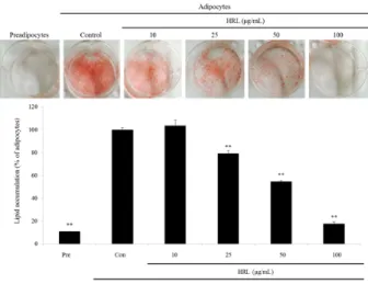
Fig. 1. Effects of HRL (extract of Hippophae rhamnoides L.
leaves) on cell viability. 3T3-L1 cells were treated with different concentrations (0 ~100 ㎍/㎖) of HRL for 24 h. Cell viability was measured by MTT assay. The viability of untreated control cells was defined as 100%. Each bar represents the mean ±
SEM (n = 3). * p < 0.05, ** p < 0.01 compared to control cells. Fig. 2. Effect of the HRL (extract of Hippophae rhamnoides L.
leaves) on the lipid accumulation in 3T3-L1 cells. Differentiation of confluent 3T3-L1 cells was initiated in DMEM containing MDI (0.5 mM IBMX, 1 μM DEX and 10 ㎍/㎖ insulin).
Following 10-day differentiation, differentiated adipocytes were fixed and stained with oil-red O in order visualize lipid droplets. Pre, preadipocyte; Con, differentiated adipocyte.
Each bar represents the mean ± SEM (n = 3). * p < 0.05, ** p <
0.01 compared to differentiated adipocyte (Con).
물의 경우에는 100 ㎍/㎖ 농도에서 89.3%의 생존율을 나타내었 으며(Lee et al., 2014), 두메부추 추출물의 100 ㎍/㎖ 농도에서 독성을 나타내지 않아(Choi and Kim, 2014a) 항비만제 및 원료 개발에 있어서 유효한 물질의 가능성을 나타내었다. 따라서 본 연구는 세포증식에 크게 영향을 미치지 않는 100 ㎍/㎖ 이하의 농도 10, 25, 50, 100 ㎍/㎖로 3T3-L1 세포에 처리하여 지방세 포 분화 억제능을 확인하였다.
중성지방 축적 억제 효과
Oil Red O 염색시약은 중성지질, 콜레스테롤만을 염색하고, 세포 내 축적된 지방구의 중성지방을 염색하여 세포의 붉은색 정도를 통해 분화 정도를 확인할 수 있다(Choi et al., 2013). 따 라서 3T3-L1 세포 분화 과정에서 HRL이 지방구 생성을 억제하 는지 확인하기 위해 Oil Red O 염색법을 이용하였다. 분화처리 군(Con)의 경우, 세포질 내 지방구의 형성이 활발하게 유도되는 것을 확인하였고, HRL을 10, 25, 50, 100 ㎍/㎖ 로 처리하였을 때 농도 의존적으로 붉은색이 적게 관찰되어 지질 축적이 감소 함을 확인하였다(Fig. 2). 이를 정량 분석한 결과, 분화처리군 (Con)에 비해 HRL 25, 50, 100 ㎍/㎖ 처리군이 각각 20.76, 45.29 및 82.25%로 중성지방 축적을 억제하는 것으로 나타났다 (Fig. 2). 이는 Yoon et al. (2010)이 보고한 목향추출물의 경우 100 ㎍/㎖에서 약 10% 축적 억제를 나타낸 연구결과와 비교하였 을 때 HRL의 우수한 지방분화능 억제 효과를 관찰할 수 있었다.
이러한 결과는 비타민나무에서 존재하는 quercetin, kaempferol 및 isorhamnetin 등의 flavonoid 성분의 영향으로 생각되며, Yang et al. (2013)의 보고에 따르면 비타민나무에서 분리된
quercetin, kamepferol 및 isorhamnetin 등의 성분이 각각 45.6, 42.2 및 44.3% (30 μM)의 중성지방 축적 억제효과를 나타 내는 것을 확인하였다. 이와 같은 결과로 HRL 처리가 지방구의 생성을 저해시켜 지방 축적을 억제하는 효과가 있음을 확인하 였다.
RT-PCR을 이용한 mRNA 발현량 측정
Adipogenesis는 지방전구세포가 지방세포로 분화되는 과정
으로, 이를 직간접적으로 조절하는 유전자 및 단백질들이 최근
다양한 연구로 인해 잘 알려져 있으며(White and Stephens,
2010), SREBP-1c, C/EBP family, PPARγ 등이 있다. SREBP-1c는
지방산이나 콜레스테롤 합성에 필수 전사인자이고(Park, 2005),
지방세포에서 PPARγ와 C/EBPα의 발현을 유도하여 지방생성
을 촉진시키며, 지방산 생성에 관여하는 효소인 FAS와 ACC 등의
발현을 조절한다(Fajas et al., 1999). PPARγ는 adipogenesis
과정에서 aP2 promoter 위치에 결합하여 adipogenesis를 조절
하고, C/EBPα는 PPARγ와의 강한 상승작용을 통해 지방전구세
포의 분화후기 과정을 촉진한다(Hauser et al., 2000; Darlington
et al., 1998). 따라서 본 연구에서는 HRL에 의한 지방전구세포
의 분화억제 효과를 확인하기 위하여 adipogenic transcription
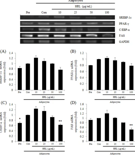
Fig. 3. Effects of HRL (extract of Hippophae rhamnoides L. leaves) on adipogenic genes expression in 3T3-L1 cells. Differentiation of confluent 3T3-L1 cells was initiated in DMEM containing MDI (0.5 mM IBMX, 1 μM DEX and 10 ㎍/㎖ insulin). Total RNA was extracted and cDNA was prepared. Equivalent amounts of cDNA were amplified using primers specific for SREBP-1c (A), PPAR- γ (B), C/EBP-α (C), FAS (D), and GAPDH. Pre, preadipocyte; Con, differentiated adipocyte. Each bar represents the mean
± SEM (n = 3) * p < 0.05, ** p < 0.01 compared to differentiated adipocyte (Con).
factor 및 관련 유전자들의 발현을 RT-PCR을 이용하여 확인하 였다(Fig. 3). 그 결과 분화를 유도한 대조군 그룹(Con)에서 SREBP-1c, PPARγ, C/EBPα 및 FAS의 mRNA발현이 증가하였 으나, 분화과정에서 HRL을 처리하여 분화를 유도한 경우 SREBP-1c, PPARγ, C/EBPα 및 FAS의 mRNA 발현이 모두 농 도 의존적으로 감소하였음을 확인하였다(Fig. 3). 특히, HRL 100 ㎍/㎖의 농도에서 SREBP-1c, PPARγ 및 C/EBPα는 분화처 리군(Con) 대비, 각각 0.87, 0.91 및 0.35배 감소하였으며(Fig.
3), 이들 전사인자들의 하위 인자인 FAS의 mRNA 발현도 0.49
배 감소하는 것을 확인하였다(Fig. 3D). 이전의 연구는 비타민
나무 잎 에탄올 추출물이 PPARα 및 CPT-1의 발현 증가에 영향
을 미쳐 지방산 베타-산화를 촉진시킴으로써 항비만 효과를 나
타낸다고 보고된 바 있고(Pichiah et al., 2012), 덖음차 추출물
식이그룹에서 PAP, β-oxidation 및 CTP의 활성을 저해시켜
fatty acid esterification pathway를 통한 혈당강하 효과를 나
타낸다고 보고된 바 있으나(Lee et al., 2011), 본 연구에서는 비
타민나무 잎 에탄올 추출물이 SREBP-1c, PPARγ, C/EBPα 및
FAS의 발현을 감소시켜 지방 합성이 억제되는 것을 확인할 수
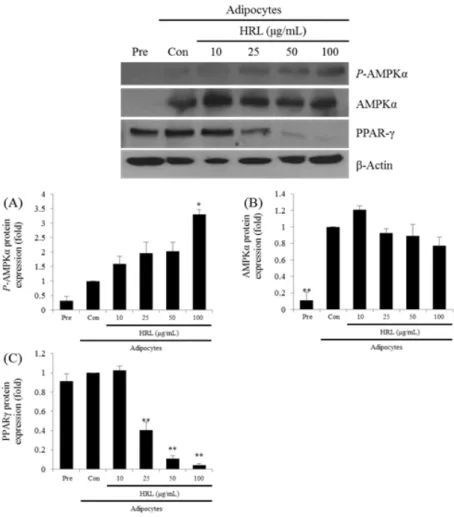
Fig. 4. Effects of HRL (extract of Hippophae rhamnoides L. leaves) on adipogenic protein expression in 3T3-L1 cells.
Differentiation of confluent 3T3-L1 cells was initiated in DMEM containing MDI (0.5 mM IBMX, 1 μM DEX and 10 ㎍/㎖ insulin).
Western blot was performed using P-AMPK α (A), AMPKα (B), PPARγ (C), and β-actin. Pre, preadipocyte; Con, differentiated adipocyte. Each bar represents the mean ± SEM (n = 3). * p < 0.05, ** p < 0.01 compared to differentiated adipocyte (Con).
있었다. Park et al. (2013)에 의하면 지방세포 분화는 adipogenic transcription factor와 지방 형성 관련 효소의 상호작용으로 조 절되고 SREBP-1c, PPARγ 및 C/EBPα의 발현 억제로 지방세포 의 분화를 억제한다고 보고된 바 있으며 이 외에도 본 연구결과 는 adipogenic transcription factor의 발현 억제를 통한 지방세 포 분화 억제를 제시한 Kim et al. (2014), Hwang et al. (2014) 및 Choi and Kim (2014b)의 보고와 일치한다. 따라서 HRL은 3T3-L1 지방전구세포에서 adipogenic transcription factor인 SREBP-1c의 발현을 억제시킴으로써 SREBP-1c의 타겟유전자 이며 지방형성 과정에 주요 인자인 PPARγ와 C/EBPα의 발현을 억제하고 지질의 합성 수송, 저장에 관여하는 FAS의 발현에 영 향을 미쳐 지방분화를 억제를 이끌어 세포 내 중성지방의 축적 이 감소되는 것으로 사료된다.
Western blot을 이용한 단백질 발현량 측정
HRL의 지방 생성 및 분해와 관련된 단백질 발현과의 연관성
을 확인하기 위해 western blot을 이용하여 AMPKα, PPARγ의
발현량을 측정하였다(Fig. 4). AMPK는 세포 내의 에너지 항상
성 유지 역할을 하는 효소로 지방의 대사조절에 중요한 역할을
한다. AMPK는 AMP가 증가되면 인산화를 통해 활성화되고 지
방산 산화를 증가시키며 지방 합성을 억제한다. AMPK는 활성
화 되어 SREBP-1c, PPARγ 및 FAS와 같은 지방세포화 인자의
발현을 억제함으로써 지방 합성을 억제시키고 ACC의 비활성화
와 CPT-1의 활성화를 통해 베타-산화를 촉진시켜 미토콘드리
아로 지방산 이동을 증가시킨다(Hardie, 2003; Foretz et al.,
1998; Fryer, 2002). HRL을 농도별로 처리한 결과, AMPK 및
PPARγ의 발현이 유의성 있는 변화를 나타냈고, P-AMPK의 발
현은 100 ㎍/㎖의 농도에서 3.5배 증가되는 것을 확인하였으며
PPARγ의 발현은 0.1배 감소되는 것을 확인하였다. 이러한 결과 는 비타민나무 잎에 존재하는 quercetin의 영향으로 생각된다.
Ahn et al. (2008)의 연구에 따르면 quercetin이 AMPK의 발현 을 증가시켜 adipogenesis를 억제시키고, PPARγ, C/EBPα, SREBP-1 및 FAS의 발현 감소로 지방전구세포에서 지방세포로 의 분화를 억제시킨다고 보고된 바 있다. 따라서 HRL은 활성화 된 AMPK의 발현을 증가시킴으로써, 지방형성 과정에 주요 인 자인 PPARγ의 발현에 영향을 준 것으로 생각되며, 이는 3T3-L1 지방전구세포에서 지방세포로의 분화 억제를 이끌어 세포 내 중성지방의 축적이 감소되는 것으로 판단된다.
적 요
본 연구는 HRL의 3T3-L1 지방전구세포의 분화과정 중에 HRL이 지방의 축적에 미치는 영향을 확인하였다. MTT assay 를 이용하여 세포 독성을 측정한 결과 100 ㎍/㎖의 농도에서도 세포증식에 영향을 미치지 않는 것을 확인하였고, 이와 같은 결 과를 토대로 Oil Red O 염색법을 이용하여 지방세포 분화 억제 능을 측정하였다. 그 결과, HRL의 경우 100 ㎍/㎖의 농도에서 82.25% 지방 축적 억제능을 나타내었다. 지방생성에 영향을 미 치는 유전자 발현량을 측정하기 위해 RT-PCR법과 western blot법을 시행하였다. HRL은 SREBP-1c, PPARγ와 C/EBPα의 mRNA 발현을 억제시켰고, 지방생성에 영향을 미치는 효소인 FAS의 생성을 조절하는 것으로 나타났다. 또한, HRL 처리로 AMPKα의 단백질 발현이 증가하였으며, PPARγ의 발현량이 감 소하는 것을 확인하였다. 이상의 결과들로부터 HRL은 AMPKα 의 활성화를 통한 지방 합성을 억제를 보유하고 있는 바, 향후 항비만 기능성 소재로 활용될 수 있을 것으로 생각한다.
사 사
본 연구는 산업통상자원부와 한국산업기술진흥원의 지역특 화산업육성사업(과제번호 R0002416)과 농촌진흥청 공동연구 어젠다사업(과제번호 PJ009859)의 지원으로 수행된 연구결과 입니다.
References