Inhibitory Effects of Loranthus Parasiticus Extract on Carbohydrate Digestive Enzymes and Postprandial Hyperglycemia
Min-Jung Park
1, Jae-Eun Park
2and Ji-Sook Han
2*
1Department of Food Science and Nutrition, Dongseo University, Busan 47011, Korea
2Department of Food Science and Nutrition, Pusan National University, Busan 46241, Korea Received October 30, 2019 /Revised December 11, 2019 /Accepted December 30, 2019
This study was designed to investigate whether Loranthus parasiticus extract (LPE) could inhibit the activities of carbohydrate digestive enzymes and alleviate postprandial hyperglycemia in diabetic mice. Lyophilized L. parasiticus was extracted with 80% ethanol and concentrated. The inhibitory ef- fects of LPE on carbohydrate digestive enzymes were evaluated by examining α-glucosidase and α- amylase, and it was seen to inhibit the activities of both enzymes in a dose-dependent manner. More specifically, the IC50 values of LPE against α-glucosidase and α-amylase were 0.121±0.007 and 0.157±
0.004 mg/ml, respectively, significantly lower than those of acarbose, showing that LPE has stronger inhibitory effects than the positive control. These results suggest that LPE strongly inhibits the activ- ities of these digestive enzymes. Blood glucose levels in the control group of diabetic mice increased to 490.00±28.52 mg/dl and 474.60±25.30 mg/dl at 60 and 120 min after a meal, respectively. However, when LPE was added to starch, postprandial blood glucose levels were significantly reduced (463.0±23.73 and 418.5±24.50 mg/dl at 60 and 120 min, respectively; p<0.05). The area under the curve also significantly decreased following administration of LPE, with no cytotoxicity. These results there- fore indicate that LPE could be used as an α-glucosidase and α-amylase inhibitor and delay carbohy- drate digestion and, thus, glucose absorption after a meal.
Key words : α-amylase, diabetic mice, α-glucosidase, Loranthus parasiticus extract, postprandial hyperglycemia
*Corresponding author
*Tel : +82-51-510-2836, Fax : +82-51-583-3648 E-mail : [email protected]
This is an Open-Access article distributed under the terms of the Creative Commons Attribution Non-Commercial License (http://creativecommons.org/licenses/by-nc/3.0) which permits unrestricted non-commercial use, distribution, and reproduction in any medium, provided the original work is properly cited.
Journal of Life Science 2020 Vol. 30. No. 1. 18~25 DOI : https://doi.org/10.5352/JLS.2020.30.1.18
Introduction
Diabetes is increasing the global health burden due to various complications [30]. An acute increase in post- prandial blood glucose is a direct and indirect acute toxicity to the vasculature, which can lead to complications of diabetes. To achieve proper glycemic control, it is necessary to reduce postprandial hyperglycemia. Various epidemio- logical studies have suggested an association between post- prandial blood sugar fluctuations and diabetes complica- tions [4].
One of the treatments for suppressing postprandial hyper- glycemia is to delay glucose absorption through inhibition of carbohydrate hydrolyzing enzymes [29]. α-Amylase and α-glucosidase were the two main hydrolytic enzymes [27].
α-Glucosidase breaks down the byproduct of starch into
glucose. Inhibitors of this enzyme are widely used for the regulation of blood glucose levels in type 2 diabetes. α- Glucosidase inhibitors block the membrane-bound intestinal α-glucosidases which hydrolyzes carbohydrates into glucose in the small intestine. Pancreatic α-amylase breaks down car- bohydrates, producing oligosaccharide, maltotriose and maltose. α-Amylase inhibitor is also considered as important factor in the development of antidiabetic drugs [34]. Saliva and pancreatic α-amylase inhibitors may inhibit post- prandial hyperglycemia by reducing the rate of digestion of carbohydrates [31].
Recent studies indicated that modulation of post prandial
blood glucose played an important role in the long term gly-
cemic control and complication prevention [9]. The most ef-
fective oral glucose-lowering drug on the market is acarbose,
which has been widely used in clinical practice as a drug
to inhibit α-glycosidase activity [25, 36]. Although acarbose
can effectively reduce the increase of postprandial blood glu-
cose, the adverse side effects are appeared simultaneously,
such as diarrhea, abdominal cramping, flatulence, and liver
disorders [14, 37]. Thus, using natural products, such as
plant extracts, to reduce hyperglycemia without causing side
effects may be a promising approach.
Loranthus parasiticus is mistletoe parasitic on mulberry.
Mistletoe, a semi-parasitic plant, is widely distributed throughout the world and has been used as an ingredient in Northeast Asian traditional medicine for centuries [16].
Loranthus parasiticus are mainly used for traditional medicine in Korea [12]. It has various beneficial effects, such as anti- cancer, antioxidant, anti-obesity, anti-inflammatory and neu- roprotective activity [19]. These effects of Loranthus para- siticus are associated with various biologically active com- pounds, including lectins, biscotoxins, phenolic compounds, sesquiterpenes lactones, triterpenes and flavonoids [39].
Nonetheless, there are few studies on the inhibitory effect of L. parasiticus on α-amylase and α-glucosidase and the reg- ulation of postprandial hyperglycemia in diabetes. There- fore, this study was conducted to determine whether L. para- siticus extract (LPE) inhibits α-amylase and α-glucosidase ac- tivities in vitro and suppresses postprandial hyperglycemia in diabetic mice in vivo.
Materials and Methods
Material and preparation of L. parasiticus extract L. parasiticus was collected from Yeongcheon, Gyeongbuk, Korea. The sample was washed with fresh water, and then freeze-dried. The lyophilized sample was homogenized with a grinder prior to extraction. The sample was extracted three times with ten volumes of 80% ethanol for 12 hr at room temperature. The L. parasiticus extract (LPE) was then evapo- rated at 40
oC using a rotary evaporator (N-1300VW, EYELA, Tokyo, Japan). After the solvent had been completely re- moved from the LPE, it was stored in a deep freezer (-80℃).
Inhibition assay for α-glucosidase activity in vitro The α-glucosidase inhibitory activity assays were carried out using readily available yeast enzymes, using the method of Watanabe et al. [35]. Yeast α-glucosidase (0.7 U, Sigma- Aldrich, St. Louis, MO, USA) was dissolved in 100 mM phos- phate buffer (pH 7.0) containing 2 g/l of bovine serum albu- min and 0.2 g/l of NaN
3and used as the enzyme test solution. Five mM p-nitrophenyl-α-D-glucopyranoside in the same buffer (pH 7.0) was used as the substrate solution. 10 μl of LPE [5 mg/ml in dimethyl sulfoxide (DMSO)] and 50 μl of enzyme solution were mixed in a well, and the absorb- ance at 405 nm was measured as time zero using a micro- plate reader. After incubation for 5 min, the substrate sol- ution (50 μl) was added, and the incubation continued for
another 5 min at room temperature. The increase in absorb- ance from the zero time point was then measured. The in- hibitory activities of varying concentrations of L. parasiticus were expressed as 100 minus the absorbance difference (%) of the test compounds relative to the absorbance change of the negative control (i.e., DMSO used as the test solution).
The measurements were performed in triplicate, and the IC
50value (i.e., the concentration of LPE that results in 50% in- hibition of maximal activity) was determined.
Inhibition assay for α-amylase activity in vitro The α-amylase inhibitory activity was analyzed in the same manner as α-glucosidase inhibition measuring method [35], except that porcine pancreatic amylase (100 U, Sigma- Aldrich, St. Louis, MO, USA) and p-nitrophenyl-α-D-malto- pentoglycoside (Sigma-Aldrich Co.) were used as the en- zyme and substrate, respectively.
Measurement of cytotoxicity
Cytotoxic cell viability was measured using the 3-(4,5-di- methylthiazol-2-yl)-2,5-diphenyltetrazolium bromide (MTT) assay, and 3T3-L1 cells were purchased from the Korean Cell Line Bank (Seoul, Korea). 3T3-L1 cells were seeded at 1×10
4cells/well in 96-well plates and pre-incubated in a humidi- fied atmosphere containing 5% CO
2at 37℃ for 24 hr. After- ward, the cells were treated with various concentrations (0.1, 0.5, 1, and 2 mg/ml) of LPE, and incubated for 20 hr. After completion of the treatment, the cells were incubated for 3 h at 37℃ with filtered MTT (Sigma-Aldrich, St. Louis, MO, USA) solution, which was added to each well at a final con- centration of 0.5 mg of MTT/ml. The supernatants were carefully aspirated, 200 μl of DMSO was added to each well, and the plates were agitated to dissolve the crystal product.
The absorbance of DMSO solution was measured spec- trophotometrically at 540 nm.
Experimental animals
Four-week-old male mice (ICR, Orient, Inc., Seoul, Korea)
were individually housed in a temperature control room
(25-30℃) with 45-55% relative humidity. Animals were ran-
domly given pellet food and tap water. After two weeks
adjustment period, streptozotocin [STZ; 60 mg/kg body
weight (b.w)] and freshly dissolved in citrate buffer (0.1M,
pH 4.5) [38]. And after 7 days, tail bleeds were performed
and animals with a blood glucose concentration above 250
mg/dL were considered to be diabetic. Mouse handling and
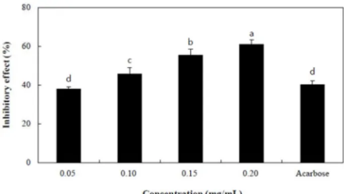
Fig. 1. α-Glucosidase inhibitory effects of L. parasiticus extract (LPE). Each value is expressed as mean ± SD in triplicate experiments. Values with different letters (a-d) are sig- nificantly different at p<0.05 as analyzed by Duncan’s multiple range test. The concentration of acarbose used as a positive control was 0.10 mg/ml.
Fig. 2. α-Amylase inhibitory effects of L. parasiticus extract (LPE).
Each value is expressed as mean ± SD in triplicate experiments. Values with different letters (a-d) are sig- nificantly different at p<0.05 as analyzed by Duncan’s multiple range test. The concentration of acarbose used as a positive control was 0.10 mg/ml.
care procedures have complied with the guidelines (NIH Guide for the Care and Use of Laboratory Animals) in com- pliance with current international laws and policies, and all procedures have been approved by the Pusan National University Animal Ethics Committee.-2018-1823.
Measurement of blood glucose levels
Both normal and STZ-induced diabetic mice were fasted overnight and randomly divided into three groups of 7 mice.
Before testing blood glucose levels, the animals were kept on an empty stomach for at least 12 hr but had free access to water. Mice were orally administered as follows: control group, mice were orally administered with starch (2 g/kg b.w); LPE, orally administered starch LPE to mice (300 mg/
kg b.w); acarbose, mice received acarbose orally adminis- tered with starch (100 mg/kg b.w). Blood samples were tak- en from the tail vein at 0, 30, 60, and 120 min. Blood glucose was measured using a glucometer (Roche Diagnostics GmbH, Mannheim, Germany). The areas under the curve (AUC) were calculated using the trapezoidal rule [17].
Data statistical analysis
Statistical analysis was performed using SAS version 9.1 (SAS Institute, Inc., Cary, NC, USA). Student's t-test was used for comparison between control and treatment groups.
Differences were assessed with one-way ANOVA followed by Duncan's multi-range test (p<0.05). Data are displayed as mean ± standard deviation (SD).
Results and Discussion
Inhibitory effect of LPE on α-glucosidase and α- amylase in vitro
The inhibitory effect of LPE on α-glucosidase activity was measured using p-nitrophenyl-α-D-glucopyranoside as a substrate and compared with the effect of acarbose, a com- mercial α-glucosidase inhibitor used as an hyperglycemic agent. LPE inhibited α-glucosidase activity in a dose-de- pendent manner by 38.11±1.09, 45.87±2.98, 55.60±2.84, and 61.12±2.15% at concentrations of 0.05, 0.10, 0.15, and 0.20 mg/mL, respectively (Fig. 1). LPE inhibited the enzyme ac- tivity by 40.38±1.81% at a concentration of 0.10 mg/dl. The α-glucosidase inhibitory activity of LPE was significantly higher than that of acarbose at the same concentration.
As shown in Fig. 2, the inhibitory effects of LPE on α- amylase were increased in a dose-dependent manner by
25.69±1.40, 38.42±2.74, 47.38±2.70, and 65.97±3.21% at con- centrations of 0.05, 0.10, 0.15, and 0.20 mg/ml, respectively.
LPE also inhibited α-amylase activity more effectively than acarbose. The IC
50values of LPE against α-glucosidase and α-amylase were 0.121±0.007 and 0.157±0.004 mg/ml, re- spectively. Its IC
50values against α-glucosidase and α-amy- lase were significantly lower than those of acarbose, indicat- ing that LPE has stronger inhibitory effects than the positive control (Table 1).
Inhibitions of α-amylase and α-glucosidase were im-
portant factors for managing postprandial blood glucose in
patients with type 2 diabetes [8]. This study investigated the
inhibitory effects of the natural product, LPE, against α-
amylase and α-glucosidase to uncover potential as a post-
prandial hyperglycemic inhibitor. LPE provided significantly
Table 1. IC50 values of the inhibitory effect of L. parasiticus ex- tract (LPE) against α-glucosidase and α-amylase activ- ities
IC50 (mg/ml)1)
α-glucosidase α-amylase LPE
Acabose
0.121±0.007*
0.130±0.008
0.157±0.004*
0.165±0.006 Each value is expressed as mean ± SD in triplicate experiments.
*Significantly different from acarbose at p<0.05.
1)IC50 value is the concentration of sample required for 50%
inhibition.
Fig. 3. Cytotoxic effects of L. parasiticus extract (LPE) in 3T3-L1 cells. 3T3-L1 cells were treated with various concen- trations (0.1, 0.2, 0.5, 1.0 and 2.0 mg/ml) of LPE for 20 hr, and cell viability was measured by MTT assay. Each value is expressed as the mean ± standard deviation (SD) of three experiments. NS: Not-significant.
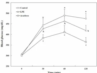
Fig. 4. Blood glucose levels after the administration of L. para- siticus extract (LPE) in streptozotocin-induced diabetic mice. Each value is expressed as mean ± SD of seven mice. Values with different letters (a-c) are significantly different at each time (p<0.05) as analyzed by Duncan’s multiple range test. Control, mice received starch orally (2 g/kg); LPE, mice received starch with Loranthus para- siticus extract orally (300 mg/kg); Acarbose, mice re- ceived starch with acarbose orally (100 mg/kg).
higher inhibitory activities against both α-amylase and α -glucosidase than acarbose, the commercial inhibitor. It also did not show any cytotoxicity (Fig. 3). The inhibitory effects of LPE on these enzymes would be attributed to the active ingredients in Loranthus parasiticus.
Loranthus parasiticus contained total phenolic compounds, flavonoids, triterpene and sesquiterpene lactones, etc. [19].
Several studies have reported the anti-diabetic effects of tri- terpenes and triterpenes-containing plant extracts [23].
Flavonoids, especially quercetin and camphorol, have been also shown to exhibit α-glucosidase inhibitory activity [18, 26]. Several beneficial flavonoids exhibited impressive hypo- glycemic effects, with significant improvement, without pro- ducing health hazards [21]. These flavonoids showed α-glu- cosidase inhibitory effect due to galloyl group and phenolic hydroxyl group, which was caused by the formation of com- plexe with the enzyme [11]. The flavonoids exerted their α- glucosidase inhibitory activities by forming complex with the enzyme through non-covalent interactions in the intes-
tine [36]. As a result of this study, LPE had inhibitory effect on α-glucosidase, which might be due to the active in- gredients contained in LP, such as flavonoids and triterpenes.
α-Amylase hydrolyses α-linked polysaccharides such as starch and glycogen. α-Amylase inhibitors block the hydrol- ysis of complex starch into oligosaccharides, reducing the rate of digestion of carbohydrates and consequently less glu- cose absorption [33]. The flavonoids such as luteolin, myr- icetin, and quercetin were potent inhibitors of α-amylase, their inhibition activities on the enzyme were related to the functional group, such as 2,3-double bond, 5-OH and the linkage of the B ring at the 3-position in the compounds [32]. LPE was known to possess a high quantity of fla- vonoids [5]. It exhibited α-amylase inhibitory effect and es- pecially the higher inhibitory effect than acarbose. Thus, the high inhibitory effects of LPE on α-glucosidase and α-amy- lase activities might be attributable to the high content of flavonoids in it.
Effects of LPE on blood glucose levels in vivo The effects of LPE on blood glucose levels after a meal were investigated in normal and STZ-induced diabetic mice.
The postprandial blood glucose levels of the LPE adminis-
tered mice were significantly lower than those of the diabetic
mice (Fig. 4). Blood glucose levels in the diabetic mice in-
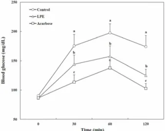
Fig. 5. Blood glucose levels after the administration of L. para- siticus extract (LPE) in normal mice. Each value is ex- pressed as mean ± SD of seven mice. Values with differ- ent letters (a-c) are significantly different at each time (p<0.05) as analyzed by Duncan’s multiple range test.
Control, mice received starch orally (2 g/kg); LPE, mice received starch with Loranthus parasiticus extract orally (300 mg/kg); Acarbose, mice received starch with acar- bose orally (100 mg/kg).
Table 2. Areas under the curve (AUC) of postprandial glucose responses in normal and streptozotocin-induced dia- betic mice
Group1) AUC (mg.h/dl)
Normal mice Diabetic mice Control
LPE Acabose
345.50±35.65a 274.05±30.35b 208.30±27.99c
901.65±56.58a 846.87±43.48b 756.65±44.96c
1)Distilled water (Control), LPE (300 mg/kg), or acarbose (100 mg/kg) was coadministered orally with starch (2 g/kg). Each value is expressed as the mean ± SD of seven mice (n=42).
a-cValues with different alphabets are significantly different at p<0.05, as analyzed by Duncan's multiple-range test. LPE:
Loranthus parasiticus extract.
creased to 443.61±31.21 mg/dl at 30 min and 490.00±28.52 mg/dl at 60 min after a meal, and then decreased to 474.60
±25.30 mg/dl at 120 min. However, when LPE was added to starch, the increases in postprandial blood glucose levels were significantly suppressed (426.75±19.80, 463.02±23.73, and 418.51±24.50 mg/dl at 30, 60, and 120 min, respectively;
p<0.05). The peak postprandial blood glucose levels also sig- nificantly decreased when the normal mice were orally ad- ministered starch with LPE (Fig. 5). The AUC for the glucose response in diabetic mice administered LPE (846.87±43.48 mg・hr/dl) was significantly lower (p<0.05) than that in dia- betic mice (901.65±56.58 mg・h/dl) (Table 2).
Postprandial hyperglycemia and fasting blood glucose control are very important in patients with type 2 diabetes.
Postprandial hyperglycemia was reported to have a stronger correlation with morbidity of diabetes complications such as cardiovascular disease than fasting hyperglycemia [22].
It was associated with glycemic variability and has been sug- gested that postprandial hyperglycemic fluctuations could contribute to the development of diabetes complications [7].
Reducing postprandial hyperglycemia is one of the im- portant diabetic treatments, which delays the absorption of glucose through the inhibition of carbohydrate hydrolase, such as α-amylase and α-glucosidase in the digestive organs.
Because α-amylase is involved in the breakdown of long chain carbohydrates, and α-glucosidase breaks down dis- accharides to glucose. In this study, LPE showed sig- nificantly higher inhibitory activities against both α-amylase and α-glucosidase than acarbose, the commercial inhibitor.
Reduction in postprandial hyperglycemia of diabetic mice treated with LPE might be due to inhibition of these enzymes. The reduction effect on postprandial hyper- glycemia of LPE was also observed in normal mice. These confirmed that LPE could inhibit the action of carbohydrate digestive enzymes and delay the absorption of glucose.
Loranthus parasiticus contained bioactive ingredients such as flavonoids, phenolic compounds, triterpene and sesqui- terpene lactones [19]. Flavonoids were demonstrated to act on biological target of type 2 diabetes such as α-glycosidase [24]. Administration of naringenin, a kind of flavonoids, pre- vented a sharp rise in blood glucose levels of diabetic rats loaded with maltose and sucrose compared to control rats.
This showed that the mechanism of action of flavonoid was associated with α-glucosidase inhibition in the intestine, thereby delaying glucose release [28]. Flavonoids also im- proved hyperglycemia in patients with type 2 diabetes [11].
Natural sesquiterpene lactones have also been reported to
attenuate hyperglycemia in streptozotocin (STZ) induced di-
abetic rats [3]. In vitro α-amylase inhibition assay showed
that sesquiterpene lactones had potent intestinal α-amylase
inhibitory activity, which has the ability to reduce starch-in-
duced postprandial blood glucose [1]. One of the anti-dia-
betic mechanisms of the triterpenes was their inhibitions
against α-amylase and α-glucosidase [2, 10]. Additionally,
some triterpenes significantly decreased the hyperglycemia
in diabetic rats by inhibiting small intestinal α-amylase, su-
crase and α-glucosidase activity [15]. The results of this
study showed that LPE could delay the digestion and ab- sorption of dietary carbohydrates in the intestine, which could suppress the rise in blood glucose levels after meals in diabetic mice. The suppression effect on postprandial hy- perglycemia of LPE was thought to be due to the active in- gredients contained in LPE, such as sesquiterpene lactones, triterpenes and flavonoids. In addition, various doses of ani- mal toxicity studies on LPE showed no mortality or morbid symptoms when administered orally up to 1,500 mg/kg [20].
Studies on the same plant leaves was administered orally up to and 827 mg/kg body weight, neither adverse bio- chemical changes nor mortality was detected [6]. While an- other study confirmed the safety of its up to 5,000 mg/kg [13]. In conclusion, LPE inhibited α-glucosidase and α-amy- lase activities and resulted in a reduction in postprandial hyperglycemia. LPE might delay the digestion and absorp- tion of dietary carbohydrates in the intestine, resulting in suppression of increased blood glucose levels after a meal.
Thus, this results suggest the possibility that LPE may be used as a natural anti-hyperglycemic food because of its in- hibitory effects on α-glucosidase and α-amylase without side effects. However, if LPE is used clinically for medical pur- poses, a special permit procedure is required, and research on intake as a functional food should be conducted in the future.
Acknowledgement
This work was supported by a 2-Year Research Grant of Pusan National University.
The Conflict of Interest Statement
The authors declare that they have no conflicts of interest with the contents of this article.
References
1. Abd-Alla, H. I., Shalaby, N. M., Hamed, M. A., El-Rigal, N. S., Al-Ghamdi, S. N. and Bouajila, J. 2016. Phytochemical composition, protective and therapeutic effect on gastric ul- cer and α-amylase inhibitory activity of Achillea biebersteinii Afan. Arch. Pharm. Res. 39, 10-20.
2. Ali, H., Houghton, P. J. and Soumyanath, A. 2006. Alpha- Amylase inhibitory activity of some Malaysian plants used to treat diabetes; with particular reference to Phyllanthus amarus. J. Ethnopharmacol. 107, 449-455.
3. Basha, R. H. and Sankaranarayanan, C. 2016. β-Caryophyllene,
a natural sesquiterpene lactone attenuates hyperglycemia mediated oxidative and inflammatory stress in experimental diabetic rats. Chem. Biol. Interact. 5, 50-58.
4. Ceriello, A. 2005. Postprandial hyperglycemia and diabetes complications: is it time to treat?. Diabetes 54, 1-7.
5. Cui, H., Lu, T., Wang, M., Zou, X., Zhang, Y., Yang, X., Dong, Y. and Zhou, H. 2019. Flavonoids from Morus alba L. leaves:
optimization of extraction by response surface methodol- ogy and comprehensive evaluation of their antioxidant, an- timicrobial, and inhibition of α-amylase activities through analytical hierarchy process. Molecules 24, E2398.
6. Edem, D. O. and Usoh, I. F. 2009. Biochemical changes in wistar rats on oral doses of mistletoe (Loranthus micranthus).
Am. J. Pharmacol. Toxicol. 4, 94-97.
7. Haller, H. 1998. The clinical importance of postprandial glucose. Diabetes Res. Clin. Pract. 40, 43-49.
8. Hanefeld, M. 1998. The role of acarbose in the treatment of non-insulin-dependent diabetes mellitus. J. Diabetes Complications 12, 228-237.
9. Herath, H. M. M., Weerarathna, T. P., Fonseka, C. L. and Vidanagamage, A. S. 2017. Targeting postprandial blood sugar over fasting blood sugar: A clinic based comparative study. Diabetes Metab. Syndr. 11, 133-136.
10. Hou, W., Li, Y., Zhang, Q., Wei, X., Peng, A., Chen, L. and Wei, Y. 2009. Triterpene acids isolated from Lagerstroemia speciosaleaves as α-glucosidase inhibitors. Phytother. Res. 23, 614-618.
11. Huang, Q., Chai, W. M., Ma, Z. Y., Ou-Yang, C. and Peng, Y. Y. 2019. Inhibitionof α-glucosidase activity and non-enzy- matic glycation by tannicacid: Inhibitory activity and molec- ular mechanism. Int. J. Biol. Macromol. 141, 358-368.
12. Hwang, K., Kim, J., Choi, Y., Choj, K. and Park, K. 2011.
One of the Korean mistletoe species, Loranthus yadoriki Sieb. exhibited potent inhibitory activities against mono- amine oxidases. Planta Med. 77, DOI:10.1055/s-0031-1282451.
13. Iwalokun, B. A., Oyenuga, A. O., Saibu, G. M., Ayorinde, J., Lagos, Y. and Polytechnic, L. S. 2011. Analyses of cyto- toxic and genotoxic potentials of Loranthus micranthus using the Allium cepa test. J. Biol. Sci. 3, 459-467.
14. Jo, S. H., Cho, C. Y., Lee, J. Y., Ha, K. S., Kwon, Y. I. and Apostolidis, E. 2016. In vitro and in vivo reduction of post-prandial blood glucose levels by ethyl alcohol and wa- ter Zingiber mioga extracts through the inhibition of carbohy- drate hydrolyzing enzymes. BMC Complement Altern. Med.
16, 111-117.
15. Khathi, A., Serumula, M. R., Myburg, R. B., Van Heerden, F. R. and Musabayane, C. T. 2013. Effects of Syzygium ar- omaticum-derived triterpenes on postprandial blood glucose in streptozotocin-induced diabetic rats following carbohy- drate challenge. PLoS One 8, e81632.
16. Kim, K. W., Yang, S. H. and Kim, J. B. 2014. Protein fractions from korean mistletoe (Viscum Album coloratum) extract in- duce insulin secretion from pancreatic beta cells. Evid. Based Complement. Alternat. Med. 2014, 703624.
17. Kim, J. S. 2004. Effect of Rhemanniae Radix on the hyper- glycemic mice induced with streptozotocin. J. Kor. Soc. Food
Sci. Nutr. 33, 1133-1138.
18. Li, Y.Q., Zhou, F. C., Gao, F., Bian, J. S. and Shan, F. 2009.
Comparative evaluation of quercetin, isoquercetin and rutin as inhibitors of α-glucosidase. J. Agric. Food Chem. 57, 11463- 11468.
19. Moghadamtousi, S. Z., Kamarudin, M. N. A., Chan, C. K., Goh, B. H. and Kadir, H. A. 2014. Phytochemistry and biol- ogy of Loranthus parasiticus Merr, a commonly used herbal medicine. Am. J. Chin. Med. 42, 23-35.
20. Mothana, R. A. A., Al-Said, M. S., Al-Rehaily, A. J., Thabet, T. M., Awad, N. A., Lalk, M. and Lindequist, U. 2012.
Anti-inflammatory, antinociceptive, antipyretic and anti- oxidant activities and phenolic constituents from Loranthus regularis Steud. ex Sprague. Food Chem. 130, 344-349.
21. Mukhopadhyay, P. and Prajapat, A. K. 2015. Quercetin in anti-diabetic research and strategies for improved quercetin bioavailability using polymer-based carriers - a review. RSC Adv. 5, 97547-97562.
22. Nalysnyk, L., Hernández-Medina, M. and Krishnarajah, G.
2010. Glycemic variability and complications in patients with diabetes mellitus: evidence from a systematic review of the literature. Diabetes Obes. Metab. 12, 288-298.
23. Nazaruk, J. and Borzym-Kluczyk, M. 2015. The roleof tri- terpenes in the management of diabetes mellitus and its complications. Phytochem. Rev. 14, 675-690.
24. Nicolle, E., Souard, F., Faure, P. and Boumendjel, A. 2011.
Flavonoids as promising lead compounds in type 2 diabetes mellitus: molecules of interest and structure-activity rela- tionship. Curr. Med. Chem. 18, 2661-2672.
25. Park, M. H., Ju, J. W., Park, M. J. and Han, J. S. 2013.
Daidzein inhibits carbohydrate digestive enzymes in vitro and alleviates postprandial hyperglycemia in diabetic mice.
Eur. J. Pharmacol. 712, 48-52.
26. Peng, X., Zhang, G., Liao, Y. and Gong, D. 2016. Inhibitory kinetics and mechanism of kaempferol on α-glucosidase.
Food Chem. 190, 207-215.
27. Poovitha, S. and Parani, M. 2016. In vitro and in vivo α- amy- lase and α-glucosidase inhibiting activities of the protein ex- tracts from two varieties of bitter gourd (Momordica char- antia L.). BMC Complement Altern. Med. 16, 185.
28. Priscilla, D. H., Roy, D., Suresh, A., Kumar, V. and Thir- umurugan, K. 2014. Naringenin inhibits alpha-glucosidase activity: A promising strategy for the regulation of post-
prandial hyperglycemia in high fat diet fed streptozotocin induced diabetic rats. Chem. Biol. Interact. 210, 77-85.
29. Saito, N., Sakai, H., Suzuki, S., Sekihara, H. and Yajima, Y.
1998. Effect of an α-glucosidase inhibitor (voglibose), in combination with sulphonylureas, on glycaemic control in type 2 diabetes patients. J. Int. Med. Res. 26, 219-232.
30. Seaquist, E. R. 2014. Addressing the burden of diabetes.
JAMA. 311, 2267-2268.
31. Selvaraj, G., Kaliamurthi, S. and Thirugnanasambandam, R.
2015. Influence of Rhizophora apiculata Blume extracts on α- glucosidase: enzyme kinetics and molecular docking studies.
Biocatal. Agric. Biotechnol. 4, 653-660.
32. Tadera, K., Minami, Y., Takamatsu, K. and Matsuoka, T.
2007. Inhibition of α-glucosidase and α-amylase by flavonoids. J. Nutr. Sci. Vitaminol. 52, 149-153.
33. Thilagam, E., Parimaladevi, B., Kumarappan, C. and Mandal, S. C. 2013. α-glucosidase and α-amylase inhibitory activity of Senna surattensis. J. Acupunct. Meridian Stud. 6, 24-30.
34. Tucci, S. A., Boyland, E. J. and Halford, J. C. 2010. The role of lipid and carbohydrate digestive enzyme inhibitors in the management of obesity: a review of current and emerging therapeutic agents. Diabetes Metab. Syndr. Obes. 3, 125-143.
35. Watanabe, J., Kawabata, J., Kurihara, H. and Niki, R. 1997.
Isolation and identification of α-glucosidase inhibitors from tochucha (Eucommia ulmoides). Biosci. Biotechnol. Biochem. 61, 177-178.
36. Zhang, B. W., Li, X., Sun, W. L., Xing, Y., Xiu, Z. L., Zhuang, C. L. and Dong, Y. S. 2017. Dietary flavonoids and acarbose synergistically inhibit alpha-glucosidase and lower post- prandial blood glucose. J. Agric. Food Chem. 65, 8319-8330.
37. Zhang, B. W., Xing, Y., Wen, C., Yu, X. X., Sun, W. L., Xiu, Z. L. and Dong, Y. S. 2017. Pentacyclic triterpenes as α -glucosidase and α-amylase inhibitors: structure-activity re- lationships and the synergism with acarbose. Bioorg. Med.
Chem. Lett. 27, 5065-5070.
38. Zheng, J., He, J., Ji, B., Li, Y. and Zhang, X. 2007. Antihyper- glycemic activity of Prunella vulgaris L. in streptozotocin-in- duced diabetic mice. Asia. Pac. J. Clin. Nutr. 16, 427-431.
39. Zorofchian-Moghadamtousi, S., Hajrezaei, M., Abdul Kadir, H. and Zandi, K. 2013. Loranthus micranthus Linn.: biological activities and phytochemistry. Evid. Based Complement.
Alternat. Med. 2013, 273712.
초록:상기생( Loranthus parasiticus ) 추출물의 탄수화물 소화 효소 및 식후 고혈당 저해 효과
박민정
1․박재은
2․한지숙
2*
(1동서대학교 식품영양학과, 2부산대학교 식품영양학과)