Blood Concentration of Polycyclic Aromatic Hydrocarbons from Non- Occupational Exposure in farming towns near metropolitan Busan, Korea : Environmental Tobacco Smoke and Roasted Food Intake as Influential Factors
부산인근 농촌지역의 비직업적 노출에 의한 혈중 다환방향족탄화수소 농도: 간접흡연과 구운음식 섭취를 중심으로
연구목적: 본 연구는 일반인의 비직업적 노출에 대한 혈 중 16종의 다환방향족탄화수소(PAHs) 의 농도르 분석하 여 PAHs의 백그라운드 노출 농도를 평가하는 것이다.
내용 및 방법: 농촌지역에 거주(비직업적 노출로서 16종 다환방향족탄화수소가 특정한 환경오염을 받지 않은 지 역에 거주)하는 156명의 연구 참여자의 혈중 16가지 다환 방향족탄화수소 농도의 기하평균치를 간접흡연군, 구운 음식물섭취군과 각대조군을 비교 하였다. 혈중 16종의 다환방향족탄화수소의 농도는 질량분석기를 장착한 가 스크로마토그라피법과 헤드스페이스 마이크로고체상 추출법에 의하여 분석하였다.
결과: 연구참여자 중, 간접흡연자들에서 4종의 다환방향 족탄화수소 아세나프틸렌(p<0.01), 아세나프텐(p<0.1), 플
로렌(p<0.01), 피렌(p=0.05)가 대조군 보다 유의하게 높았 다. 또한 구운육류와 생선 섭취군에서는 벤조(a)피렌
(p<0.1)이 대조군 보다 유의하게 높았다.
결론: 간접흡연과 구운음식물 섭취는 비직업적 노출에 서 다환방향족탄화수소 노출원의 가능성이 높다고 추정 된다. 혈중 다환방향족탄화수소는 한국일반인의 비직업 적 노출 농도로 사용 가능하다고 생각된다.
문찬석1‡ㆍ조은미2ㆍ이채관3ㆍ김정만2ㆍ홍영습2ㆍ손부순4ㆍ백종민1
1부산가톨릭대학교 산업보건학과
2부산 동아대학교 예방의학교실
3부산 인제대학교 산업의학연구소
4순천향대학교 환경보건학과
Chan-Seok Moon
1‡ㆍEun Mi Jo2ㆍChae-Kwan Lee3ㆍJung Man Kim2ㆍYoung Seoub Hong2ㆍBu-Soon Son4ㆍJong-Min Paik11
Dept. of Industrial Health, College of Applied Science, Catholic University of Pusan, Busan, Korea
2
Dept. of Preventive Medicine, Dong-A University, Busan, Korea
3
Institute of Industrial Medicine, Inje University College of Medicine, Busan, Korea
4
Dept. of Environment Health Science, Soonchumhyang University, Chungnam, Korea
접수일 : 2010년 4월 21일, 채택일 : 2010년 6월 18일
‡ 교신저자 : 문찬석( 부산광역시 금정구 부곡3동 9번지 부산가톨릭대학교 산업보건학과,
Tel. : +82-51-510-0633 , Fax.: +82-51-510-0638, E-Mail: [email protected])
Non-occupational exposure, polycyclic aromatic hydrocarbons, blood, environmental tobacco smoking, roasted food intake
Key word :
Ⅰ. Introduction
Polycyclic aromatic hydrocarbons (PAHs) are widely-distributed and hazardous environmental pollutants that are discharged into the environment from the versatile sources such as motor vehicle emissions, fossil fuels, industrial plants. They are produced during the incomplete combustion of coal, gas, oil, and wood, and can also be found in tobacco smoke, charbroiled meats, and smoked foods (International Agency for Research on Cancer, 1985; International Agency for Research on Cancer, 1987). Industries in exposure to PAHs is likely to occur include those using coke ovens and coal tar, iron and steel works, aluminum works, foundries, carbon electrode and carbon black manufacture, asphalt manufacture and use, etc.
(Redmond et al., 1976; Palmer and Scott, 1981; Armstrong et al., 1986; NIEHs, 2000; Campo et al., 2006; Unwin et al., 2006; Buratti et al., 2007). Many of these PAHs shown carcinogenic activity, proven in animals, and are thought to have a similar impact on humans (Boffetta et al., 1997; International Agency for Research on Cancer, 1973; International Agency for Research on Cancer, 1983;
International Agency for Research on Cancer, 1984; International Agency for Research on Cancer, 1984a; National Toxicology Program, 2001). Background exposures to PAHs for the general population typically come from sources such as tobacco smoke or environmental tobacco smoke (ETS), charbroiled or smoked foods, and automobile exhaust fumes (WHO, 1984; Grimmer et al., 1987;
Lioy et al., 1988; Scherer et al., 1990; Scherer et al., 2000; Kim et al., 2001; Hu et al., 2006).
As representative biological exposure marker, urinary 1- hydroxypyrene (1-OHP) for environmental PAHs exposure has been somewhat limitation for the explanation of environmental PAHs exposure (WHO, 1984). Moreover, the marker is not mostly influenced by respiratory intake but by food intake and tobacco smoking among general population (Grimmer et al., 1987; Lioy et al., 1988; Scherer et al., 1990; Jongeneelen et al., 2001). In these sense, urinary 1-OHP is total exposure marker for pyrene exposure via respiration and food intake so that PAHs exposure sources are not clear whether exposure source is from ambient air or from food intake. More effective exposure markers than urinary 1-OHP are necessary to PAHs exposure via respiratory intake. Blood PAHs are, therefore, thought to more effective exposure marker than 1-OHP as urinary metabolite.
A venous blood sample is a most effective and sensitive biological matrix in cases of low-level exposure to volatile organic compounds (Kawai et al., 1992; Kawai et al., 1992a; Kawai et al., 1994; Kawai
et al., 1996; Ikeda, 1999). PAHs in blood have been used as exposure markers for long-term low-level exposure. However, few reports are available for the evaluation of the PAHs in blood for non- occupational exposure. Moreover, a large-scale national field survey (cohort study) for the health effects on residents living near industrial sites was conducted in six Korean industrial areas every year for 20 years, in order to clearify the exposure to PAHs from the combustion of coal, gas, oil etc. The detection of PAHs in blood is required a sensitive and rapid method to handle a large number of blood samples in a very short detection period. The present study was initiated to examine the background levels of US EPA 16 PAHs (naphthalene, acenaphthylene, acenaphthene, fluorene, phenanthrene, anthracene, fluoranthene, pyrene, benzo(a)anthracene, chrysene, benzo(b)fluoranthene, benzo(k)fluoranthene, benzo(a)pyrene, indeno(1,2,3-cd)pyrene, dibenz(a,h)anthracene, benzo(g,h,i)perylene) in venous blood samples. The blood concentration of 16 PAHs samples were compared exposed group to ETS and nonsmoker group, and group of roasted food intake with non-intake group of these foods.
Ⅱ. Materials and Methods
1
1.. S Su urrvve eyy p pa arrttiic ciip pa an ntts s a an nd d s sttu ud dyy d de es siig gn n
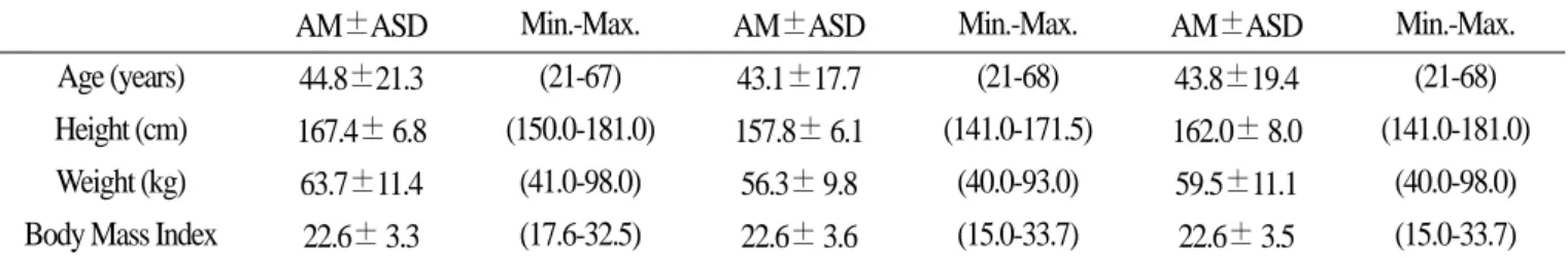
This survey was conducted from February to April 2008 in several farming towns near metropolitan Busan, Korea, where there is no known industrial pollution from PAHs. The participants were 158 healthy non-smoking participants (68 man and 90 women), with an age range of 21-68 years (mean ± SD, 43.8±19.4), (Table 1).
The subjects were divided into subgroups of nonsmokers exposed to ETS and non-smoker, or intake of roasted meats or fish and non- intake of these foods. Then, significant increases in 16 PAH concentrations in blood samples were evaluated.
In selecting the group of nonsmokers exposed to ETS, the
participants were asked to describe their exposure to ETS in terms of
minutes per day, and were accepted if they had an ETS exposure of
up to 1 minute a day. Roasted meats or fish involved charbroiled,
briquette-roasted, butane gas-roasted, city gas (LNG)-roasted or
electronic grill-roasted meats or fish. Moreover, the questionnaires
were fully explained the participants.
2
2.. S Sa am mp plle e c co olllle ec cttiio on n a an nd d a an na allyys siis s o off tth he e 1 16 6 P PA AH Hs s
Venous blood samples and questionnaires were collected from the participants with informed consents provided in writing.
Heparinized vacuum tubes for blood sampling (Vacutainer, Becton Dickinson, N.J., USA) were employed (Moon et al., 1995) with due caution to avoid the contamination of PAHs. Then, each blood sample was stored at -18 ℃ until it was analyzed.
PAHs in blood were analyzed using headspace-solid phase microextraction (CTC CombiPAL automated sample injectors for
gas chromatography, Agilent Technologies, Inc., USA) coupled with gas chromatography-mass spectrometry [HS-SPME/GC-MS (GCMS-QP2010, Shimadzu, Japan)] according to published methods (Aguinaga et al., 2007; Campo et al., 2006), with slight modifications made. Whole blood (1 ml) was transferred to a vial containing 100 mg of NaCl and K2CO3 and 1 ppm of internal standard solution (phenanthrene-d10 and pyrene-d10) was added.
Analytes were extracted from the headspace of the samples using a 65 μm PDMS/DVB SPME fiber (Supelco) with agitation. To increase absorption effency to the fiber, an absorption temperature of
Table 1. The general characteristics of the study participants *
Man (68)
AM±ASD Min.-Max. AM±ASD Min.-Max. AM±ASD Min.-Max.
Women (90) Total (158)
†Age (years) Height (cm) Weight (kg) Body Mass Index
44.8±21.3 167.4± 6.8 63.7±11.4 22.6± 3.3
(21-67) (150.0-181.0)
(41.0-98.0) (17.6-32.5)
43.1±17.7 157.8± 6.1 56.3± 9.8 22.6± 3.6
(21-68) (141.0-171.5)
(40.0-93.0) (15.0-33.7)
43.8±19.4 162.0± 8.0 59.5±11.1 22.6± 3.5
(21-68) (141.0-181.0)
(40.0-98.0) (15.0-33.7)
* healthy non-smoker.
†number of participants
Table 2. Analytical condition of subjected16 PAHs
Retention time
(min)
Selected ion (m/z)
Detection limit (㎍/l) Regression parameter*
α β
Naphthalene Acenaphthylene Acenaphthene Fluorene Phenanthrene Anthracene Fluoranthene Pyrene
Benzo(a)anthracene Chrysene
Benzo(b)fluoranthene Benzo(k)fluoranthene Benzo(a)pyrene Indeno(1,2,3-cd)pyrene Dibenz(a,h)anthracene Benzo(g,h,i)perylene
10.111 14.214 14.690 16.003 18.441 18.560 21.478 22.026 25.078 25.172 27.650 27.683 28.367 30.864 30.895 30.926
128, 129, 127 152, 151, 153 154, 153, 152 166, 165, 167 178, 179, 176 178, 176, 179 202, 101, 203 202, 200, 203 228, 229, 226 228, 226, 229 252, 253, 125 252, 253, 125 252, 253, 125 276, 138, 227 278, 139, 279 276, 138, 277
0.0064 0.0025 0.0179 0.0216 0.0104 0.0984 0.0563 0.0319 0.0296 0.0568 - 0.0172
0.0772 0.0327 - 0.0054 - 0.0045 - 0.0183
0.0983 0.0107 0.0989 0.0127 0.0579 0.0507 0.0147 0.0193 0.0218 0.0286 0.0107 0.0319 0.0133 0.0074 0.0231 0.0178
0.8 0.7 0.8 0.7 0.9 0.9 0.8 0.9 0.8 1.0 0.9 0.9 0.9 1.1 1.3 1.3
* αand βare parameters of a regression line of Y= α+ βX, where X is the blood concentration (㎍/l ) and Y is the value for mass chromatogram.
r > 0.99 in all of 16 PAHs.
90 ℃ for 50 min was applied. The analytes were then desorbed from the fiber for 5 min in the injector, operating in a splitless mode at a temperature of 250 ℃, equipped with an inlet liner for SPME (internal diameter 0.75mm, Supelco).
The GC conditions used were as follows: HP-5 capillary GC column (30 m, 0.25 μm inner diameter, 0.25 μm film thickness), helium carrier gas at a flow rate of 1 ml/min; gas chromatograph oven temperature programmed from 40 ℃ (2 min initial hold) to 230 ℃ at 15℃/min (then 2 min. hold), then to 230 ℃ at 5 ℃/min (then 6 min hold), and finally to 300 ℃ at 10 ℃/min (then 15 min hold). The MS conditions were 280 ℃ in transfer line temperature and 200 ℃ in ion source temperature. The detector was operated in the selected ion monitoring mode (SIM) for nominal molecular ions (M/Z) (Table 2).
3
3.. S Stta attiis sttiic ca all a an na allyys siis s
The statistical analysis was performed using SPSS 17.0 for Windows. Arithmetic means (AM) and arithmetic standard deviations (ASD) were employed for age, height, weight and body mass index (BMI) because these measures were assumed to be
distributed normally. A log-normal distribution was assumed for the 16 PAHs (naphthalene, acenaphthylene, acenaphthene, fluorene, phenanthrene, anthracene, fluoranthene, pyrene, benzo(a)anthracene, chrysene, benzo(b)fluoranthene, benzo(k)fluoranthene, benzo(a)pyrene, indeno(1,2,3-cd)pyrene, dibenz(a,h)anthracene, and benzo(g,h,i)perylene), so that geometric means (GMs) and geometric standard deviations (GSD) were taken to express the distribution. Student’s t-test was employed for comparison between the groups of man and women, nonsmokers exposed to ETS and the control group, and intake of roasted meat or fish and the control group.
To calculate the GM and GSD, the value below the detection limit (DL) was assumed as one-half of the DL. DLs in values from GC/MS ranged from 0.7 to 1.3 ㎍/l for the 16 PAHs.
Ⅲ. Results
1
1.. G Ge eo om me ettrriic c m me ea an ns s ((G GM Ms s)) a an nd d m ma axxiim mu um m o off tth he e 1
16 6 P PA AH Hs s iin n b bllo oo od d
Table 3. Geometric mean and maximum concentration of the 16 PAHs in blood samples (㎍/l)
Man
(No. 68)
Women (No. 90)
Total [maximum]
(No. 158) Naphthalene
Phenanthrene Anthracene Acenaphthylene Pyrene
Benz(a)anthacene Chrysene
Dibenzo(a,h)anthracene Benzo(g,h,i)perylene Fluoranthene Acenaphthene Benzo(a)pyrene Benzo(k)fluoranthene Fluorine
Indeno(1,2,3-cd)pyrene Benzo(b)fluoranthene
27.66 4.08 4.26 3.80 1.72 1.44 1.91 2.35 1.98 1.71 1.35 1.32 1.98 0.97 1.16 -
23.26 4.61 4.16 3.10 1.82 1.39 1.49 2.85 2.71 1.76 1.48 1.36 1.67 0.99 1.07 -
25.20 [918.45]
4.36 [64.66]
4.21 [61.65]
3.41 [325.44]
1.77 [75.08]
1.41 [65.89]
1.67 [24.08]
2.60 [26.15]
2.34 [17.22]
1.76 [150.31]
1.42 [5.98]
1.34 [34.64]
1.81 [242.61]
0.98 [227.97]
1.11 [68.83]
-
Student t-test between man and women were not statistical significant with all of 16 analytes (p>0.05).
The 16 PAHs measured in the blood samples of the study participants are summarized in Tables 3. GMs of 16 PAHs between man and women were not significantly different (p>0.05). GMs of naphthalene in blood were 25.20 ㎍/l in total, 27.66 ㎍/l in man, and 23.26 ㎍/l in women, respectively. That of Phenanthrene in blood were 4.46 ㎍/l in total, 4.08 ㎍/l in man, and 4.61 ㎍/l in women.
That of Anthracene were 4.21 ㎍/l in total, 4.26 ㎍/l in man, and 4.16 ㎍/l in women. Acenaphthylene were 3.41 ㎍/l in total, 3.80
㎍/l in man and 3.10 in women, respectively. Most of the 16 PAHs
analytes showed low blood concentration, however, maximums of naphthalene (918.45 ㎍/l), acenaphthylene (325.44 ㎍/l), fluoranthene (150.31 ㎍/l), benzo(k)fluoranthene (242.61 ㎍/l), fluorine (227.97 ㎍/l) shown to high levels of blood concentration.
Pyrene, benzo(a)pyrene, indeno(1,2,3-cd)pyrene as pyrenes were 1.77 ㎍/l (GM in total), 75.08 ㎍/l (maximum) in pyrene, 1.34 ㎍/l (GM in total), and 34.64 ㎍/l (maximum) in benzo(a)pyrene, and 1.11 ㎍/l, 68.83 ㎍/l (maximum) in indeno(1,2,3-cd)pyrene. These 3 PAHs analytes were not showed to high blood concentration
2
2.. B Bllo oo od d P PA AH H c co on nc ce en nttrra attiio on n a an nd d n no on ns sm mo ok ke errs s e
exxp po os se ed d tto o E ET TS S..
In environmental tobacco smoking (ETS), survey participants were evaluated for blood PAH concentrations, as shown in Fig.1.
The numbers for nonsmokers exposed to ETS, and nonsmokers (control group) were 18 and 138, respectively. Among the 16 PAHs, 4 PAHs analytes [acenaphthylene (p<0.01), acenaphthene (p<0.1),
fluorene (p<0.01), pyrene (p<0.05)] in GM of blood PAHs were higher in the group with exposure to ETS than in the control group.
Anthracene, benzo(a)pyrene, and benz(a)anthracene also had higher GMs in the group exposed to ETS, but with no statistical significance.
3
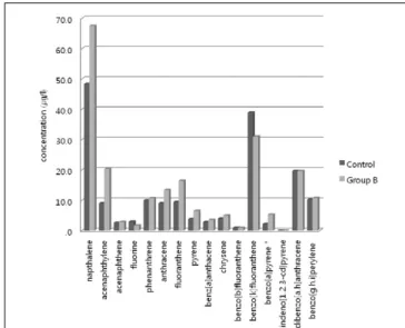
3.. B Bllo oo od d P PA AH H c co on nc ce en nttrra attiio on n a an nd d iin ntta ak ke e o off rro oa as stte ed d m
me ea atts s o orr ffiis sh h
Participants (69) had consumed roasted meats or fish in the previous three days (Group B), and 19 participants had not (control) intake these roasted foods. Figure 2 highlights the comparison between Group B and the control group for blood concentrations of the 16 PAHs. Among the geometric means of the 16 PAHs, only benzo(a)pyrene was significantly higher in Group B (intake of roasted meats or fish in the previous three days) than in the control group (p<0.1). Acenaphthylene, phenanthrene, anthracene, fluoranthene, pyrene, benz(a)anthracene, and chrysene were higher in Group B than in the control group, however, there was no statistical significance (p>0.1).
Ⅳ. Discussion
In the study, very low level 16 PAHs in blood could efficiently detected for the evaluation of non-occupational exposure. The head space-solid phase microextraction (HS-SPME) method was applied
Fig. 1 The comparison of GM of bloodconcentration between the group of ETS smoker (Group A), and control group (Control)
Fig. 2 The comparison of GM of blood concentration between the group of intake of roasted meats or fish for the past 3 days (Group B), and control group (Control)
for the detection of 16 PAHs in blood. This method did not require large volumes of organic solvents for extraction, which can be time consuming and may involve a multi-step process that could result in the loss of some analytes (King et al., 2003). Moreover, simplicity, low cost and high sensitivity make this method very useful for quantitative analysis of volatile or semi-volatile compounds (Cam et al., 2004; Ahn et al., 2001). By using the head space analysis method, the GC column is influenced by the direct injection of the sample to the GC injection port, rather than by the sample contents, so that a large number of samples provide efficient qualitative analysis. Moreover, the solvent-extraction and concentration required by existing methods requires a longer experimental time, a large amount of solvents and man-power for the experiment, so that the method is thought to be very limited in cases with large samples and reduced analytical working periods.
GM, and the maximum of PAHs in blood are elucidated in Tables 3. The GM of Naphthalene was 25.20 ㎍/l in total among the PAH analytes. However, there was no significant variation in the GM for blood concentration between man and women. The maximum was 1118.49 ㎍/l for man and women combined. If the participant had not been exposed to smoking or ETS or motor vehicle emissions, or any other occupational exposure sources, the other source of exposure to naphthalene for non-smokers is likely to be mothballs (used in toilets and to protect clothing), which represents a continuous exposure. More studies are necessary to evaluate the impact of indoor air pollutants on the general Korean population.
One of the main sources of 16 PAHs exposure for the general population is cigarette smoke (Hu et al., 2006; Ding et al., 2006).
This study showed that there was an effect of ETS in the passive smoking group because 4 analytes, acenaphthylene (p<0.01), acenaphthene (p<0.1), fluorene (p<0.01), and pyrene (p<0.05), among the 16 PAHs showed the significant increase in the blood samples. The participants showed the significant increase in the 4 PAHs were mainly exposed a particulate phase of the ETS. In the report from Lu and Zhu (2007), the particulate phase showed fluorene, phenanthrene, benzo(g,h,i)perylene at high concentrations.
On the other hand, the vapor phase showed high concentrations of acenaphthene, acenaphthylene, fluorene, phenanthrene, fluoranthene, and pyrene (Lu and Zhu, 2007). Therefore, as a result of this study, 4 PAHs from the blood regarded as an indicator of the exposure of ETS. Benzo(a)pyrene was related to smoking or to ETS (Fagundes et al., 2006). The results of the study, however, showed a very low detection percentage (12.7 % in total). Further studies of dose-response relations between blood PAHs and exposure to ETS
are needed to clarify the exposure source and route for the general Korean population.
Participants who had consumed roasted meats or fish showed a significant increase in benzo(a)pyrene levels (Fig. 2). Reinik et al., 2007 reported that the maximum acceptable concentration for benzo(a)pyrene of 5 ㎍/kg was exceeded in 3.4 % of samples.
These samples included commercial, cured meat products, and home grilled meats in Estonia. In non-smoking Japanese university students, pyrene, benzo(k)fluoranthene, and benzo(a)pyrene levels primarily came from food intake (Suzuki and Yoshinaga, 2007).
Moreover, benzo(a)pyrene was identified as a principal dietary source in the adult Spanish population (Ibanez et al., 2005). When the results of the current study were compared to recent reports, blood benzo(a)pyrene was closely related to meat intake or the method used for cooking the meat (Sinha et al., 2005; Kazerouni et al., 2001; Phillips, 1999). Thus it is assumed that levels of benzo(a)pyrene in the blood are influenced by exposure to roasted meats or fish. No trials were possible in the present study, however, to make quantitative assessment of roasted food intake. Further studies are necessary to identify such factors quantitatively as being influential sources of PAHs exposure to general population.
Ⅴ. Conclusion
The blood PAHs were efficiently detected by GC-MS in non- occupational exposed subjects. ETS is an influential factor, which it could affect the increase of blood PAHs [i.e., four PAHs, acenaphthylene (p<0.01), fluorene (p<0.01), acenaphthene (p<0.1), and pyrene(p=0.1)]. The roasted meat and fish ingestion was also found to be influential factors, which were increased the concentrations of benzo(a)pyrene in the blood. The concentrations of blood PAHs in this study are regarded some of the background exposure level in the general Korean population. In relation to the dietary ingestion of 16 PAHs, the further study of Korean food is needed to clarify the daily exposure and the dose-response relationship in background exposure.
Ⅵ. Acknowledgments
This work was supported by the Korea Research Foundation Grant funded by the Korean Government (MOEHRD) (KRF-2007- 331-E00055).
´~
REFERENCES
Aguinaga N, Campillo N, Vinas P, Hernandez-Cordoba M.
Determination of 16 polycyclic aromatic hydrocarbons in milk and related products using solid-phase microextraction coupled to gas chromatography-mass spectrometry. Analytica Chemica Acta 2007;596: 285-290
Ahn Y-G, Seo J-B, Hong J. Comparison solid phase microextraction with Purge & trap on the GC/MS analysis of volatile organic compounds in biota samples. Analytical Science & Technology 2001;145: 392-399 (in Korean)
Armstrong BG, Tremblay CG, Cyr D, Theriault GP. Estimating the relationship between exposure to tar volatiles and the incidence of bladder cancer in aluminum smelter workers. Scandinavian Journal of Work Environmental & Health 1986; 12: 486-493 Boffetta P, Jourenkova N, Custavsson P. Cancer risk from
occupational and environmental exposure to polycyclic aromatic hydrocarbons. Cancer Causes & Control 1997; p. 444- 472
Buratti M, Campo L, Fustinoni S, Cirla PE, Martinotti I et al.
Urinary hydrocylated metabolites of polycyclic aromatic hydrocarbons as biomarkers of exposure in asphalt workers.
Biomarkers 2007;123: 221-239
Cam D, Gagni S, Lombardi N, Punin MO. Solid-phase microextraction and gas chromatography-mass spectrometry for the determination of polycyclic aromatic hydrocarbons in environmental solid matrices. Journal of Chromatographic Science 2004;426: 329-35
Campo L, Addario L, Buratti M, Scibetta L, Longhi O et al.
Biological monitoring of exposure to polycyclic aromatic hydrocarbons by determination of unmatabolized compound in urine. Toxicology Letters 2006;162: 132-138
Ding YS, Yan XJ, Jain RB, Lopp E, Tavakoli A et al. Determination of 14 polycyclic aromatic hydrocarbons in mainstream smoke from U.S. brand and non-U.S. brand cigarettes. Environmental Science & Technology 2006;404: 1133-1138
Fagundes R B, Abnet C C, Strickland P T, Kamangar F, Roth M J et al. Higher urinary 1-hydroxy glucuronide (1-OHPG) is associated with tobacco smoke exposure and drinking mate in healthy subjects from Rio Grande do Sul, Brazil. BMC Cancer 2006;6: 1-7
Gimmer G, Naujack K W, Dettbarn G. Gas chromatographic determination of polycyclic aromatic hydrocarbons, aza- arenens, aromatic amines in the particle and vapor phase of
mainstream and side stream smoke of cigarettes. Toxicology Letters 1987;35: 117-124
Hu Y, Zhou Z, Xue X, Li X, Fu J et al. Sensitive biomarker of polycyclic aromatic hydrocarbons (PAHs): urinary 1- hydroxypyrene glucuronide in relation to smoking and low ambient levels of exposure. Biomarkers 2006;11: 306-318 Ibanez R, Agudo A, Berenguer A, Jakszyn P, Tormo M J et al.
Dietary intake of polycyclic aromatic hydrocarbons in a Spanish population. Journal of Food Protection 2005;68: 2190- 2195
Ikeda M. Solvents in urine as exposure markers. Toxicology Letters 1999;108: 99-106
International Agency for Research on Cancer (). Polynuclear aromatic compounds, Part 4: Bitumens, coal-tars and derived products, shale-oils and soots. In IARC monographs on the evaluation of carcinogenic risks to humans. Lyon, France:
International Agency for Research on Cancer; 1985;Vol. 35: p.
271
International Agency for Research on Cancer . Polynuclear aromatic compounds, Part 2 Carbon Blacks, mineral oils (lubricant base oils and derived products) and some nitroarenes. In: IARC Monographs on the evaluation of carcinogenic risks to humans). Lyon, France: International Agency for Research on Cancer; 1984.; Vol. 33: p.245
International Agency for Research on Cancer. Certain polycyclic aromatic hydrocarbons and heterocyclic compounds. In: IARC monographs on the evaluation of carcinogenic risks to humans Vol. 3 Lyon, France: International Agency for Research on Cancer; 1973. p. 271
International Agency for Research on Cancer. Monographs of the evaluation of the carcinogenic risk of chemicals to humans.
Polycyclic aromatic hydrocarbons. Part 1. Chemical, environmental and experimental data, In: IARC monographs on the evaluation of carcinogenic risks to humans Vol. 32.
Lyon, France: International Agency for Research on Cancer;
1983
International Agency for Research on Cancer. Overall evaluation of carcinogenic risks to humans. In IARC monographs on the evaluation of carcinogenic risks to humans. Supplement 7.
Lyon, France: International Agency for Research on Cancer;
1987
International Agency for Research on Cancer. Polynuclear aromatic compounds, Part 3 Industrial exposures in aluminium production, coal gasification, coke production, and iron and
´~