돼지 자궁 세포의 3차원 배양이 Plasminogen 활성과 수정란 발육에 미치는 영향
이상희1․황보 용1․차혜진1․김수지1․김민경1․정희태2․양부근1․박춘근1,†
1강원대학교 동물생명과학대학, 2강원대학교 수의과대학
Effect of Three Dimensional Culture of Porcine Endometrial Cells on Their Plasminogen Activity and Pre-implantation Embryo Development after Co-culture
Sang-Hee Lee
1, Yong HwangBo
1, Hye-Jin Cha
1, Su-Ji Kim
1, Min-Gyeong Kim
1, Hee-Tae Cheong
2, Boo-Keun Yang
1and Choon-Keun Park
1,†1
College of Animal Life Sciences, Division of Applied Animal Science, Kangwon National University, Chuncheon 200-701, Korea
2
College of Veterinary Medicine, Kangwon National University, Chuncheon 200-701, Korea
ABSTRACT
Three-dimensional (3D) culture system is useful technique for study of in vivo environment and it was used various experiments. This study was investigated to establish of embryo co-culture system and changes of PAs activity in 3D cultured endometrial cells of pigs. In results, growth of stromal cells into gel matrix were detected only with endometrial and myometrial cells. The most rapid growth of stromal cells were confirmed in 2.5×10⁵ cells/ml and gel matrix containing 15% FBS. Expression of urokinase-PA (uPA) after treatment of hCG (0.5, 1.0, 1.5 and 2.0 IU/ml) were higher than without hCG, but, there are not significant difference among the treatment. On the other hand, expression of uPA after treatment of IL-1β (0.1, 1, 10 and 100 ng/ml) were higher than without IL-1β, but, there are not significant difference. Expression of uPA after treatment of estrogen (0.2, 2, 20 and 200 ng/ml) were not difference, but PA activity was significantly decreased (p<0.05). Blastocyst was producing in PZM-3 medium containing FBS and endometrial cells were grown in PZM-3 medium. When embryos development with cultured endometrial cells, cleavage rates were not significant difference and blastocyst were not produced in co-culture with stromal cells and 3D culture system. 3D culture system had similar activity to in vivo tissue and these features are very useful for study of in vivo physiology. Nevertheless 3D culture system was not proper in embryo co-culture system. Therefore, we suggest that 3D culture system with embryo co-culture need continuous research.
(Key words : 3-dimensional culture system, embryo, co-culture, plasminogen activator, implantation)
* 본 연구는 한국연구재단(2010-0021580)의 지원에 의해 수행되었습니다.
†
Correspondence : [email protected]
서 론
포유동물에 있어 임신과 분만은 종족 번식을 위한 매우 중 요한 과정이다. 임신은 난소에서 배란된 자성 생식 세포인 난 자와 웅성 생식 세포인 정자의 결합으로 생성된 수정란이 자 궁에 착상되고 발육되는 과정으로, 수정란의 착상 시 모체는 임신을 인지하게 된다(Gadella와 Evans, 2011). 수정란이 자궁 에 정상적으로 착상하기 위해 자궁에서 다양한 생리학적 및 형태학적 변화가 일어나게 되는데, 황체에 의한 혈관 내의 프 로게스테론의 증가(McNeill 등, 2006), 혈관 신생 작용(Demir 등, 2010)으로 인한 혈류량의 증가(Nardo, 2005), 자궁 내막
두께의 증가(Baker 등, 1998) 및 자궁 내막 분비샘의 증가 (Hempstock 등, 2004) 등이 대표적인 현상이다.
이러한 자궁 변화에 영향을 미치는 인자로써 interleukin-1 beta(IL-1β)는 돼지에서 모체의 임신 인지에 관련된 cytokine 으로 자궁 내막 세포에 존재하는 특이적 수용체인 interleukin-1 receptor(IL-1R)와 결합하며, lutealizing hormone(LH)는 황체 를 형성시킬 뿐만 아니라 LH receptor(LHR)와 결합을 한다.
그 후 PG-endoperoxide synthase 2(PTGS2)의 발현을 증가시켜 prostaglandins(PGs) 합성에 관여한다는 보고가 있으며(Herrler 등, 2003; Burton 등, 2007), 이러한 PGs 합성을 통해 자궁의 구조를 변화시켜 성공적인 착상과 임신의 확립을 준비하는데,
이러한 과정을 자궁 내막의 황체기화(lutealization)라 한다(Fa- dhillah 등, 2014; Sugino, 2014).
Plasminogen activators(PAs)는 비활성 plasminogen을 활성 plasmin으로 전환시키는 serine계 단백질 분해 효소로서, uro- kinase-type PA(uPA)와 tissue-type PA(tPA)로 나누어진다(
Makarova 등, 2011; Asuthkar 등, 2013). 일반적으로 포유동물 에 있어서 PAs는 섬유소 용해 작용(Leibundgut 등, 2012; Luc- king 등, 2013; Takabayashi 등, 2013)과 혈관 신생 작용(Teng 등, 2012; Yip 등, 2013) 같은 생리학적인 역할을 수행한다.
특히, uPA는 세포외 기질(Extracellular matrix) 분해 작용 및 세포의 이동에 관여하며(Carriero 등, 2011), 이러한 특징에 따 라 포유동물의 자궁에서는 자궁 세포의 이동과 조직 재구성 에 영향을 미쳐, 발정 주기에 따른 자궁의 생리학적 및 형태학 적 변화를 유도한다(Bazer, 2013). 이러한 PA와 포유동물 자 궁과의 관계에 대한 연구는 사람(Koh 등, 1992; Estelle 등, 2003), 쥐(Sappino 등, 1989) 및 소(Tanikawa 등, 2009) 등에서 다양한 연구가 이루어지고 있지만, 돼지에서는 그 연구가 미 미한 실정이다.
생체 내의 변화를 알아보기 위해 2차원 세포 배양 시스템 을 구현하여 세포 기작을 연구하는 방법을 많이 사용하는데, 이러한 방법은 세포의 활발한 증식으로 인한 세포의 확보가 용이하다는 점이 있지만, in vivo 상태에서의 세포의 기능과 구조적인 형태를 잃어버린다는 단점이 있다. 또한 세포는 조 직 안에서 세포외 기질 구조와 함께 여러 종류의 세포가 여러 층을 이루며 상호작용을 하는 반면, 2차원 세포 배양 체계는 extracellular matrix(ECM) 구조가 없는 단일 층으로 세포가 성장하기 때문에, in vivo에서 일어나는 세포의 정확한 기작을 연구하기 힘들다는 보고가 있다(White 등, 2014). 생체 내를 직접적으로 연구하는 in vivo assay는 정확한 결과를 확인할 수 있지만, 살아있는 생체 또는 조직을 이용한 연구는 시간과 공간에 제한적이며, 낮은 재현성과 조작이 어렵다는 단점이 있다. 따라서 이러한 문제점을 해결하기 위해 인위적인 세포 외 기질을 이용하여 세포를 성장시키는 3차원 배양 시스템을 사용하고 있다(Ip와 Darcy, 1996).
체외수정은 체내에서 생산하기 힘든 수정란을 체외에서 생 산하는 기술로써, 산자 생산과 불임의 치료 목적 외에도 체내 에서 이루어지는 수정란과 자궁 또한 그 둘 사이에 작용하는 세포 신호를 연구하는데 유용한 기술이다. 이러한 체외수정은 수정란 이식과 동물생명공학산업의 발전과 함께 우수한 형질 을 가진 개체 및 형질 전환 동물을 생산하는데도 이용되고 있 으며, 가축 육종 시간 단축 및 동물을 이용한 생의약품 생산 에도 이용된다(Hansen, 2014). 이러한 기술은 현재 사람(Cobo 등, 2012), 쥐(Tsunoda 등, 2012), 토끼, 양(Fidanza 등, 2014), 돼지(Tanihara 등, 2013) 및 소(Sugimura 등, 2012) 등 다방면
으로 연구가 이루어지고 있다. 하지만 체외수정은 체내의 환 경을 모방하여 구현한 것으로, 체내 환경에 비하여 물리적, 화 학적 충격을 받게 되어 수정란 생산 효율이 낮아지며, 이러한 요소들 때문에 정확한 세포 신호를 연구하는데 문제점이 발 생한다. 이러한 원인으로는 인큐베이터의 불안정한 대기 조 성, 온도, pH, medium 조성의 변화 및 체외에서의 수정란 조 작 등 체내 환경과 다른 다양한 화학적 및 물리적인 피해로 볼 수 있다(Van Loendersloot 등, 2010). 특히, 돼지 체외수정 기 법은 다른 포유동물의 경우보다 연구가 다소 늦게 시작되었 기 때문에, 일반적인 체외수정 기법을 이용한 양질의 수정란 생산에 많은 한계점이 있어, 체외수정의 효율을 높이기 위한 배양액의 조성 변화(Yoshioka, 2011), 당류의 첨가(Oliveira 등, 2014), 항산화제 첨가, 비타민의 첨가(Tareq 등, 2012) 및 체세포와의 공동 배양(Bui 등, 2012)을 실시하여 체외수정의 효율을 증진시키는 연구가 활발히 진행되고 있다. 일반적으로 체외수정 효율 증진을 위한 공동 배양 시스템으로는 난관 또 는 자궁의 상피 세포와 간질 세포를 단일로 이용한 2차원 배 양 조건하에서 수정란과 함께 공동 배양하는 방식을 많이 이 용하고 있다(Bui 등, 2012). 하지만 2차원 배양된 자궁 세포의 기능은 실제 자궁 환경과 달리 형태학적 및 생리학적으로 차 이가 있을 것이라 예상된다. 그렇기 때문에 실제 자궁 조직과 유사한 구조를 만들어 배양한 3차원 배양 시스템은 2차원 배 양된 세포와의 공동 배양에 비하여 수정란이 잘 발달할 수 있 는 환경을 제공해 줄 수 있을 것이라 예상되며, 여러 연구를 통하여 세포의 3차원 배양은 2차원 배양에 비하여 실제 조직 과 형태 및 생리학적 기능과 분비되는 인자들의 발현이 유사 하다는 보고(Schneider 등, 2010; Sart 등, 2013; Tseng 등, 2014; White 등, 2014)가 있기 때문에 이러한 기술을 돼지 수 정란 발달에 적합한 3차원 배양 시스템으로 구현시키는 연구 가 필요하다.
따라서 3차원 배양 시스템의 효율을 증진시키기 위한 연구 를 진행하여 3차원 자궁 세포 공동 배양 시스템을 확립 후, hCG와 IL-1β에 따른 자궁 내막 세포의 lutealization을 확인하 였으며, 또한 돼지의 임신 인지 인자를 lutealization된 3차원 자궁 세포 공동 배양 시스템에 처리하여 착상 과정 동안의 자 궁 내막과 PAs 사이에 작용하는 관계를 규명하였다. 최종적 으로 3차원 자궁 세포 공동 배양 시스템과 수정란의 공동 배 양을 위한 배양액의 최적의 조건을 확립하여, 돼지 자궁의 3 차원 공동 배양 시스템에서의 체외수정 효율을 증진시키는데 있다.
재료 및 방법
1. 실험 설계
1) 자궁 내막 세포의 효율적인 3차원 배양 체계 확립 자궁 내막 세포를 2차원 배양 후 3차원 배양에 이용하는 방 법은 시간이 오래 걸린다는 단점을 가지고 있다. 이러한 단점 을 개선하기 위하여 자궁 내막 조직과 근육 조직을 3차원 배양 체계에 이용하였다. 조직을 이용한 3차원 배양 시스템을 확립 하기 위한 방법으로는 자궁 내막 및 근육 조직을 1 × 1 cm로 자른 후 바로 이용, 세절한 후 tris-NH4를 이용하여 혈액 성분 을 제거한 후 이용하고, 조직에 collagenase Ⅳ(Worthington, USA)를 처리하여 회수한 세포를 이용하여 3차원 배양하였다.
또한 3차원 배양 시스템 환경에서 최적의 세포 농도를 확립하 기 위해 자궁 내막 간질 세포를 2.5 × 10⁵, 5 × 10⁵, 7.5 × 10⁵ 및 10 × 105 cells/ml의 농도로 나누어 배양하였고, gel 조성을 확인하기 위하여 다른 FBS 농도(10%, 15%) 및 200 mM CaCl2 를 첨가하여 3차원 배양하였다.
2) 3차원 배양된 자궁 내막 세포의 인위적인 발정 주기 조절
3차원 배양에 사용된 자궁 내막 세포는 난포기의 자궁으로
부터 채취하였으며, 이러한 자궁 내막 세포를 인위적으로 황 체기 상태를 유도하기 위하여 확립된 3차원 배양 시스템에 hCG(0, 0.5, 1.0, 1.5 및 2 IU/ml)와 IL-1β(0, 0.1, 1, 10 및 100 ng/ml)를 처리한 후, 24시간 동안 38℃, 5% CO2 조건에서 배 양하였다. 또한 착상 시 환경을 만들기 위하여 인위적으로 황 체기로 유도된 자궁 내막 세포에 E2(0, 0.2, 2, 20 및 200 ng/ml) 처리한 후 24시간 동안 배양한 뒤, 배양액과 세포를 회수하여 실험 전까지 —80℃에서 보관하였다.
3) 3차원 배양된 자궁 내막 세포와 돼지 수정란의 공동 배양 체계 확립
3차원 배양 시스템과 돼지 수정란의 공동 배양 체계를 확립
하기 위하여 먼저, 돼지 수정란과 자궁 내막 세포가 같이 자랄 수 있는 배양액 조건을 확인하였다. 수정란의 발달을 확인하 기 위하여 체외수정 48시간 후 다른 조건의 배양액의 조건은 Table 1과 2에 나타내었으며, 수정 168시간 후 분할률과 배반 포 형성률을 확인하였다. 그 뒤 최적의 배양액 조건을 확립한 뒤, 수정 후 48시간 배양된 돼지 수정란을 2차원 및 3차원 배 양된 자궁 내막 세포와 공동 배양하여 수정 168시간 후 분할 률과 배반포 형성율을 확인하였다.
2. 자궁 내막 세포의 배양
도축장으로부터 난포에 황체가 전혀 형성되지 않은 난포기 의 돼지 자궁을 1∼1.5시간 이내에 실험실로 운반하였으며, Hank's Balanced Salt Solution(HBSS)을 이용하여 자궁 외부를 세척한 후 난소와 자궁간막을 제거하고, 자궁각 부분만을 때어 내었다. 그 후 자궁강 내에 HBSS를 관류시켜 세척하고, 자궁
Table 1. Medium condition for in vitro culture of porcine embryo
Medium Treatment ratio
1PZM-3 : 2D/F-12
PZM-3 10 7 5 0
D/F-12 0 3 5 10
1 PZM-3: Porcine zygote medium-3.
2 D/F-12: Dulbecco’s modified Eagle’s medium/nutrient mix- ture F-12 ham.
Table 2. Medium conditions by different FBS concentrations for
in vitro culture of porcine embryo
Medium Concentrations of FBS (%)
1TCM-199 0 2.5 5 10
TCM-199+hypotaurine 0 2.5 5 10
2PZM-3 0 2.5 5 10
1 TCM-199: Tissue culture medium-199.
2 PZM-3: Porcine zygote medium-3.
각을 세로축으로 절개한 뒤 메스를 이용하여 자궁 내막에서 상 피층을 긁어내었다. 그 다음 Dulbecco's Modified Eagle's Medi- um(DMEM, Invitrogen, USA)에 회수한 후 collagenase Ⅳ를 66 U/ml의 농도로 처리하여 진탕 배양기에서 배양하였다(1 h, 38℃, 120 rpm). 배양 후 cell strainer(SPL, Korea)를 이용하여 걸러낸 후 원심분리(5 min, 1,200 rpm)하여 상층액을 제거한 뒤 Tris-NH4를 이용하여 혈액 성분을 제거하였고, HBSS로 세 척하고 10% FBS(Invitrogen, USA), 0.2% amphotericin B와 0.5
% Antibiotic-Antimycotic(ABAM, Invitrogen, USA)을 포함한 DMEM과 Ham’s F-12가 1:1로 섞인 D/F12 (Invitrogen, USA) 배양을 이용해 38℃, 5% CO2조건에서 배양하였으며, 48시간 마다 배양액을 교체하였다.
3. 자궁 내막 세포의 3차원 배양
인위적으로 세포외 기질을 만들기 위하여 Collagen gel(Cell matrix type Ⅰ, Nitta gelatin, Japan), FBS를 포함한 5배로 농축 된 D/F-12(5xD/F-12) 및 buffer(0.05 N NaOH, 2.2% NaHCO3, 200 mM HEPES)를 7:2:1의 비율로 혼합하여 gel solution을 만 들었으며, 소량의 gel solution을 dish 바닥에 분주 후 37℃에 서 30분 동안 굳혀주어 인위적으로 기저층을 만들어주었다. 2 차원 배양된 자궁 내막 간질 세포는 0.25% trypsin-EDTA를 처리한 뒤 38℃에서 4분간 배양하여, 세포를 회수하여 원심분 리 후 DMEM으로 세척하였다. 그 후 trypan blue를 이용하여 세포 수를 측정한 뒤 gel solution과 혼합하고, 미리 준비한 기
Table 3. Primer conditions of PCR
Gene Primer sequence Product size (bp) Tem. (℃) Cycles
uPA F:CCTACAAGTACTTCTC
R:GCAAACCAAGGCTGGTTTCTC 460 55 45
GAPDH F:TCGGAGTGAACGGATTTG
R:CCTGGAAGAGATGGTGATGG 200 59 30
저층 위에 분주하여 만든 3차원 gel matrix를 38℃, 5% CO2
조건에서 10일간 배양하였다. 간질 세포와 마찬가지로 0.25%
trypsin-EDTA를 처리하여 상피 세포를 회수한 뒤 3 × 10⁵ cells/
ml의 농도로 3차원 gel matrix 위에 분주하였다. 상피 세포를 분주 후 24시간 뒤 상피 세포의 성장을 확인한 후 실험에 사 용하였다.
4. 난자의 체외성숙, 수정 및 발육
도축장으로부터 채취한 난소에서 2∼6 mm 직경의 난포로 부터 18-gauge 주사기를 이용하여 미성숙 난자를 채취하였다.
채취한 미성숙 난자는 난구 세포가 3겹 이상 조밀하게 붙어있 고, 난자의 세포질이 균질한 것만을 선발하여 실험에 사용하 였다. 미성숙 난자는 10% porcine follicle fluid(pFF), 1% hCG, 0.1% FSH, LH 및 epidermal growth factor(EGF)를 포함한 Tissue Culture Medium-199(TCM-199, Invitrogen, USA) 배양 액을 이용하여 38.5℃, 5% CO2 조건에서 22시간 동안 성숙 배양 후, 10% pFF와 0.1% EGF를 포함한 TCM-199 배양액을 이용해 같은 조건으로 22시간 동안 배양하였다.
성숙 배양 후 난구 세포가 확장된 난자는 0.1% hyaluroni- dase를 처리한 후 체외수정 배양액인 2 mg/ml BSA가 포함된 mTBM 배양액 50 µl 소적으로 옮겼다. 수정에 사용된 정액은 1,500 rpm에서 5분간 원심분리 후 상층액을 제거하였으며, Modena B를 이용하여 2회 세척하였고, 4 mg/ml caffeine이 첨 가된 mTBM 배양액에 6 × 10⁵ sperms/ml의 농도로 희석하여 난자가 있는 소적에 50 µl씩 분주하여 38.5℃, 5% CO2 조건에 서 6시간 동안 배양하였다.
체외수정 6시간 후 수정된 난자의 난구 세포와 정자를 제 거한 후 3 mg/ml BSA가 첨가된 Porcine Zygote Medium-3 (PZM-3) 배양액 100 µl 소적으로 옮겨 48시간 배양하였고, 그 후 본 실험에서 사용된 체외배양 배양액으로 옮겨 4-well dish 에서 168시간 동안 배양 후 분할률과 배반포 형성률을 확인 하였다.
5. RT-PCR
배양된 자궁 내막 세포로부터 RNA를 추출하기 위해 RNAiso plus(TAKARA, Japan)을 400 µl를 처리하여 50분 동안 교반한
뒤 chloroform을 100 µl 처리하고 5분 동안 교반하였다. 그 후 4℃, 12,000 rpm 조건 하에 5분 동안 원심분리 후 RNA 층만을 분리하여 isopropyl alcohol을 200 µl 첨가하고, 다시 5분 동안 교반한 뒤 4℃, 12,000 rpm 조건 하에 10분 동안 원심분리 후 상층액을 제거하고 무균 실험대에서 건조하였다. 추출한 RNA 는 DEPC-DW와 혼합한 뒤 Nano drop 200(Thermo, USA)을 이용하여 정량하였고, Maxime RT Premix(Intronbio, Korea)을 이용하여 cDNA로 합성하였다. 합성된 cDNA는 uPA 및 tPA 와 GAPDH에 대한 primer(Table 3)를 이용하여 증폭시켰으며, 증폭된 산물은 Ethidium bromide(EtBr, Bioneer, Korea)을 포함 한 2% Agarose gel을 사용하여 전기영동으로 확인하였다.
6. PA 활성도 측정
PA 활성을 측정하기 위하여 보관해 둔 샘플과 standard buffer를 96-well microplate에서 plasminogen working solution (Sigma, USA)과 함께 38℃에서 1시간 동안 배양하였다. 그 후 substrate buffer[0.18 mM Z-L-LYS-SBzl hydrochloride, 0.22 mM 5,5’-ditho-bis(2-nitrobenxoic acid), 0.01% Triton-X100, Sigma, USA]를 첨가하여 38℃에서 1시간 동안 배양한 뒤 microplate reader를 이용하여 405 nm 파장에서 흡광도를 측 정하였다.
7. 통계 처리
실험에서 얻어진 결과는 SAS 9.2를 이용하여 최소 유의차 검정(Least Significant Different test; LSD test)과 General Linear model(GLM)을 적용하여 Duncan의 multiple range test에 의하 여 유의차(P<0.05)를 검정하였다.
결 과
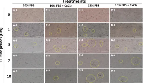
1. 자궁 내막 세포의 효율적인 3차원 배양 체계 확립 효율적인 자궁 내막 세포의 3차원 배양 체계를 확립하기 위 하여 자궁 내막 조직과 세포(Fig. 1) 및 근층 조직과 세포(Fig.
2)를 3차원 배양 체계에 이용하였다. 배양 후 7일 및 14일 때 확인한 결과, 내막조직(Fig. 1 A, B, D, E) 및 근층 조직(Fig.
2 A, B, D, E) 배양하였을 때, gel solution 쪽으로 세포가 자라
Fig. 1. The morphology of porcine endometrial tissue mass (A, D), endometrial tissue smear (B, E), endometrial tissue with collagenase
Ⅳ (C, F) into three dimensional collagen gel (green arrow: tissue mass; blue arrow: matrix gel; yellow arrow: stromal cell; scale bar: 200 µm).
Fig. 2. The morphology of myometrial tissue mass (A, D), myometrial tissue smear (B, E) and myometrial tissue with collagenase Ⅳ (C, F) into three dimensional collagen gel (green arrow: tissue mass; blue arrow: matrix gel; yellow arrow: stromal cell; scale bar: 200 µm).
Fig. 3. The morphology of porcine uterine stromal cell in three-dimensional culture according to different cell concentration (yellow circle:
stromal cells; scale bar: 200 µm).
지 않았으나, 내막 조직 및 근층에서 채취한 세포(Fig. 1 C, F 와 Fig. 2 C, F)를 이용하였을 때, gel solution 내에서 간질 세 포가 성장하는 것을 확인하였으며, 이차원 배양된 세포와 다 르게 gel solution 내에서 방사형으로 성장하는 것을 확인하였
다(Fig. 3). 본 결과를 통하여 조직보다 세포가 3차원 배양 체 계를 확립하는데 더 적합하다는 것을 확인하였다. 다음으로 3 차원 배양에 적합한 조건을 확립하기 위하여 각기 다른 농도 의 간질 세포(Fig. 4)와 다른 gel solution 조건(Fig. 5)을 이용
Fig. 4. Comparison of porcine uterine stromal cells growth in three-dimensional culture according to different gel condition (yellow circle:
stromal cells. scale bar: 200 µm).
Fig. 5. Morphology of two dimensional cultured porcine uterus epithelial (A), stromal (B) cells and stromal cells in the three dimensional culture system (C: 0 days; D: 1 days; E: 5 days; F: 7 days) (yellow arrow: stromal cells; scale bar: 200 µm).
하였다. 그 결과, 2.5 × 10⁵ cells/ml의 세포 농도에서 가장 많 이 성장하는 것을 확인하였고, 15% FBS를 포함한 gel solution 에서 간질 세포가 더 크게 성장하며 오랜 기간 배양할 수 있 는 것을 확인하였다. 하지만 gel solution 내의 CaCl2 첨가는 세 포 성장에 영향을 미치지 않았다. 본 결과를 통하여 최적의 3 차원 배양 체계를 확립하였다.
2. 3차원 배양된 자궁 내막 세포의 인위적인 발정 주기 조절
3차원 배양된 자궁 내막 세포의 환경을 조절하기 위하여
hCG와 IL-1β를 처리하여 uPA mRNA의 발현 변화를 확인하 였다. 서로 다른 농도의 hCG를 처리한 결과, 유의적인 차이는 보이지 않았지만, 농도의존적으로 uPA mRNA의 발현이 증가 하는 것을 확인하였다(Fig. 6 A). 또한 IL-1β를 처리하였을 때 는 uPA mRNA의 발현량이 유의적 차이는 보이지 않았으나 대조군에 비하여 발현량은 증가하였고, IL-1β의 농도가 증가 할수록 uPA mRNA가 감소하였다(Fig. 6 B). 따라서 3차원 배
양된 자궁 세포의 인위적 발정 조절을 위해서 hCG가 적합하 다는 것을 확인하였다. 또한 인위적으로 착상 시기의 환경을
Fig. 6. Effects of concentration of hCG (A) and IL-1β (B) on expression of urokinase-type PA (uPA) and β-actin in porcine three-dimensional cultured uterine cells.
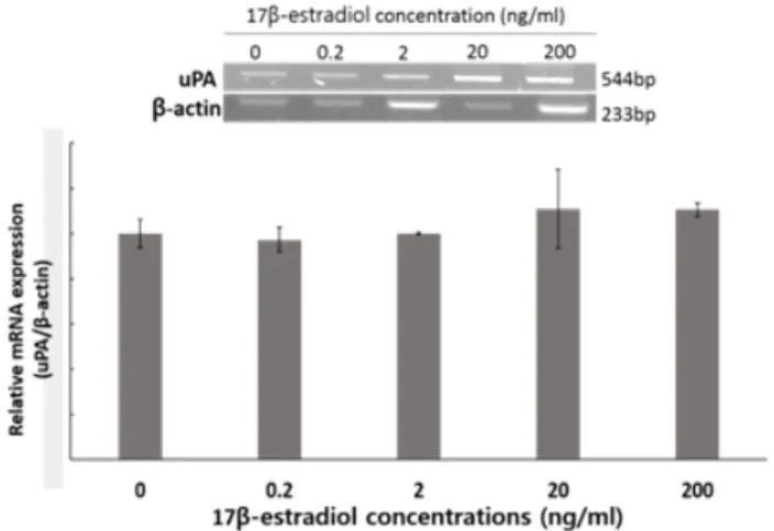
만들기 위해 E2를 처리하여 uPA mRNA(Fig. 7)와 PA activity (Fig. 8)를 확인한 결과, uPA mRNA의 발현량은 변화가 없었 으나, PA activity는 E2 처리구(2, 20, 200 ng/ml)에서 유의적 으로 감소하는 것을 확인하였다(P<0.05).
3. 3차원 배양된 자궁 내막 세포와 돼지 수정란의 공동 배양 체계 확립
3차원 배양된 자궁 세포와 수정란의 공동 배양 체계를 확
립하기 위하여 수정란의 발달이 가능한 배양액을 확인하였다.
수정란 배양액인 PZM-3와 세포 배양액인 D/F-12를 혼합하여 사용하였을 때 모든 처리구에서 분할율의 유의적인 차이는 보이지 않았지만, PZM-3만 이용한 대조군에서만 배반포 형성 을 확인할 수 있었다. TCM-199와 항산화제인 hypotaurine을 포함한 TCM-199 배양액에 FBS를 첨가하여 사용한 결과, 이 전 실험보다 분할률은 향상되었지만, 모든 처리구에서 배반포 가 형성되지 않는 것을 확인하였다(Fig. 9). PZM-3 배양액에
Fig. 7. Effects of 17β-estradiol (E2) on expression of urokinase-type PA (uPA) mRNA in three-dimensional culture.
Fig. 8. Effects of exposure to different concentration of 17β-estradiol (E2) on plasminogen activators (PAs) activity in porcine uterine three-dimensional culture (p<0.05).
Fig. 9. Comparison of cleavage rate according to different medium condition on porcine embryo development after 168 h of
in vitro fertilization.
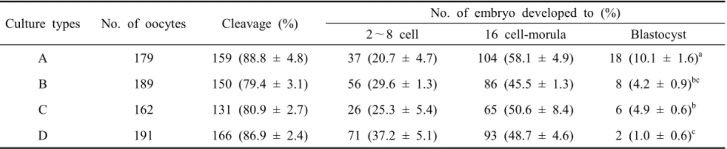
다른 농도의 FBS를 첨가하여 사용한 결과(Table 4), 분할률은 유의적 차이를 보이지 않았지만 FBS를 첨가하지 않은 대조구 에서 배반포 형성률이 유의적으로 높게 나타났으며(P< 0.05),
10% FBS를 첨가한 처리구에서 유의적으로 낮게 나타났다
(P<0.05). 수정란의 발달을 확인한 뒤 PZM-3를 이용한 배양액 에서 세포의 성장을 확인한 결과(Fig. 10), 배양 1일 후 모든 처리구에서 세포가 자라는 것을 확인하였지만, 3일 후 FBS를 첨가하지 않은 배양액에서는 세포가 자라지 못하는 것을 확인 하였다. 본 결과를 통하여 수정란과 자궁 세포의 공동 배양을 위한 배양액으로 2.5% FBS를 포함한 PZM-3 배양액을 선발 하였다. 선발된 배양액을 이용하여 수정란 단독 배양, 2차원 배양된 자궁상피, 자궁간질 세포와 공동 배양, 3차원 배양된 자궁 세포와 공동 배양한 결과(Table 5), 분할율은 처리구간 유 의적 차이는 보이지 않았지만, 수정란을 단독 배양한 처리구에 서 배판포 형성률이 유의적으로 높게 나타났으며(P<0.05), 간 질 세포와 3차원 배양된 자궁 세포와 공동 배양 처리구에서는 배반포가 형성되지 않는 것을 확인하였다.
고 찰
현재 3차원 배양 시스템을 확립하는 방법은 조직에서 세포 를 분리하여 2차원 배양 뒤 다시 3차원 배양에 이용을 하는 방법으로 세포를 분리하고, collagen 등을 이용하여 만든 인위 적인 세포외 기질에 넣어주어 배양해야 하기 때문에 시간과 비용이 많이 소모되며, 실제로 조직 자체를 배양하는 연구도 다양한 방면에서 이루어지고 있는 실정이다(Badylak 등, 2011).
따라서 자궁 내막 조직을 자체적으로 사용하여 3차원 배양 시 스템의 효율을 높일 필요성이 있다. 효율적인 3차원 배양 시 스템의 확립을 위하여 자궁 내막과 근층 조직 및 조직으로부 터 회수한 세포를 이용하였고, 최적의 배양 조건을 확립하기 위하여 세포의 농도와 gel solution의 조성을 확인하였다. 본 실험 결과, collagen type의 cell matrix를 이용한 3차원 배양
Table 4. Comparison of cleavage rate according to FBS rate of PZM-3 on porcine embryo development after 168h of in vitro fertilization
Culture types No. of oocytes Cleavage (%) No. of embryo developed to (%)
2∼8 cell 16 cell-morula Blastocyst A 179 159 (88.8 ± 4.8) 37 (20.7 ± 4.7) 104 (58.1 ± 4.9) 18 (10.1 ± 1.6)a B 189 150 (79.4 ± 3.1) 56 (29.6 ± 1.3) 86 (45.5 ± 1.3) 8 (4.2 ± 0.9)bc C 162 131 (80.9 ± 2.7) 26 (25.3 ± 5.4) 65 (50.6 ± 8.4) 6 (4.9 ± 0.6)b D 191 166 (86.9 ± 2.4) 71 (37.2 ± 5.1) 93 (48.7 ± 4.6) 2 (1.0 ± 0.6)c Means ± SEM are presented.
a∼d Values with different super scripts in the same column were significantly different.
A: PZM-3 + 0.3% BSA, B: PZM-3 + 0.3% BSA containing 2.5% FBS, C: PZM-3 + 0.3% BSA containing 5% FBS, D: PZM-3 + 0.3% BSA containing 10% FBS, n=3.
Fig. 10. The morphology of porcine endometrial cells in different medium condition of PZM-3 containing FBS (scale bar=300 µm).
시에는 조직보다는 분리된 세포를 이용하는 것이 접합하며, 일반 세포 배지에 높은 농도의 혈청을 이용할 때 효율적인 3 차원 배양 시스템을 확립할 수 있었다.
Human chorionic gonadotropin(hCG)는 LH와 receptor를 공 유하기 때문에 lutealization을 유도하기 위하여 쥐(Guo 등, 2011), 돼지(Bołzan 등, 2013), 토끼(Balazi 등, 2012) 및 소 (Zhang 등, 2011) 등 in vitro 연구에 많이 이용하고 있으며, in
vivo
측면에서는 가축의 발정과 배란을 유도하기 위해 돼지(Wongkaweewit 등, 2012; Tomás 등, 2013; Misumi 등, 2014) 및 소(Rostami 등, 2011; Giordano 등, 2012)에서 많이 이용된 다. 황체기화 된 자궁은 황체에서 분비되는 progesterone에 의 하여 자궁 내벽을 두껍게 유지하며, 배아를 인지할 수 있는 상 태를 갖추게 된다(Bazer 등, 2010). 이후 배아는 모체가 임신 을 인지할 수 있도록 물질을 분비하게 되는데, 소의 경우에는 interferon tau(INF-τ)(Shirasuna 등, 2011), 돼지의 경우에는
estrogen(E2)(Bazer, 2013) 등과 같은 물질을 분비하여 자궁 내 막의 염증을 유도한다(Ziecik 등, 2011). 임신 인지 후 모체의 자궁 내막에서는 PG를 생산하여 lutealizaion에 의해 변화된 자 궁 내막을 유지시켜 임신이 지속될 수 있도록 한다(Bazer 등, 2010; Ziecik 등, 2011). 또한 PAs는 배란(Brännström 등, 2010), 자궁 상피 세포의 염증 및 세포외 기질 분해 및 수정란 착상 (Grullón 등, 2013; Martinez-Hernandez 등, 2011) 등 다양한 번식학적 현상에 관여하는 것으로 알려져 있다. 따라서 본 연 구에서 3차원 배양된 자궁 내막 세포를 인위적으로 황체기화 시키기 위해 hCG와 IL-1β를 처리하였으며, 착상 시기의 자궁 을 구현하기 위하여 황체기화 된 3차원 배양 시스템에 E2를 처리한 후 PA activity의 변화를 확인하였다. hCG 농도에 따른 uPA의 발현 변화는 유의적 차이는 보이지 않았으며, 농도의 존적으로 발현이 증가하는 경향을 보였고, IL-1β의 처리는 대 조군에 비하여 증가하였지만 농도의존적으로 uPA의 발현이
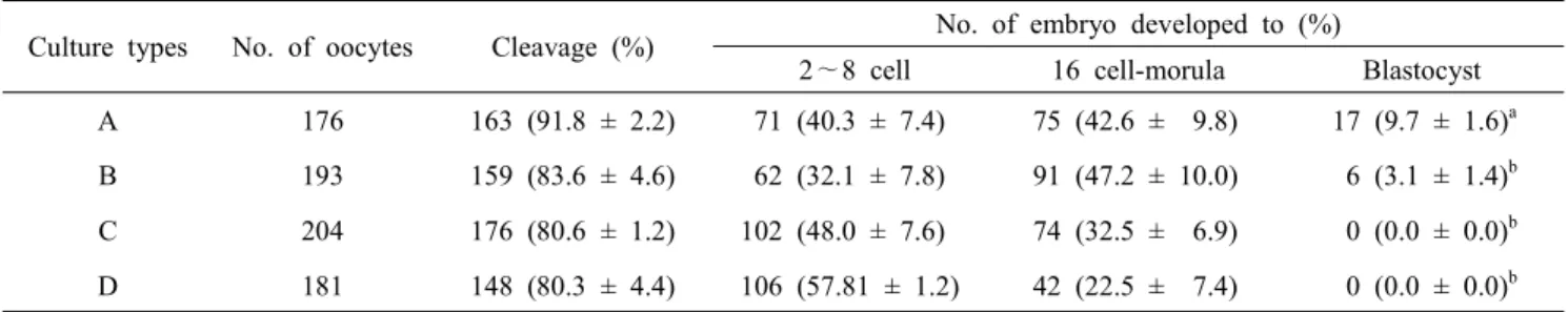
Table 5. Comparison of cleavage rate according to different culture condition using established 2.5 % FBS medium on porcine embryo development after 168h of in vitro fertilization
Culture types No. of oocytes Cleavage (%) No. of embryo developed to (%)
2∼8 cell 16 cell-morula Blastocyst A 176 163 (91.8 ± 2.2) 71 (40.3 ± 7.4) 75 (42.6 ± 9.8) 17 (9.7 ± 1.6)a B 193 159 (83.6 ± 4.6) 62 (32.1 ± 7.8) 91 (47.2 ± 10.0) 6 (3.1 ± 1.4)b C 204 176 (80.6 ± 1.2) 102 (48.0 ± 7.6) 74 (32.5 ± 6.9) 0 (0.0 ± 0.0)b D 181 148 (80.3 ± 4.4) 106 (57.81 ± 1.2) 42 (22.5 ± 7.4) 0 (0.0 ± 0.0)b Means ± SEM are presented.
a∼d Values with different super scripts in the same column were significantly different.
A: IVC with drop culture, B: IVC with porcine uterine epithelial cells, C: IVC with stromal cells, D: IVC with 3D culture system, n=3.
감소하는 것을 확인하였다(Fig. 6).
Kim 등(2011)의 연구에서 황체기 자궁에서 난포기 자궁보다 uPA의 발현이 증가한다는 보고가 있었으며, 이는 hCG를 처 리하였을 때 uPA 발현이 증가하는 것과 일치하였다. 따라서
3차원 배양 시스템의 인위적인 발정 주기 조절이 가능하다는
것을 확인하였다. 착상 시기의 환경을 구현하기 위하여 hCG를 처리하여 황체기화 된 3차원 배양 시스템에 E2를 처리한 결과, uPA 발현에는 영향을 미치지 않았지만(Fig. 7) PAs의 활성은 유의적으로 감소(P<0.05)하는 것을 확인하였다(Fig. 8). Cha 등(2013)의 연구에서 3차원 배양된 자궁 내막 세포는 실제 조 직과 PAs 활성이 차이가 나지 않는다는 것을 보여주며, 이를 통하여 돼지에서 착상 시 PAs의 발현은 변화시키지 않지만,
PAs의 활성을 억제함으로써 성공적인 착상에 영향을 미치는
것을 예상할 수 있었다.
3차원 배양 시스템에 이용되는 배양액 조성은 일반 체세포
를 배양할 때 사용하는 Modified Eagle Medium(MEM)(Charo- enpanich 등, 2011), Dulbecco's MEM(DMEM)(Eiraku와 Sasai, 2012), medium-199(Rezavand 등, 2013), DMEM: Nutrient Mixture F-10(D/F10)(Merrick 등, 2010) 및 DMEM: Nutrient Mixture F(D/F12)(Vidi 등, 2013)에 Fetal bovine serum(FBS) 와 같은 혈청을 첨가하여 배양하는 방식을 많이 사용하고 있 다. 하지만 돼지 수정란 배양을 위한 배양액으로는 porcine zygote medium-3(PZM-3)(Jia 등, 2014), PZM-5(Mito 등, 2012), North Carolina State University-23(NCSU-23)(Hiraga 등, 2013) 및 NCSU-37(Haraguchi 등, 2014)을 이용한 연구가 주로 이루 어져 있다. 또한 일반적인 체세포 배양 및 3차원 배양 시스템 에서와 다르게 돼지 수정란 배양액에는 주로 Bovine serum
albumin을 첨가하여 배양하는 방식을 많이 사용하고 있다
(Suzuki and Yoshioka, 2006). 이렇듯 앞서 언급한 돼지 수정 란 배양액을 이용한 체세포 배양 및 3차원 배양 시스템에 대
한 연구도 거의 이루어지지 않았기 때문에, 체외 수정 효율 증 진을 위해 수정란과 체세포 공동 배양 시스템을 확립하기 위 해서는 공동 배양액에 적절한 배양액 조건을 확립할 필요가 있 다. 공동 배양액 조건을 확립하기 위하여 다양한 배양액(Table 1, 2)를 사용한 결과, D/F-12와 TCM-199가 포함된 배양액에 서 수정란이 분할하는 것은 확인하였지만, 배반포의 형성을 확 인할 수 없었다(Fig. 9). 위 결과를 바탕으로 수정란 배양액은 PZM-3에 FBS를 첨가하여 체외배양을 실시한 결과, 모든 처 리구에서 배반포의 형성을 확인할 수 있었으며(Table 4), 자궁 세포 역시 성장이 가능하지만 FBS를 첨가하지 않은 배양액에 서는 장기간 배양이 불가능한 것을 확인하였다(Fig. 10). 본 실 험을 통하여 수정란과 3차원 배양 시스템의 공동 배양에는 수 정란 배양액에 FBS를 첨가하여 쓰는 것이 적합하다는 것을 확 인하였으며, 확립한 배양액을 이용하여 2, 3차원 배양된 자궁 세포와 공동 배양한 결과, 수정란 단독 배양과 자궁 상피 세 포와의 배양은 배반포의 형성을 확인할 수 있었지만, 간질 세 포와 3차원 배양 시스템과의 공동 배양에서는 확인할 수 없었 다(Table 5). 간질 세포와의 공동 배양에서 분할률은 유의적 차이를 보이지 않았지만, 배반포 형성이 없는 것은 간질 세포 에서 분비되는 미지의 인자 때문으로 생각되며, 3차원 배양 시 스템 역시 많은 간질 세포를 포함하고 있어 수정란의 발달을 저해한 것으로 생각된다.
따라서 3차원 배양 시스템은 자궁 환경을 인공적으로 구현 하여 in vivo와 유사한 생리활성을 구현할 수 있으나, 체외수정 의 효율을 개선하는 데는 많은 문제점을 가지고 있으며, 이를 개선하기 위해선 앞으로 더 많은 연구가 필요하다.
참 고 문 헌
Asuthkar S, Stepanova V, Lebedeva T, Holterman AL, Estes
N, Cines DB, Rao JS and Gondi CS. 2013. Multifunctional roles of urokinase plasminogen activator (uPA) in cancer stemness and chemoresistance of pancreatic cancer. Mol.
Biol. Cell. 24:2620-2632.
Badylak SF, Taylor D and Uygun K. 2011. Whole-organ tissue engineering:Decellularization and recellularization of three- dimensional matrix scaffolds. Annu. Rev. Biomed. Eng. 13:
27-53.
Baker VL, Draper M, Paul S, Allerheiligen S, Glant M, Shifren J and Jaffe RB. 1998. Reproductive endocrine and endome- trial effects of raloxifene hydrochloride, a selective estrogen receptor modulator, in women with regular menstrual cycles 1. J. Clin. Endocr. Metab. 83:6-13.
Balazi A, Sirotkin AV and Chrenek P. 2012. Activators of pro- tein kinase A and oxytocin affect rabbit reproduction. Cent.
Eur. J. Biol. 7:973-979.
Bazer FW. 2013. Pregnancy recognition signaling mechanisms in ruminants and pigs. J. Anim. Sci. Biotechnol. 4:23.
Bazer FW, Wu G, Spencer TE, Johnson GA, Burghardt RC and Bayless K. 2010. Novel pathways for implantation and establishment and maintenance of pregnancy in mammals.
Mol. Hum. Reprod. 16:135-152.
Bołzan E, Andronowska A, Bodek G, Morawska-Pucińska E, Krawczyński K, Dąbrowski A and Zięcik A. 2013. The novel effect of hCG administration on luteal function maintenance during the estrous cycle/pregnancy and early embryo de- velopment in the pig. Pol. J. Vet. Sci. 16:323-332.
Brännström M, Lind AK and Dahm-Kähler P. 2010. Ovulation:
A molecular view. Reproductive Endocrinology and Infer- tility. Springer, New York, pp. 119-132.
Bui X, Nguyen T, Nguyen V, Kikuchi K, Nagai T, Nguyen T, Nguyen T, Nguyen T, Nguyen V and Nguyen T. 2012.
Effects of co-culture with fibroblasts and oviduct cells on
in vitro production of porcine embryos. Reproduction, Fer-
tility and Development 25:219-220.Burton G, Jauniaux E and Charnock-Jones D. 2007. Human early placental development:potential roles of the endo- metrial glands. Placenta 28:S64-S69.
Carriero MV, Franco P, Votta G, Longanesi-Cattani I, Vento MT, Masucci MT, Mancini A, Caputi M, Iaccarino I and Stoppelli MP. 2011. Regulation of cell migration and inva- sion by specific modules of uPA: Mechanistic insights and specific inhibitors. Curr. Drug Targets 12:1761-1771.
Cha HJ, Lee SH, Cheong HT, Yang BK and Park CK. 2013.
Analysis of plasminogen activators activity and three di- mensional (3D) culture of endometrial cells in pigs. J. Emb.
Trans. 28:273-280.
Charoenpanich A, Wall ME, Tucker CJ, Andrews DM, Lalush DS and Loboa EG. 2011. Microarray analysis of human adipose-derived stem cells in three-dimensional collagen culture:osteogenesis inhibits bone morphogenic protein and Wnt signaling pathways, and cyclic tensile strain causes upregulation of proinflammatory cytokine regulators and angiogenic factors. Tissue Eng. Part A 17:2615-2627.
Chen J, Dosier CR, Park JH, De S, Guldberg RE, Boyan BD and Schwartz Z. 2013. Mineralization of three dimensional osteoblast cultures is enhanced by the interaction of 1α, 25di- hydroxyvitamin D3 and BMP2 via two specific vitamin D receptors. J. Tissue. Eng. Regen. M.
Cobo A, Bellver J, de los Santos MJ and Remohí J. 2012.
Viral screening of spent culture media and liquid nitrogen samples of oocytes and embryos from hepatitis B, hepatitis C, and human immunodeficiency virus chronically infected women undergoing in vitro fertilization cycles. Fertil. Steril.
97:74-78.
Demir R, Yaba A and Huppertz B. 2010. Vasculogenesis and angiogenesis in the endometrium during menstrual cycle and implantation. Acta. Histochem. 112:203-214.
Eiraku M and Sasai Y. 2012. Mouse embryonic stem cell culture for generation of three-dimensional retinal and cortical ti- ssues. Nat. Protoc. 7:69-79.
Estelle JG, Estelle A, Gilabert J, Castello R, EspanÄ F, Falco C, Romeu A, Chirivella M, Zorio E and Aznar J.
2003. Expression of several components of the plasminogen activator and matrix metalloproteinase systems in endome- triosis. Hum. Reprod. 18:1516-1522.
Fadhillah, Yoshioka S, Nishimura R and Okuda K. 2014. Hy- poxia promotes progesterone synthesis during luteinization in bovine granulosa cells. J. Reprod. Develop. 60:194-201.
Fidanza A, Toschi P, Zacchini F, Czernik M, Palmieri C, Sca- polo P, Modlinski JA, Loi P and Ptak GE. 2014. Impaired placental vasculogenesis compromises the growth of sheep embryos seveloped in vitro. Biol. Reprod. 113:113-902.
Gadella BM and Evans JP. 2011. Membrane fusions during mammalian fertilization. Cell Fusion in Health and Disease.
Springer, Netherlands, pp. 65-80.
Giordano J, Wiltbank M, Guenther J, Ares M, Lopes Jr G, Herlihy M and Fricke P. 2012. Effect of presynchronization
with human chorionic gonadotropin or gonadotropin-releasing hormone 7 days before resynchronization of ovulation on fertility in lactating dairy cows. J. Dairy. Sci. 95:5612- 5625.
Grullón LA, Gadea J, Mondéjar I, Matás C, Romar R and Coy P. 2013. How is plasminogen/plasmin system contributing to regulate sperm entry into the oocyte. Reprod. Sci. 20:
1075-1082.
Guo B, Wang W, Zhang L, Tian XC, Wang ST, Zhang XM, Liu JX and Yue ZP. 2011. Differential expression and re- gulation of angiopoietin-2 in the mouse ovary during sexual maturation and luteal development. Biol. Reprod. 85:693.
Hansen PJ. 2014. Current and future assisted reproductive tech- nologies for mammalian farm animals. Current and Future Reproductive Technologies and World Food Production.
Springer, New York, pp. 1-22.
Haraguchi S, Dang-Nguyen T, Kikuchi K, Tanihara F, Bodo S, Somfai T, Akagi S, Hirao Y, Watanabe S and Nagai T.
2014. 157 Leukemia inhibitory factor improves oocyte matu- ration and developmental competence in pigs. Reproduction, Fertility and Development 26:192-192.
Hempstock J, Cindrova-Davies T, Jauniaux E and Burton GJ.
2004. Endometrial glands as a source of nutrients, growth factors and cytokines during the first trimester of human pregnancy:a morphological and immunohistochemical study.
Reprod. Biol. Endocrinol. 2:1-14.
Herrler A, von Rango U and Beier HM. 2003. Embryo-maternal signalling:how the embryo starts talking to its mother to accomplish implantation. Reprod. Biomed. Online 6:244- 256.
Hiraga K, Hoshino Y, Tanemura K and Sato E. 2013. Selection of in vitro-matured porcine oocytes based on localization patterns of lipid droplets to evaluate developmental compe- tence. J. Reprod. Develop. 59:405-408.
Ip MM and Darcy KM. 1996. Three-dimensional mammary primary culture model systems. J. Mammary. Gland. Biol.
1:91-110.
Jia B, Wu G, Fu X, Mo X, Du M, Hou Y and Zhu S. 2014.
Trans 10, cis 12 conjugated linoleic acid enhances in vitro maturation of porcine oocytes. Mol. Reprod. Dev. 81:20- 30.
Kim KH, Lee YS, Gu HN, Yang BK, Cheong HT and Park CK. 2011. Changes in plasminogen activity in uterus tissue during the estrous cycle in the pigs. Reprod. Dev. Biol.
35:463-468.
Koh SCL, Wong PC, Yuen R, Chua SE, Ng BL and Ratnam SS. 1992. Concentration of plasminogen activators and in- hibitor in the human endometrium at different phases of the menstrual cycle. J. Reprod. Fertil. 96:407-413.
Leibundgut G, Arai K, Orsoni A, Yin H, Scipione C, Miller ER, Koschinsky ML, Chapman MJ, Witztum JL and Tsimikas S.
2012. Oxidized phospholipids are present on plasminogen, affect fibrinolysis, and increase following acute myocardial infarction. J. Am. Coll. Cardiol. 59:1426-1437.
Lucking AJ, Gibson KR, Paterson EE, Faratian D, Ludlam CA, Boon NA, Fox KA and Newby DE. 2013. Endogenous tissue plasminogen activator enhances fibrinolysis and limits thrombus formation in a clinical model of thrombosis. Arte- riosclerosis, Thrombosis, and Vascular Biology 33:1105- 1111.
Makarova AM, Lebedeva TV, Nassar T, Higazi AA-R, Xue J, Carinato ME, Bdeir K, Cines DB and Stepanova V. 2011.
Urokinase-type plasminogen activator (uPA) induces pul- monary microvascular endothelial permeability through low density lipoprotein receptor-related protein (LRP)-dependent activation of endothelial nitric-oxide synthase. J. Biol. Chem.
286:23044-23053.
Martinez-Hernandez MG, Baiza-Gutman LA, Castillo-Trápala A and Armant DR. 2011. Regulation of proteinases during mouse peri-implantation development:urokinase-type plas- minogen activator expression and cross talk with matrix metalloproteinase 9. Reproduction 141:227-239.
McNeill R, Sreenan J, Diskin M, Cairns M, Fitzpatrick R, Smith T and Morris D. 2006. Effect of systemic progesterone concentration on the expression of progesterone-responsive genes in the bovine endometrium during the early luteal phase. Reproduction, Fertility and Development 18:573- 583.
Merrick D, Chen H-C, Larner D and Smith J. 2010. Adult and embryonic skeletal muscle microexplant culture and isolation of skeletal muscle stem cells. J. Vis. Exp. 43:2051.
Misumi K, Hirayama Y, Suzuki M, Nakai M, Kaneko H, No- guchi J and Kikuchi K. 2014. Production of middle white piglets after transfer of embryos produced in vitro. J. Reprod.
Dev. 60:246-249.
Mito T, Yoshioka K, Yamashita S, Suzuki C, Noguchi M and Hoshi H. 2012. Glucose and glycine synergistically enhance the in vitro development of porcine blastocysts in a chemi-
cally defined medium. Reproduction, Fertility and Develop- ment 24:443-450.
Nardo LG. 2005. Vascular endothelial growth factor expression in the endometrium during the menstrual cycle, implantation window and early pregnancy. Current Opinion in Obstetrics and Gynecology 17:419-423.
Oliveira RJ, Pesarini JR, Salles MJS, Kanno TYN, Lourenço ACDS, Leite VdS, Silva AFD, Matiazi HJ, Ribeiro LR and Mantovani MS. 2014. Effects of β-glucan polysaccharide revealed by the dominant lethal assay and micronucleus assays, and reproductive performance of male mice exposed to cyclophosphamide. Genet. Mol. Biol. 37:111-119.
Rezavand N, Khazaei M, Oliapanah E, Nikzad H and Khazaei MR. 2013. Low doses of celecoxib stimulate human en- dometrium growth in a three-dimensional culture model.
International Journal of Fertility & Sterility 7:7-12.
Rostami B, Niasari-Naslaji A, Vojgani M, Nikjou D, Amanlou H and Gerami A. 2011. Effect of eCG on early resumption of ovarian activity in postpartum dairy cows. Anim. Reprod.
Sci. 128:100-106.
Sappino AP, Huarte J, Belin D and Vassalli JD. 1989. Plasmi- nogen activators in tissue remodeling and invasion:mRNA localization in mouse ovaries and implanting embryos. J.
Cell Biol. 109:2471-2479.
Sart S, Tsai AC, Li Y and Ma T. 2013. Three-dimensional agg- regates of mesenchymal stem cells:cellular mechanisms, biological properties, and applications. Tissue Eng. Part B:
Rev.
Schneider RK, Puellen A, Kramann R, Raupach K, Bornemann J, Knuechel R, Pérez-Bouza A and Neuss S. 2010. The os- teogenic differentiation of adult bone marrow and perinatal umbilical mesenchymal stem cells and matrix remodelling in three-dimensional collagen scaffolds. Biomaterials 31:
467-480.
Shirasuna K, Matsumoto H, Jiemtaweeboon S, Kobayashi A, Haneda S, Matsui M, Shimizu T, Bollwein H and Miyamoto A. 2011. Recruitment of neutrophils within the pregnant corpus luteum in response to interferon tau at the maternal recognition period in the cow. Biol. Reprod. 85:89-89.
Sugimura S, Akai T, Hashiyada Y, Somfai T, Inaba Y, Hiraya- ma M, Yamanouchi T, Matsuda H, Kobayashi S and Aikawa Y. 2012. Promising system for selecting healthy in vitro- fertilized embryos in cattle. PLoS One 7:e36627.
Sugino N. 2014. Molecular mechanisms of luteinization. Obs-
tetrics & Gynecology Science 57:93-101.
Suzuki C and Yoshioka K. 2006. Effects of amino acid su- pplements and replacement of polyvinyl alcohol with bovine serum albumin in porcine zygote medium. Reproduction, Fertility and Development 18:789-795.
Takabayashi T, Kato A, Peters AT, Hulse KE, Suh LA, Carter R, Norton J, Grammer LC, Cho SH and Tan BK. 2013.
Excessive fibrin deposition in nasal polyps caused by fibri- nolytic impairment through reduction of tissue plasminogen activator expression. Am. J. Resp. Crit. Care. 187:49-57.
Tanihara F, Nakai M, Kaneko H, Noguchi J, Otoi T and Kiku- chi K. 2013. Evaluation of zona pellucida function for sperm penetration during in vitro fertilization in pigs. J. Reprod.
Dev. 59:385-392.
Tanikawa M, Kim TS, Okuda K, Ryoo ZY, Park SB, Shin JH, Park CK and Lee DS. 2009. Cell-type specificity of inter- leukins 1α and 1β on prostaglandin and plasminogen acti- vator production in bovine endometrial cells. Anim. Reprod.
Sci. 114:32-42.
Tareq K, Akter QS, Khandoker MY and Tsujii H. 2012. Se- lenium and vitamin E improve the in vitro maturation, fertilization and culture to blastocyst of porcine oocytes. J.
Reprod. Dev. 58:621-628.
Teng H, Chopp M, Hozeska-Solgot A, Shen L, Lu M, Tang C and Zhang ZG. 2012. Tissue plasminogen activator and plasminogen activator inhibitor 1 contribute to sonic hedge- hog-induced in vitro cerebral angiogenesis. PLoS One 7:
e33444.
Tomás C, Blanch E, Cebrián B and Mocé E. 2013. In vivo fertilising ability of frozen-thawed boar sperm treated with cholesterol-loaded cyclodextrins prior to cryopreservation.
Anim. Reprod. Sci. 140:77-82.
Tseng H, Balaoing LR, Grigoryan B, Raphael RM, Killian T, Souza GR and Grande-Allen KJ. 2014. A three-dimensional co-culture model of the aortic valve using magnetic levita- tion. Acta. Biomater. 10:173-182.
Tsunoda S, Kawano N, Miyado K, Kimura N and Fujii J. 2012.
Impaired fertilizing ability of superoxide dismutase 1-defi- cient mouse sperm during in vitro fertilization. Biol. Reprod.
87:121, 1-6.
Van Loendersloot L, Van Wely M, Limpens J, Bossuyt P, Repping S and Van Der Veen F. 2010. Predictive factors in in vitro fertilization (IVF):a systematic review and meta- analysis. Hum. Reprod. Update. 00:1-13.
Vidi PA, Bissell MJ and Lelièvre SA. 2013. Three-dimensional culture of human breast epithelial cells:the how and the why. Epithelial Cell Culture Protocols. Springer, Humana Press, pp. 193-219.
White EA, Kenny HA and Lengyel E. 2014. Three-dimensional modeling of ovarian cancer. Advanced Drug Delivery Rev.
Wongkaweewit K, Prommachart P, Raksasub R, Buranaamnuay K, Techakumphu M, De Rensis F and Tummaruk P. 2012.
Effect of the administration of GnRH or hCG on time of ovulation and the onset of estrus-to-ovulation interval in sows in Thailand. Trop. Anim. Health. Pro. 44:467-470.
Yip HK, Sun CK, Tsai TH, Sheu JJ, Kao YH, Lin YC, Shiue YL, Chen YL, Chai HT and Chua S. 2013. Tissue plasmino- gen activator enhances mobilization of endothelial progenitor cells and angiogenesis in murine limb ischemia. Int. J.
Cardiol. 168:226-236.
Yoshioka K. 2011. Development and application of a chemi- cally defined medium for the in vitro production of porcine embryos. J. Reprod. Dev. 57:9-16.
Zhang Z, Yu D, Yin D and Wang Z. 2011. Activation of PI3K/mTOR signaling pathway contributes to induction of vascular endothelial growth factor by hCG in bovine de- veloping luteal cells. Anim. Reprod. Sci. 125:42-48.
Ziecik A, Waclawik A, Kaczmarek M, Blitek A, Jalali BM and Andronowska A. 2011. Mechanisms for the establish- ment of pregnancy in the pig. Reprod. Domest. Anim.
46:31-41.
Received August 14, 2014, Revised September 6, 2014, Accepted September 17, 2014