807
Copyright © 2016 The Korean Society of Fisheries and Aquatic Science pISSN:0374-8111, eISSN:2287-8815
서 론
특발폐섬유증
(idiopathic pulmonary fibrosis, IPF)
은아직밝 혀지지않은원인에의해폐포,
모세혈관,
세기관지,
림프관등 간질조직에염증이발생하여점차조직이딱딱하게굳어지는 섬유화가특징적인통상성간질성폐렴(usualinterstitial pneu- monia, UIP)
을보이는질환이다(King et al., 2000).
최근에는 여러가지실험과연구를통해반복된자극으로인하여폐가조 금씩손상을받으면상처회복(wound healing)
이지연되면서섬 유화가되어염증질환이아닌섬유성질환이유도된다는기전 이제시되었다(Ahluwalia et al., 2014).
주로섬유모세포(fibro- blast)
와근섬유모세포(myofibroblast)
에의해생성되는세포외 기질(extracellular matrix: ECM)
의과도한축적으로폐의정상구조를파괴하여폐기능부전이유도된다
(Camelo et al., 2014;
Cottin, 2016).
특발폐섬유증의조직학적소견인통상성간질성 폐렴은초기변화로섬유모세포집단병소(fibroblastic foci)
형 성이특징이다(King et al., 2011; Raghu et al., 2006).
집단병소 는폐포상피세포밑의간질에위치하는섬유모세포/
근섬유모 세포의집합체로폐섬유화과정에서주요한위치를차지하는 것으로알려져있다(Moore and Herzog, 2013).
따라서집단병 소가특발폐섬유증의중요한진단기준이되었고병리학적으로 도의미있는소견으로인정되고있다.
폐에서의섬유화증은반 복적이고지속적인폐손상에대한비정상적인상처치유의결 과로서,
정상적으로는자극에의한폐포상피세포(alveolar epi- thelial cells)
의활성은혈관누출과혈관외응고;
자연면역반응 활성화;
섬유세포침윤,
증식그리고활성화;
세포외기질의합후코이단에 의한 인간 폐 섬유모세포의 활성 억제 효과
임미진·이대성·최그레이스·이정민·최일환
1*
국립해양생물자원관 천연물연구팀, 1인제대학교 의과대학 미생물학교실
Inhibitory Effect of Fucoidan on TGF-β1-Induced Activation of Human Pulmonary Fibroblasts
Mi-Jin Yim, Dae-Sung Lee, Grace Choi, Jeong Min Lee and Il-Whan Choi
1
*Natural Products Research Team, National Marine Biodiversity Institute of Korea, Seochun 33662, Korea
1
Department of Microbiology and Immunology, College of Medicine Inje University, Busan 47392, Korea
Fucoidan, one of the dominant sulfated polysaccharides extracted from brown seaweed, possesses a wide range of biological activities. Transforming growth factor-β (TGF-β) plays a pivotal role in the pathogenesis of pulmo- nary fibrosis, by stimulating the synthesis of profibrotic factors. In this study, we investigated the in vitro effects of fucoidan on collagen synthesis, α-smooth muscle actin (α-SMA) expression, and interleukin (IL)-6 production in TGF-β-stimulated human pulmonary fibroblasts. The expression of type I collagen and α-SMA was detected by Western blot, and the production of IL-6 by enzyme-linked immunosorbent assay. TGF-β1 treatment of pulmonary fibroblasts enhanced the expression of α-SMA, type I collagen, and IL-6 whereas these effects were inhibited in cells pretreated with fucoidan. The activation of Smad2/3, p38 mitogen-activated protein kinases (MAPKs), and Akt was also inhibited in fucoidan-pretreated, TGF-β1-stimulated human pulmonary fibroblasts. These data demonstrate the anti-fibrotic potential of fucoidan in TGF-β-induced human pulmonary fibroblasts, via the inhibition of Smad2/3, p38 MAPKs, and Akt phosphorylation. Our results suggest the therapeutic potential of fucoidan in the prevention or treatment of pulmonary fibrosis.
Key words: Fucoidan, Pulmonary fibrosis, Transforming growth factor-β, α-smooth muscle actin, Type I collagen
This is an Open Access article distributed under the terms of the Creative Commons Attribution Non-Commercial Licens (http://creativecommons.org/licenses/by-nc/3.0/) which permits unrestricted non-commercial use, distribution, and reproduction in any medium, provided the original work is properly cited.
http://dx.doi.org/10.5657/KFAS.2016.0807 Korean J Fish Aquat Sci 49(6) 807-814, December 2016
Received 6 October 2016; Revised 3 November 2016; Accepted 7 November 2016
*Corresponding author: Tel: +82. 51. 890. 6461 Fax: +82. 51. 890. 6004
E-mail address: [email protected]
에서지속적인섬유세포의활성화와기질생성을하게되면폐 섬유화에따른폐기능부전을유도하게된다
(Ahluwalia et al.,
2014).
이과정에서수많은사이토카인이분비되는데,
이중에서전환성장인자
(transforming growth factor-β, TGF-β)
가중 요한역할을하게된다(Blobe et al., 2008). TGF-β1
는상처치유 와폐섬유화에중요한섬유화인자이며근섬유모세포의활성 과분화와같은섬유화기전에연관되어있다.
폐섬유증환자의 폐조직에서TGF-β1
의발현이증가되어있고(Broekelmann et al., 1991; Khalil et al., 1991),
폐섬유증동물모델에서TGF-β1
의활성을억제하면collagen
의간질축적(interstitial accumu- lation)
을억제한다는보고가있다(Wang et al., 1999).
Fucoidan
은미역,
톳,
감태,
대황,
다시마등의갈조류의세포 벽 구성 성분으로부터분리한 고분자황산화다당류(sulfated polysaccharides)
로서점성이낮고용해성이우수한특징을가 지고있다(Park et al., 2007). Fucoidan
은세포내골지체에서 합성되어세포간조직에존재하는다당류로서L-fucose
골격 에에스테르화황산을주성분으로하여mannose, glucose, ga- lactose, xylose, glucuronic acid, rhamnose
등이결합되어있 다(Park et al., 2010). Fucoidan
은항산화,
항염증,
항응고,
항 알레르기,
항바이러스,
항암등의다양한생리활성기능을갖고 있는것으로알려져있다(Kuznetsova et al., 2014; Cho et al., 2015).
또한림프구,
대식세포,
자연살해세포,
수지상세포와같 은다양한면역세포에대해면역조절기능을가지고있는것으 로알려져있다(Jin et al., 2014).
또한fucoidan
은의약품,
기능 성식품,
화장품의기초원료와첨가제로이용되고있다(Ahmed et al., 2014).
이전연구에의하면
fucoidan
이상처치유효능을가지는것 으로보고되어지지만,
폐섬유증에관련한효능연구는보고된 바없다(O'Leary et al., 2004).
본연구에서는fucoidan
이특발폐섬유증치료에효능이있는지를알아보기위해
TGF-β1
으로자극한인간섬유모세포
(Human pulmonary fibroblast)
에서섬 유화에밀접히관련되어있는type I collagen
과alpha smooth muscle actin (α-SMA)
의발현조절효능과조절기전을살펴 보고자한다.
재료 및 방법
재료
실험에사용된인간재조합
TGF-β1
은R&D system
사(Min- neapolis, USA)
의제품을사용하였고,
세포독성테스트를위해 사용된Cell Counting Kit-8
은Dojindo
사(Kumamoto, Japan)
의 제품을 사용하였다. Fucoidan
은Sigma (St. Louis, MO, USA)
에서구입하여사용하였다. Western blot
실험에사용된 일차항체는α-Smooth muscle actin (α-SMA)
와collagen-1 (Abcam, Cambridge, UK), p-P38, p-Akt, t-Akt, p-Smad2,
USA), P38 (Santa Cruz, CA, USA), Actin (BD Biosciences, CA, USA)
이며,
이차항체는horseradish peroxidase
가결합된goat-α-rabbit
과goat-α-mouse (santacruz biotechnology Inc, Texas, USA)
를사용하였다.
세포배양
인간섬유모세포를
Promo Cell (Germany)
에서구입하여5%
우태아혈청이포함된섬유모세포성장배지
(fibroblast growth medium 2, Promo Cell, Germany)
에서5% CO
2incubator
에 서배양하였다.
세포독성테스트
인간섬유모세포를
8×10
4cell/mL
로분주하고16
시간동안 우태아혈청이없는배지로serum starvation
시킨후fucoidan
을농도별로처리하여30
분동안배양후TGF-1
을처리하여24
시간동안배양하였다. CCK-8
를각각10 L
씩처리하고37℃
에 서1
시간동안반응시킨후450 nm
파장에서SpectraMax M2 (Molecular Devices, CA, USA)
를사용하여분석하였다. Collagen gel contraction 분석
Collagen gel contraction
은Luo et al. (2014)
의방법에준하 여분석하였다. 2 mg/mL Rat tail type I Collagen (BD biosci- ences, CA, USA)
과2×10
5cells/mL
의섬유모세포를섞은섬 유모세포성장배지를12 well plate
에500 μL
씩분주하였다. 37℃
에서30
분동안굳힌후fucoidan
이포함된500 μL
의배 지를TGF-β1
를처리하기1
시간전에첨가하여24
시간경과후 의gel
의크기를National Institutes of Health (NIH) ImageJ software
프로그램을사용하여측정하였다.
IL-6 발현 분석
인간섬유모세포를
8×10
4cell/mL
로분주하고16
시간동안 우태아혈청이없는배지로serum starvation
시킨후fucoidan
을1
시간전처리한후TGF-β1
를처리하여24
시간동안배양하 였다. BioLegend ELISA kit (San Diego, CA, USA)
를사용하 여IL-6
를측정하였고, 450 nm
파장에서microplate reader
를 사용하여분석하였다.
Western blot 분석
인간섬유모세포를
PBS
로3
번세척후cell lysis buffer (Mam- malian Cell-PE LB, G Biosciences, St. Louis, MO, USA)
를 사용하여단백질을추출하였다.
동량의단백질을10% SDS–
polyacrylamide minigel
을사용하여분리시키고nitrocellulose
membrane
으로옮긴다.
이후membrane
은적절한primary an-
tibody
로4℃
에서overnight
반응시키고secondary antibody
로 실온에서1
시간반응시킨후ECL detection system (Thermo
Fisher Scientific Inc, MA, USA)
을사용하여확인하였다.
후코이단의 항섬유화 효과
809
Smad2/3 siRNA를 이용한 Western blot 분석 8×10
4cell/mL
의인간섬유모세포를분주후24
시간동안배 양한후, 100 L
의transfection medium (Santa Cruz, CA, USA)
에20 nM
과40 nM
의Smad2/3 siRNA (Santa Cruz, CA, USA)
와6 L
의transfection reagent (Santa Cruz, CA, USA)
를넣은100 L
의transfection medium
혼합액을세포에넣어주었다. 6
시간경과후2
배의우태아혈청이포함된1 mL
의섬유모세포 성장배지를넣어주어15
시간배양하고,
무혈청배지환경에서1 ng/mL
의TGF-β1
로자극후24
시간동안배양하였다. 통계 분석
실험결과는평균
±
표준오차로나타내었으며,
통계는Graph- ad Prism
의unpaired t-test
를사용하여분석하였으며,
대조군과비교하여
P<0.05
일때통계적으로유의하다고판정하였다.
결과 및 고찰
특발폐섬유증은원인모르게폐조직의심한섬유화가진행 되며점차폐기능이저하되어사망하게되는질환이다
.
진단후 평균생존기간이3-5
년정도되는예후가매우나쁜질병이다(Tanaka et al., 2010).
최근연구되고있는특발폐섬유증에대한 치료에는항섬유화제제,
항산화제,
항응고제,
사이토카인등을사용하고있으나현재까지뚜렷하게효과가있는약제는보고 된바없다
(Antoniou et al., 2013).
이에본연구에서저자들은 다양한생리활성을보유한fucoidan
을이용하여특발폐섬유증 에대한치료및억제효능이있는지확인하고자하였다.
따라 서인간섬유모세포를이용하여collagen
젤의수축성분석(Rat tail type I Collagen gel contraction), IL-6
발현분석, α-SMA
발현분석, collagen-1
발현분석및억제기전을분석함으로써 Fig. 1. The effect of fucoidan on Human pulmonary fibroblastsviability. The cells were treated with various fucoidan concentra- tions (1-20 μg/mL) for 24 h. Cell viability was assessed using the CCK-8 assay, and the results are expressed as the percentage of surviving cells relative to the fucoidan untreated cells. Each value indicates the mean±S.D. and is representative of results obtained from three independent experiments.
0 20 40 60 80 100 120
C ell v iab ilit y (%)
Fucoidan (mg/mL)
0 1 5 10 20
No cell NT TGF-β-
TGF-β/F1 TGF-β/F5 TGF-β/F10
0 20 40 60 80 100 120
G el a rea (%)
* *
TGF-β (1 ng/mL) Fucoidan (mg/mL)
-
1 5 10
0
+ 0
+ +
+ A)
B)
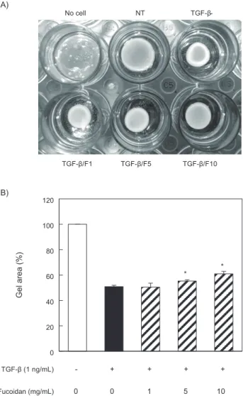
Fig. 2. Effect of fucoidan on TGF-β1-induced fibroblasts contrac- tile activity in rat tail type-1 collagen gel. (A) Human pulmonary fibroblasts (2×10⁵ cells/mL) were cultured in rat tail type I collagen gels (2 mg/mL) and pre-treated with fucoidan for 24 h. Cells were exposed to TGF-β1 (1 ng/mL). TGF-β1 treatment decreased the size of the gel while fucoidan pre-treatment blocked the reduction in gel size. (B) A summation of the percentage of gel surface area in each well of Fig. 2A to the control gel surface area. All values are mean±S.D. for three separate experiments, each performed in triplicate. *P<0.05; compared with the values of TGF-β1 treated group.
0 20 40 60 80
C ell v iab ilit y (%)
Fucoidan (mg/mL)
0 1 5 10 20
No cell NT TGF-β-
TGF-β/F1 TGF-β/F5 TGF-β/F10
0 20 40 60 80 100 120
G el a rea (%)
* *
TGF-β (1 ng/mL) Fucoidan (mg/mL)
-
1 5 10
0
+ 0
+ +
+ A)
B)
fucoidan
의섬유화억제효능을평가하였다.
먼저본연구에들어가기앞서
,
인간섬유모세포에서의fucoi- dan
의세포독성을측정하였다. Fig. 1
에서와같이fucoidan
은20 g/mL
농도에서도독성이없어서이후실험은20 g/mL
이하 의농도를실험에사용하였다.
다음으로, fucoidan
에의한수축 능억제효능을분석을하였다(Fig. 2).
근섬유모세포의과도한 수축능은심장,
폐,
간,
콩팥,
피부등의여러장기에서섬유화를 유발한다(Hinz, 2007).
근섬유모세포의과도한수축능은응축 섬유(stress fibers)
의특징인분화된근섬유모세포의α-SMA
에 의해유도된다(Tomasek et al., 2002).
현재,
근섬유모세포의과 도한수축능은collagen gel
수축분석법을이용한다. TGF-β1
를처리한gel
에서는처리하지않은대조군보다수축이많이되 는것을확인할수있었다.
이러한gel
의수축정도는fucoidan
의처리에의해억제되었고이억제수준은fucoidan
의농도에 비례하여증가하였다.
이러한결과는fucoidan
이폐의섬유화 억제후보물질로서의가능성을가지는것을의미한다.
TGF-β
는TGF-β1, -β2, -β3
의아형이있으며이중TGF-β1
이섬유화기전에가장연구가많이되어있다
. TGF-β
는다기능적사이토카인으로세포증식및분화
,
세포자살에작용하고,
세 포외기질단백질분해를억제하는PAI-1 (plasminogen acti- vator inhibitor-1)
활성과Type І collagen
과같은세포외기질 단백질축적을증가시켜섬유화유도에핵심인자로작용한다(Guo et al., 2005; Leask and Abraham, 2004). TGF-β1
은정 상폐조직의기관지상피세포와섬유모세포에서생산되는주 된사이토카인이다(Coker et al., 1996). TGF-β1
는섬유모세포 를근섬유모세포로의 분화를촉진하고,
분화된근섬유모세포 의지표인α-SMA
을발현을증가시킨다(Hu et al., 2003).
따 라서,
폐섬유모세포에서의α-SMA
와collagen-1
의발현표현 조절기전을이해하면폐섬유화의발생과진행을억제할수있 는치료법을제시할수있을것이다. TGF-β1
에의한폐섬유모 세포의섬유화유도유무와fucoidan
에의한섬유화억제효과 를확인하고자α-SMA
와collagen-1
의단백질발현정도를확 인하였다.
인간섬유모세포에fucoidan
을30
분동안전처리하 고TGF-β1
의자극후24
시간동안배양한세포로부터α-SMA
와collagen-1
항체를이용하여Western blot
방법으로발현억 제효과를확인하고분석하였다. Fig. 3A
에서와같이TGF-β1
IL-6 (pg/ml)
0 200 400 600 800 1000 1200 1400 1600
**
** **
TGF-β (1 ng/mL) Fucoidan (mg/mL)
-
1 5 10
0 + 0
+ + + α-SMA
Collagen-1
Actin
Fucoidan (mg/mL) 0 0 1 5 10
0 0.5 1 1.5
0 0.4 0.8 1.2
a-SMACollagen-1
TGF-β (1 ng/mL) Fucoidan (mg/mL)
- 1 5 10
0 +0
+ + +
**
**
**
Relative intensity (%) control
TGF-β (1 ng/mL) Fucoidan (mg/mL)
-
1 5 10
0 + 0
+ +
+
t-Smad2 p-Smad2 p-Smad3
t-Smad3
A)
TGF-β (1 ng/mL) Fucoidan (mg/mL)
-
1 5 10
0 + 0
+ + + 0
0.4 0.8 1.2
*
** **
p-Smad3
0 0.4 0.8 1.2
** *
p-Smad2
Relative intensity (%) control
TGF-β (1 ng/mL) Smad 2/3 siRNA (nM)
-
20 40
0 + 0
+ +
Collagen-1 α-SMA
Actin
B)
*
**
0 0.5 1
0 0.5 1
Collagen-1α-SMA
TGF-β (1 ng/mL) Smad 2/3 siRNA (nM)
-
20 40
0 + 0
+ +
*
Relative intensity (%) control
A)
p-AKT
t-AKT p-P38
t-P38 TGF-β (1 ng/mL) Fucoidan (mg/mL)
-
1 5 10
0 + 0
+ + +
**
**
**
0 0.4 0.8 1.2
0 0.4 0.8 1.2
TGF-β (1 ng/mL) Fucoidan (mg/mL)
-
1 5 10
0 + 0
+ + +
p-AKTp-P38
Relative intensity (%) control
Collagen-1
Actin α-SMA- TGF-β (1 ng/mL) SB203538 (mM)
-
1 10 0
0 + 0
+ + +
0 0 1
0 0
LY294002 (mM)
0 +
10
B)
*
**
*
**
0 0.4 0.8 1.2
0 0.4 0.8 1.2
TGF-β (1 ng/mL) SB203538 (mM)
-
1 10 0
0 + 0
+ + +
0 0 1
0 0
LY294002 (mM)
0 + 10 α-SMACollagen-1
** *
** **
Relative intensity (%) control
0 100 200 300 400 500 600
IL-6 (pg/mL)
#
**
**
**
TGF-β (1 ng/mL) SB203538 (mM)
-
1 10 0
0 + 0
+ + +
0 0 1
0 0
LY294002 (mM)
0 +
10
C)
Fig. 3. The effect of fucoidan on α-SMA, collagen-1, and IL-6 production by TGF-β1-stimulated human pulmonary fibroblasts.
The Cells were seeded at 8×10⁴ cells/mL and incubated with vari- ous fucoidan concentrations (1, 5, and 10 µg/mL) for 1 h prior to TGF-β1 stimulation (1 ng/mL) (A) After treating with TGF-β1 for 24 h, the cell lysates were resolved on SDS-polyacrylamide gels, transferred to nitrocellulose membranes, and probed using antibodies against α-SMA and collagen-1. Actin was used as an internal control for the western blot analyses. (B) After incubating for 24 h, the culture supernatant was collected, and the quantity of IL-6 was measured using an ELISA. Each value indicates the mean ± S.D. and is representative of results obtained from three independent experiments. (#P<0.05 vs. control group; **P<0.01 vs. TGF-β1 group). Control, untreated cells.
후코이단의 항섬유화 효과
811
에의해
α-SMA
와collagen-1
의단백질의발현이증가되었고, fucoidan
에의해농도의존적으로그발현이감소되었다.
인터루킨
-6 (interleukin-6, IL-6)
는면역및염증반응의조절 을포함한여러기능을갖는다면발현성사이토카인이다. IL-6
는혈관내피세포,
단핵구,
대식세포및섬유모세포등에서생 성되어급성염증반응을촉진하거나억제시킨다(Fries et al., 1994).
또한,
폐섬유모세포의증식을유도하고,
폐섬유증에서 분비가증가된다고알려져있다(Pedroza et al., 2011). TGF-β1
로자극한인간섬유모세포에서fucoidan
의IL-6
분비억제효 능을알아보기위하여, 1
시간전처리한후, 1 ng/mL
의TGF-β1
를24
시간동안처리하였다. TGF-β1
자극에의해분비가증가된
IL-6
는fucoidan
의 처리농도상승에의존적으로 그분 비량이유의하게감소하는것을확인하였다(Fig. 3B).
따라서,
TGF-β
수용체이후의신호전달체계를조절할수있으면폐섬유화의발생과진행을차단할수있는가능성이있다
. TGF-β
의수용체를매개로하는생물학적활성은Smad
의존 적인활성과Smad
신호기전에비의존적인extracellular signal- regulated kinase (ERK), c-Jun-N-terminal kinase (JNK), p38 MAPK
와 같은mitogen activated protein (MAP) kinases
와phosphoinositide 3-kinase/Akt (PI3K/Akt)
세포내신호전달 경로를통해조절된다(Pan et al., 2016).
저자들은fucoidan
이 위와같은세포내신호전달경로를차단하는효능이있는지IL-6 (pg/ml)
0 200 400 600 800 1000
1200 **
** **
TGF-β (1 ng/mL) Fucoidan (mg/mL)
-
1 5 10
0 + 0
+ + + α-SMA
Collagen-1
Actin
0 0.5 1 1.5
0 0.4 0.8 1.2
a-SMACollagen-1
TGF-β (1 ng/mL) Fucoidan (mg/mL)
- 1 5 10
0 +0
+ + +
**
**
**
Relative intensity (%) control
TGF-β (1 ng/mL) Fucoidan (mg/mL)
-
1 5 10
0 + 0
+ +
+
t-Smad2 p-Smad2 p-Smad3
t-Smad3
A)
TGF-β (1 ng/mL) Fucoidan (mg/mL)
-
1 5 10
0 + 0
+ + + 0
0.4 0.8 1.2
*
** **
p-Smad3
0 0.4 0.8 1.2
** *
p-Smad2
Relative intensity (%) control
TGF-β (1 ng/mL) Smad 2/3 siRNA (nM)
-
20 40
0 + 0
+ +
Collagen-1 α-SMA
Actin
B)
*
**
0 0.5 1
0 0.5 1
Collagen-1α-SMA
TGF-β (1 ng/mL) Smad 2/3 siRNA (nM)
-
20 40
0 + 0
+ +
*
Relative intensity (%) control
A)
p-AKT
t-AKT p-P38
t-P38 TGF-β (1 ng/mL) Fucoidan (mg/mL)
-
1 5 10
0 + 0
+ + +
**
**
**
0 0.4 0.8 1.2
0 0.4 0.8 1.2
TGF-β (1 ng/mL) Fucoidan (mg/mL)
-
1 5 10
0 + 0
+ + +
p-AKTp-P38
Relative intensity (%) control
Collagen-1
Actin α-SMA- TGF-β (1 ng/mL) SB203538 (mM)
-
1 10 0
0 + 0
+ + +
0 0 1
0 0
LY294002 (mM)
0 +
10
B)
*
**
*
**
0 0.4 0.8 1.2
0 0.4 0.8 1.2
TGF-β (1 ng/mL) SB203538 (mM)
-
1 10 0
0 + 0
+ + +
0 0 1
0 0
LY294002 (mM)
0 + 10 α-SMACollagen-1
** *
** **
Relative intensity (%) control
0 100 200 300 400 500 600
IL-6 (pg/mL)
#
**
**
**
TGF-β (1 ng/mL) SB203538 (mM)
-
1 10 0
0 + 0
+ + +
0 0 1
0 0
LY294002 (mM)
0 +
10
C)
Fig. 4. The effect of fucoidan on Smad2/3 signaling pathway. The Cells were seeded at 8×10⁴ cells/mL and incubated with various fucoidan concentrations (1, 5, and 10 µg/mL) for 1 h prior to TGF-β1 stimulation (1 ng/mL). (A) After treating with LPS for 30 min, the cell lysates were resolved on SDS-polyacrylamide gels, transferred to nitrocellulose membranes, and probed using antibodies against Smad2 and Smad3. (B) The production of α-SMA and collagen-1 were blunted by siRNA-Smad2/3 transfection in TGF-β1-induced human pulmonary fibroblasts. siRNA-Smad2/3 transfected and non-transfected human pulmonary fibroblasts were stimulated with TGF-β1 (1 ng/mL) for 24 h. Each value indicates the mean±S.D. and is representative of results obtained from three independent experiments. (*P<0.05 and **P<0.01 vs. TGF-β1 group).
임미진
ㆍ
이대성ㆍ
최그레이스ㆍ
이정민ㆍ
최일환812
IL-6 (pg/ml)
0 200 400 600 800 1000
** **
TGF-β (1 ng/mL) Fucoidan (mg/mL)
-
1 5 10
0 + 0
+ + + Collagen-1
Actin
0 0.5 1 1.5
0 0.4 0.8 1.2
a-SMACollagen-1
TGF-β (1 ng/mL) Fucoidan (mg/mL)
- 1 5 10
0 +0
+ + +
**
**
**
Relative intensity (%) control
TGF-β (1 ng/mL) Fucoidan (mg/mL)
-
1 5 10
0 + 0
+ +
+
t-Smad2 p-Smad2 p-Smad3
t-Smad3
A)
TGF-β (1 ng/mL) Fucoidan (mg/mL)
-
1 5 10
0 + 0
+ + + 0
0.4 0.8 1.2
*
** **
p-Smad3
0 0.4 0.8 1.2
** *
p-Smad2
Relative intensity (%) control
TGF-β (1 ng/mL) Smad 2/3 siRNA (nM)
-
20 40
0 + 0
+ +
Collagen-1 α-SMA
Actin
B)
*
**
0 0.5 1
0 0.5 1
Collagen-1α-SMA
TGF-β (1 ng/mL) Smad 2/3 siRNA (nM)
-
20 40
0 + 0
+ +
*
Relative intensity (%) control
A)
p-AKT
t-AKT p-P38
t-P38 TGF-β (1 ng/mL) Fucoidan (mg/mL)
-
1 5 10
0 + 0
+ + +
**
**
**
0 0.4 0.8 1.2
0 0.4 0.8 1.2
TGF-β (1 ng/mL) Fucoidan (mg/mL)
-
1 5 10
0 + 0
+ + +
p-AKTp-P38
Relative intensity (%) control
Collagen-1
Actin α-SMA- TGF-β (1 ng/mL) SB203538 (mM)
-
1 10 0
0 + 0
+ + +
0 0 1
0 0
LY294002 (mM)
0 +
10
B)
*
**
*
**
0 0.4 0.8 1.2
0 0.4 0.8 1.2
TGF-β (1 ng/mL) SB203538 (mM)
-
1 10 0
0 + 0
+ + +
0 0 1
0 0
LY294002 (mM)
0 + 10 α-SMACollagen-1
** *
** **
Relative intensity (%) control
0 100 200 300 400 500 600
IL-6 (pg/mL)
#
**
**
**
TGF-β (1 ng/mL) SB203538 (mM)
-
1 10 0
0 + 0
+ + +
0 0 1
0 0
LY294002 (mM)
0 +
10
C)
Fig. 5. The effect of fucoidan on P38 MAPK and Akt pathway activation induced by TGF-β1. (A) Human pulmonary fibro- blasts were pretreated with fucoidan and then cultured in 1 ng/mL TGF-β1 for 1 h. The levels of p-P38 and p-Akt were determined by Western blot analysis. Cells were pretreated with SB203580 (1 or 10 µM) and LY294002 (1 or 10 µM) 30 min prior to TGF-β1 stim- ulation. (B) After incubating for 24 h, the production of α-SMA and collagen-1 were determined by western blot analysis. (C) After incubating for 24 h, the culture supernatant was collected, and the IL-6 quantity was measured using an ELISA. (#P<0.05 vs. con- trol group; *P<0.05 and **P<0.01 vs. TGF-β1 group). Control, untreated cells.
를조사하였다
.
인간섬유모세포에fucoidan
을1
시간동안전 처리하고TGF-β1
의자극을준후30
분이경과한세포로부터Smad2
와Smad3
의인산화신호기전을Western blot
방법을이용하여확인하였다
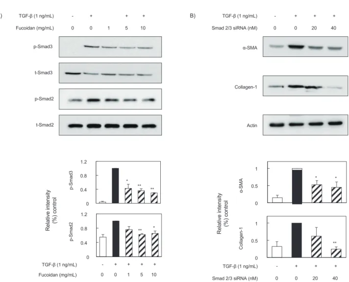
(Fig. 4A). TGF-β1
의자극에의해Smad2
와Smad3
의인산화가촉진되었고,
증가된인산화는fucoidan
에의해감소되었다. Fucoidan
의이러한세포내신호전달경로차단효능을직접적으로검증하기위하여
small interfering RNA (siRNA)
를 사용해Smad
유전자를knock-down
시킨 후α-SMA
와collagen-1
의발현변화를관찰하였다(Fig. 4B).
siRNA
도입후α-SMA
와collagen-1
단백질발현이감소한것 을확인하였다.
또한, Smad
신호기전에비의존적인세포내신 호전달경로인MAPK
와Akt
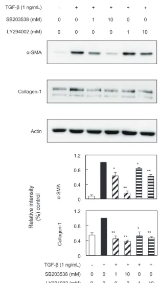
를확인하였다. Fig. 5A
에서와같 이,
인간섬유모세포에TGF-β1
의처리로P38
과Akt
의인산화 가유도되었다.
증가된P38
과Akt
의인산화는fucoidan
의처리 에의해감소되는것을확인할수있었다.
또한, P38
과Akt
특 이억제제는α-SMA
와collagen-1
단백질의발현과(Fig. 5B)
세포밖으로분비된
IL-6
단백질의발현양을감소시키는것을확인할수있었다
(Fig. 5C).
따라서,
본연구에서fucoidan
의생 리활성효능은Smad
의존적인활성과Smad
신호기전에비의 존적인p38 MAPK
과Akt
신호전달경로를통해조절됨을확 인하였다.
이와같이, TGF-β1
의세포내신호전달경로에대한fucoidan
의차단효능은폐섬유화증억제에유용한치료법이될수있음을제시한다
.
이상의결과에서
fucoidan
이TGF-β1
으로자극한인간폐섬 유모세포에서폐섬유증표지단백질(α-SMA, collagen-1, IL-
6)
발현과세포내신호전달경로를차단하는효능을보유하고있음을확인하였다
.
따라서,
본실험을통하여fucoidan
은 폐섬유화증을차단할수있는항-
섬유화물질로서의가능성 을제시하였다.
추가적으로폐섬유화증실험동물을이용하여fucoidan
의효능을검증하여야할것이다.
사 사
본논문은
2011
년도인제대학교학술연구조성비보조에의한것임
.
References
Ahluwalia N, Shea BS and Tager AM. 2014. New therapeutic targets in idiopathic pulmonary fibrosis. Aiming to rein in runaway wound-healing responses. Am J Respir Crit Care Med 190, 867-878. http://dx.doi.org/10.1164/rccm.201403- 0509PP.
Ahmed AB, Adel M, Karimi P and Peidayesh M. 2014. Pharma- ceutical, cosmeceutical, and traditional applications of ma- rine carbohydrates. Adv Food Nutr Res 73, 197-222. http://
dx.doi.org/10.1016/B978-0-12-800268-1.00010-X.
Antoniou KM, Margaritopoulos GA and Siafakas NM. 2013.
Pharmacological treatment of idiopathic pulmonary fibro- sis: from the past to the future. Eur Respir Rev 22, 281-291.
http://dx.doi.org/10.1183/09059180.00002113.
Blobe GC, Schiemann WP and Lodish HF. 2000. Role of transforming growth factor beta in human disease. N Engl J Med 342, 1350-1358. http://dx.doi.org/10.1056/
NEJM200005043421807.
Broekelmann TJ, Limper AH, Colby TV and McDonald JA.
1991. Transforming growth factor beta 1 is present at sites of extracellular matrix gene expression in human pulmonary fibrosis. Proc Natl Acad Sci U S A 88, 6642-6646.
Camelo A, Dunmore R, Sleeman MA and Clarke DL. 2014. The epithelium in idiopathic pulmonary fibrosis: breaking the- barrier. Front Pharmacol 4, 173. http://dx.doi.org/10.3389/
fphar.2013.00173.
Cho Y, Yoon JH, Yoo JJ, Lee M, Lee DH, Cho EJ, Lee JH, Yu SJ, Kim YJ and Kim CY. 2015. Fucoidan protects hepato- cytes from apoptosis and inhibits invasion of hepatocellu- lar carcinoma by up-regulating p42/44 MAPK-dependent NDRG-1/CAP43. Acta Pharm Sin B 5, 544-553. http://
dx.doi.org/10.1016/j.apsb.2015.09.004.
Coker RK, Laurent GJ, Shahzeidi S, HernandezRodriguez NA, Pantelidis P, du Bois RM, Jeffery PK and McAnulty RJ.
1996. Diverse cellular TGF-β1 and TGF-β3 gene expression in normal human and murine lung. Eur Respir 9, 2501-2507.
Cottin V. 2016. Idiopathic interstitial pneumonias with connec- tivetissue diseases features: A review. Respirology 21, 245- 258. http://dx.doi.org/10.1111/resp.12588.
Fries KM, Felch ME and Phipps RP. 1994. Interleukin-6 is an autocrine growth factor for murine lung fibroblast sub- sets. Am J Respir Cell Mol Biol 11, 552-560. http://dx.doi.
org/10.1165/ajrcmb.11.5.7946384.
Guo B, Inoki K, Isono M, Mori H, Kanasaki K, Sugimoto T, Akiba S, Sato T, Yang B, Kikkawa R, Kashiwagi A, Haneda M and Koya D. 2005. MAPK/AP-1-dependent regulation of PAI-1 gene expression by TGF-beta in rat mesangial cells.
Kidney Int 68, 972-984. http://dx.doi.org/10.1111/j.1523- 1755.2005.00491.x.
Hinz B. 2007. Formation and function of the myofibroblast during tissue repair. J Invest Dermatol 127, 526-537. http://
dx.doi.org/10.1038/sj.jid.5700613.
Hu B, Wu Z and Phan SH. 2003. Smad3 mediates transform- ing growth factor-β-induced α-smooth muscle actin expres- sion. Am J Respir Cell MolBiol 29, 397-404. http://dx.doi.
org/10.1165/rcmb.2003-0063OC.
Jin JO, Zhang W, Du JY, Wong KW, Oda T and Yu Q. 2014.
Fucoidan can function as an adjuvant in vivo to enhance dendritic cell maturation and function and promote antigen- specific T cell immune responses. PLoS One 9, e99396.
http://dx.doi.org/10.1371/journal.pone.0099396.
Khalil N, O’Connor RN, Unruh HW, Warren PW, Flanders KC, Kemp A, Bereznay OH and Greenberg AH. 1991. Increased production and immunohistochemical localization of trans- forming growth factor-beta in idiopathic pulmonary fibrosis.
Am J Respir Cell Mol Biol 5, 155-162.
King TE, Costabel U, Cordier JF, Dopoco GA, Bois RM, Lynch D, Myers J, Panos R, Raghu G, Schwartz D and Smith CM. 2000. American Thoracic Society. Idiopathic pulmonary fibrosis: diagnosis and treatment. International
and the European Respiratory Society (ERS). Am J Respir Crit Care Med 161, 646-664. http://dx.doi.org/10.1164/
ajrccm.161.2.ats3-00.
King TE Jr, Pardo A and Selman M. 2011. Idiopathic pulmonary fibrosis. Lancet 378, 1949-1961. http://dx.doi.org/10.1016/
S0140-6736(11)60052-4.
Kuznetsova TA, Besednova NN, Somova LM and Plekhova NG. 2014. Fucoidan extracted from Fucus evanescens pre- vents endotoxin-induced damage in a mouse model of endo- toxemia. Mar Drugs 12, 886-898. http://dx.doi.org/10.3390/
md12020886.
Leask A and Abraham DJ. 2004. TGF-beta signaling and the fibrotic response. FASEB J 18, 816-827. http://dx.doi.
org/10.1096/fj.03-1273rev.
Luo F, Zhuang Y, Sides MD, Sanchez CG, Shan B, White ES and Lasky JA. 2014. Arsenic trioxide inhibits transforming growth factor-β1-induced fibroblast to myofibroblast dif- ferentiation in vitro and bleomycin induced lung fibrosis in vivo. Respir Res 15,51. http://dx.doi.org/10.1186/1465- 9921-15-51.
Moore MW and Herzog EL. 2013. Regulation and Relevance of Myofibroblast Responses in Idiopathic Pulmonary Fibrosis.
Curr Pathobiol Rep 1, 199-208. http://dx.doi.org/10.1007/
s40139-013-0017-8.
O'Leary R, Rerek M and Wood EJ. 2004. Fucoidan modulates the effect of transforming growth factor (TGF)-beta1 on fibroblast proliferation and wound repopulation in in vitro models of dermal wound repair. Biol Pharm Bull 27, 266- 270.
Pan R, Zhang Y, Zang B, Tan L and Jin M. 2016. Hydroxysafflor yellow A inhibits TGF-β1-induced activation of human fetal lung fibroblasts in vitro. J Pharm Pharmacol 68, 1320-1330.
http://dx.doi.org/10.1111/jphp.12596.
Park KY, Back JH, Hur W and Lee SY. 2007. In vitro glucose and bile acid retardation effect of fucoidan from Laminaria japonica. Kor J Biotechnol Bioeng 4, 265-269.
Pedroza M, Schneider DJ, Karmouty-Quintana H, Coote J, Shaw S, Corrigan R, Molina JG, Alcorn JL, Galas D, Ge- linas R and Blackburn MR. 2011. Interleukin-6 contributes to inflammation and remodeling in a model of adenosine mediated lung injury. PLoS ONE 6, e22667. http://dx.doi.
org/10.1371/journal.pone.0022667.
Raghu G, Weycker D, Edelsberg J, Bradford WZ and Oster G.
2006. Incidence and prevalence of idiopathic pulmonary fibrosis. Am J Respir Crit Care Med 174, 810–816. http://
dx.doi.org/10.1164/rccm.200602-163OC.
Tanaka K, Ishihara T, Azuma A, Kudoh S, Ebina M, Nukiwa T, Sugiyama Y, Tasaka Y, Namba T, Ishihara T, Sato K, Mizushima Y and Mizushima T. 2010. Therapeutic ef- fect of lecithinized superoxide dismutase on bleomycin- induced pulmonary fibrosis. Am J Physiol Lung Cell Mol
plung.00289.2009.
Tomasek JJ, Gabbiani G, Hinz B, Chaponnier C and Brown RA.
2002. Myofibroblasts and mechano-regulation of connec- tive tissue remodelling. Nat Rev Mol Cell Biol 3, 349-363.
http://dx.doi.org/10.1038/nrm809.
Wang Q, Wang Y, Hyde DM, Gotwals PJ, Koteliansky VE, Ryan ST and Giri SN. 1999. Reduction of bleomycin in- duced lung fibrosis by transforming growth factor beta solu- blereceptor in hamsters. Thorax 54, 805-812.
Park KH, Cho EH, Kim NC and Chae HJ. 2010. Production of Fucoidan Using Marine Algae. KSBB Journal 25, 223-229.