Osteoblastogenic Activity of Locusta migratoria Ethanol Extracts on Pre-Osteoblastic MG-63 Cells
Minhee Baek
†, Minchul Seo
†, Joon Ha Lee, In-Woo Kim, Mi-Ae Kim and Jae-Sam Hwang*
Department of Agricultural Biology, National Institute of Agricultural Sciences, Rural Development Administration, Wanju-gun, Jeonbuk 55365, Korea
Received September 10, 2018 /Revised November 6, 2018 /Accepted November 7, 2018
Insects have been investigated as a novel source of food and biomaterial in several recent studies.
However, their osteoblastogenic cell activity has not been sufficiently researched and so, to investigate the potential of this natural material for promoting osteoblastogenesis, we studied the activity of Locusta migratoria ethanol extract (LME) on MG-63 pre-osteoblast cells. The cytotoxicity and pro- liferation effects of LME on MG-63 cells were measured by MTS assay, and there was no cytotoxicity up to 1,000 μg/ml. With LME treatment of 500 and 1,000 μg/ml for 48 hr, cell proliferation increased to 105% and 116% versus control, respectively. The osteoblastogenic activity of the LME was meas- ured through alkaline phosphatase (ALP) staining at three and five days. As a result, both 500 and 1,000 μg/ml LME concentrations were seen to increase ALP activity by more than three times com- pared with control at three and five days. In addition, the expression level of the osteogenic markers ALP and RUNX2 was markedly increased after LME treatment. These results demonstrate that Locusta migratoria ethanol extract promotes osteoblastogenesis as evidenced by the increased osteogenic mark- ers and suggest that LME may be a potential agent for bone formation and osteoporosis prevention.
Key words : Alkaline phosphatase, bone formation, Locusta migratoria, MG-63, osteoblastogenesis
†Authors contributed equally.
*Corresponding author
*Tel : +82-63-238-2974, Fax : +82-63-238-3833
*E-mail : [email protected]
This is an Open-Access article distributed under the terms of the Creative Commons Attribution Non-Commercial License (http://creativecommons.org/licenses/by-nc/3.0) which permits unrestricted non-commercial use, distribution, and reproduction in any medium, provided the original work is properly cited.
Journal of Life Science
2018 Vol. 28. No. 12. 1448~1454 DOI : https://doi.org/10.5352/JLS.2018.28.12.1448서 론
뼈는 체내의 장기, 뇌 및 척추를 보호하고 몸의 형태를 유지 하는 역할을 하며 칼슘과 인의 혈중 농도 조절 및 조혈작용에 관여하는 조직이다. 이러한 기능들은 일정한 주기를 통해 일 어나는 골 재형성에 의해 수행되며 이 과정에 의해 골 항상성 이 유지된다[10]. 골 재형성에는 조골세포(osteoblast)와 파골 세포(osteoclast)가 관여하며 칼슘과 인을 분비하여 새로운 뼈 를 형성하는 조골세포의 역할과 노화된 뼈로부터 골 기질을 흡수하는 파골세포의 역할이 균형을 맞춰 조절되며 균형이 깨지는 경우에 골 대사 질환이 발생한다[4, 37]. 골다공증은 가장 흔한 골 대사 질환으로 골 질량의 감소 및 골 파열 위험의 증가가 나타나며 주로 폐경기를 겪는 여성에게서 에스트로겐 결핍에 의해 발생하는 질환이다. 에스트로겐이 부족한 경우 파골세포의 활성이 증가함에 따라 골 흡수가 촉진되어 골다공 증이 유발되는 것으로 보고되고 있다[15, 22].
현대사회에서는 폐경기 여성뿐 아니라 젊은 층 및 남성에게 도 골다공증이 유발되는 것으로 보고되었으며, 요인으로는 체 내 칼슘 결핍, 운동 부족, 유전적 요인, 호르몬의 영향, 약물 복용, 흡연 및 알코올 섭취 등 다양하게 나타난다[31]. 이러한 요인들에 의한 골다공증 발병은 그 원인이 조골세포의 기능저 하에 있는지, 또는 파골세포의 활성 증가에 있는지 정확하게 밝혀지지 않는 상황임에도 불구하고 현재는 에스트로겐, 비스 포스포네이트와 같은 골 흡수 억제제만이 골다공증 치료에 사용되고 있다[26]. 이와 같은 약제를 이용하면 골 소실을 줄일 수 있지만 이미 진행된 골 소실에 대한 회복 효과는 없어 골다 공증의 완전한 예방 및 치료는 불가능한 실정이다[19, 20]. 따 라서 골다공증 예방 및 치료를 위해 골 형성을 증가시키는 연구에 대한 관심이 증가하는 추세이며 복분자, 황금 및 가시 오가피와 같은 천연물을 이용하여 조골세포 활성화에 미치는 효능 및 효과에 대한 연구가 진행되었다[20, 24, 31].
조골세포는 골수의 간엽줄기세포(mesenchymal stem cell)
로부터 기원한 것으로, 지방세포, 근원세포, 연골세포 및 섬유
아세포로 분화되기도 한다[29]. 조골세포는 뼈의 세포 외 기질
을 합성하고 칼슘 침착 및 미네랄화를 조절하며 기계적 자극
에 반응하는 기능을 함으로써 뼈의 형성 및 재형성 과정에서
가장 중요한 역할을 한다[12, 16]. 따라서 골다공증 예방 및
치료 소재 개발을 위해서는 조골세포를 이용한 연구가 필요할
것으로 사료된다. 조골세포는 골의 표면에 근접해 분포하며
세포질 내에는 과립형질 내 세망이 발달하였고, 세포막에는
당단백 효소인 alkaline phosphatase (ALP)가 존재하는데 이 효소는 기질 특이성과 염기성 pH에서 최적의 활성을 나타내 고, 석회화 과정 동안 무기인산의 운반, 세포분열 및 분화의 조절자로서 세포의 외막과 석회화 조직의 기질 소포에서 높은 농도로 발견이 되며 이를 통해 골세포 분화의 표지인자로 사 용되고 있다[13, 14, 32].
현재 지구상에 존재하는 곤충은 130만 종으로 알려져 있으 나 그에 비해 곤충을 이용한 소재 개발은 미비한 실정이다.
과거 동의보감, 본초강목을 통해 다양한 효능이 보고되었으 며, 극한 외부 환경 조건에서 살아가는 곤충의 특성 상 체내에 기능성을 가진 2차 대사산물을 함유하고 있을 것으로 추정되 고 있다[18]. 뿐만 아니라 미래 인구 증가에 따른 식량 부족에 대한 대안으로써 식용곤충이 각광받고 있으며, 현재 국내에 7종의 식용곤충이 일반식품으로 식품공전에 등록되어 있다 [2]. 최근에는 다양한 곤충 유래의 생리활성 물질에 대한 연구 가 활발히 진행되고 있으며 곤충의 기능성 연구를 통해 식용 및 약용으로 곤충의 이용이 증가할 것으로 사료된다[9].
풀무치(Locusta migratoria)는 메뚜기목(Orthoptera) 메뚜기 과(Acrididae)의 곤충으로 우리나라를 포함하여 아시아, 아프 리카, 오스트레일리아, 유럽 등 전세계적으로 분포한다[5, 25].
크기는 4-6 cm 정도이고, 하루에 100 km를 이동할 수 있을 정도로 이동성이 강한 것으로 알려져 있다[21]. 풀무치는 사료 용, 식·약용 등으로 사용되는 유용한 곤충 종 중 하나로 다양한 목적으로의 연구를 위하여 실험실에서 흔하게 사육되고 있고, 체코, 폴란드, 터키 등 유럽 국가에서는 수출용으로 사육되고 있다[11]. 현재까지 풀무치를 이용한 연구로는 집단형 풀무치 에 대한 방제 연구[21], 국내 풀무치의 계통 분석[17], 풀무치 유래 펩타이드 및 GST의 구조 및 기능 분석[23, 27] 등이 진행 되었으나 풀무치를 식·약용 소재로써 이용하기 위한 연구로는 풀무치 물 추출물을 이용하여 유전독성을 확인한 것이 유일하 다[33]. 따라서 본 연구에서는 풀무치 추출물이 MG-63 조골세 포 증식 및 활성에 미치는 영향을 분석하고자 하였으며, 이를 통해 풀무치 추출물의 골다공증 개선 가능성을 확인하고자 하였다.
재료 및 방법
풀무치 추출물 제조
본 연구에 사용된 풀무치(Locusta migratoria)는 국립농업과 학원 곤충산업과에서 실내 계대 사육한 해남 6세대를 사용하 였다. 실험 곤충은 밀을 먹이로 사용하였으며 30℃, 65% R.
H., 9L/15D, 1,800 Lux의 조건에서 사육하였다. 풀무치 추출 물은 상기 풀무치를 동결건조 한 후 분말화 하여 Baek 등[2]
방법에 따라 추출하였다. 풀무치 동결건조 분말을 70% 에탄올 과 혼합하여 초음파 처리(250 J, 10 s, twice)한 후 30분간 실온 에서 방치하였다. 혼합액의 상등액을 걸러 centrifugal evapo-
rator (EYELA, Tokyo, Japan)를 이용하여 완전히 건조한 후 사용 전까지 -20℃에서 냉동 보관하였다.
세포주
조골세포(human osteoblast-like cell, MG-63)는 ATCC (VA, USA)에서 구입하였으며 10% FBS (fetal bovine serum) (Hyclone, UT, USA)와 1× penicillin-streptomycin (Hyclone) 이 첨가된 Dulbecco’s Modified Eagle Medium (DMEM) (Hyclone)을 사용하여 배양하였고 세포는 37℃, 5% CO
2의 배 양조건을 유지하였다. 세포가 충분히 성장하는 2-3일 간격으 로 계대 배양하며 실험을 진행하였다.
세포 생존율 측정
풀무치 추출물이 MG-63의 세포 생존율에 미치는 영향은 Chao 등[6]의 방법을 응용하여 확인하였다. MG-63에 대한 풀 무치 추출물의 세포독성 및 세포증식을 확인하기 위하여 MG- 63을 96-well plate (SPL life sciences, Korea)에 5.0×10
3cells/
well로 분주하여 약 80%의 confluency에 도달할 때까지 10%
FBS (Hyclone)가 들어있는 배지에서 약 24시간 동안 배양하였 다. 그 후 풀무치 추출물을 100, 500 및 1,000 μg/ml의 농도로 처리하여 24시간 및 48시간 동안 추가 배양한 후 MTS (3-(4,5- dimethylthiazol-2-yl)-5-(3-carboxymethoxyphenyl)-2- (4-sulfophenyl)-2H-tetrazolium) reagent (Sigma, MO, USA) 를 사용하여 세포 생존율을 측정하였다.
ALP (Alkaline phosphatase) 효소 활성 측정
ALP (염기성 인산 분해 효소) 효소 활성 검사는 p-nitro-
phenyl phosphate (p-NPP) (Sigma)의 가수분해 반응에 ALP
가 촉매로 작용하는 원리를 이용하여, 가수분해 산물인 p-ni-
trophenol의 양을 측정함으로써 ALP의 활성을 간접적으로 확
인하는 방법이다. 풀무치 추출물 처리 시 조골세포의 분화 정
도 비교를 위해 positive control로서 50 μg/ml ascorbic acid
(Sigma)와 10 mM β-glycerophosphate (Sigma)가 함유된 조
골세포 분화배지(DM)를 함께 처리하였다[28]. MG-63세포를
96-well plate (SPL life sciences)에 1.5×10
4cells/well 농도로
분주하여 24시간 배양한 다음, 농도별 풀무치 추출물(100, 500,
1,000 μg/ml) 및 조골세포 분화 배지로 교환하여 2일마다 배지
를 갈아주며 3일 및 5일간 배양하였다. 배양한 세포는 PBS
(Caisson laboratories, UT, USA)로 세척하고 0.1% Triton
X-100 (Sigma)을 20 μl씩 분주하여 37℃, 5% CO
2조건에서
30분간 lysis 한 후, 100 μl의 p-NPP (Sigma)을 분주하여 30분
간 반응시켰다. 반응 후 3 M NaOH (Sigma)로 반응을 정지한
후 405 nm에서 흡광도를 측정하여 ALP 효소에 의해 p-nitro-
phenol로 전환된 양을 측정하였고 이를 통해 ALP 효소 활성
을 산출하였다.
Table 1. The sequences of primers for RT-PCR
Primer Sequence
GAPDH F: 5’-GAAGGTGAAGGTCGGAGTC-3’
R: 5’-GAAGATGGTGATGGGATTTC-3’
ALPL F: 5’-AAACCGAGATACAAGCACTC-3’
R: 5’-TCCGTCACGTTGTTCCTGTTCAG-3’
Runx2 F: 5’-GCCTTCAAGGTGGTAGCCC-3’
R: 5’-CGTTACCCGCCATGACAGTA-3’
ALP (Alkaline phosphatase) staining
ALP (염기성 인산 분해 효소) 활성 및 조골세포의 분화 정 도를 확인하기 위하여 ALP staining을 통해 염색 정도를 확인 하였다. MG-63세포를 96-well plate (SPL life sciences)에 1.5×
10
4cells/well 농도로 분주하여 24시간 배양한 다음, 농도별 풀무치 추출물(50, 100, 500, 1,000 μg/ml) 및 조골세포 분화배 지로 교환하여 2일마다 배지를 갈아주며 5일간 배양하였다.
배양된 세포는 PBS (Caisson laboratories)로 세척하였고 10%
formalin (Biosesang, Korea)을 사용하여 세포를 고정한 후, BCIP/NBT substrate solution (Sigma)을 분주하여 빛이 없는 조건에서 1-2시간 동안 방치하였다. 세포에 푸르게 염색된 정 도를 통해 조골세포의 분화 및 ALP 활성을 확인하였다.
RT-PCR
MG-63 세포를 3×10
5cells/well의 농도로 6-well plate (SPL life sciences)에 분주하여 24시간 배양하였다. 그 후 농도별 풀무치 추출물(50, 100, 500, 1,000 μg/ml) 및 조골세포 분화배 지로 교환하여 2일마다 배지를 갈아주며 3일 및 5일간 배양하 였다. TRIzol reagent (Ambion Life Technology, MA, USA)를 이용하여 RNA를 분리하였으며 RNA의 정량은 50배 희석한 후 UV/vis 분광광도계(Beckman coulter, CA, USA)를 이용하 여 260 nm에서 흡광도를 측정하였다. 정량한 RNA는 cDNA reverse transcription kit (Thermo Fisher Scientific, MA, USA)를 이용하여 cDNA를 합성하였고, qPCR Green Mix (ENZO, NY, USA)와 Table 1에 제시한 primer (Bioneer, Korea)를 사용하여 Real-time PCR 반응을 수행하였다. 측정하 고자 하는 목적 유전자의 발현은 Gapdh의 발현 양을 이용하 여 정량화 하였다.
Western blot
MG-63 세포를 3×10
5cells/well의 농도로 6-well plate (SPL life sciences)에 분주하여 24시간 배양하였다. 그 후 농도별 풀무치 추출물(50, 100, 500, 1,000 μg/ml) 및 조골세포 분화배 지로 교환하여 2일마다 배지를 갈아주며 5일간 배양하였다.
배양이 끝나는 날, 세포를 PBS (Caisson laboratories)로 세척 한 뒤 protease와 phosphatase inhibitor cocktail가 함유된 RIPA buffer를 사용하여 세포를 lysis 하고 원심분리하여 상등 액을 얻은 후 BCA를 이용하여 단백질을 정량하였다. 그 후
SDS-PAGE gel에서 전기영동을 통해 단백질을 분자량에 따라 분리하고, PVDF membrane으로 transfer 하였다. Membrane 을 5% skim milk 용액으로 blocking 한 후 rabbit-anti-ALP 및 rabbit-anti-RUNX2 항체를 4℃에서 overnight으로 반응시 켰으며, 그 후 각 항체에 대한 2차 항체를 반응시킨 후 ECL kit를 이용하여 각 band를 관찰하였다. Western blot에 사용한 시약은 Thermo fisher (MA, USA)로부터 구입하여 사용하였 으며, 1차 항체는 Cell signaling (MA, USA), 2차 항체는 Promega (WI, USA)에서 구입하여 사용하였다.
통계처리
모든 실험 결과는 3회 반복하여 평균과 표준편차(mean±
SD)로 나타냈다. 실험군 간의 유의성은 Student’s t-test를 통 해 검정하였고, p<0.05일 때 군 간의 차이가 유의적인 것으로 판단하였다.
결과 및 고찰
조골세포 증식 및 독성 평가
MG-63 세포는 인간 골육종으로부터 추출된 세포로 Alkaline phosphatase (ALP), Runt-related transcription factor 2 (Runx 2) 및 Osteoprotegerin (OPG) 등 다양한 골 형성 관련 인자들 이 발현되어 골 형성 연구에서 primary osteoblast cell을 대체 하여 폭넓게 사용되고 있다[8]. 풀무치 추출물이 MG-63 조골 세포의 증식 및 독성에 미치는 영향을 확인하기 위해서 추출 물을 24시간 및 48시간 동안 처리한 후 세포 생존율을 측정하 였다. 세포 생존율 측정 결과, LME는 1,000 μg/ml의 범위에서 MG-63 세포 독성을 유발하지 않았으며, 48시간 동안 배양한 경우, 500 μg/ml과 1,000 μg/ml 농도에서 유의적으로 세포 생존율이 증가함에 따라 풀무치 추출물의 세포증식 효능을 확인하였다(Fig. 1). 따라서 이후의 실험은 LME의 안전한 농도 인 1,000 μg/ml 이하의 농도에서 수행하였다.
Alkaline phosphatase (ALP) 활성에 미치는 영향 염기성 인산 분해 효소(alkaline phosphatase, ALP)는 거의 모든 조직에 분포하며 특히 골 조직에서는 골 성장이 일어날 때 그 활성이 증가하는 것으로 알려져 있다[3]. 따라서 조골세 포의 활성을 나타내는 표지인자로써 조골세포에서의 ALP 활 성을 측정하여 풀무치 추출물이 조골세포의 골 성장 및 골 세포 분화 활성에 미치는 영향을 확인하였다.
조골세포의 ALP 활성은 시료처리를 하지 않은 control (CTR) 군에 대한 비율로 나타내었으며 그 결과는 Fig. 2와 같 다. 풀무치 추출물을 농도별로 처리한 결과 100 μg/ml 농도부 터 활성이 유의적으로 증가하였고 1,000 μg/ml 농도에서는 CTR군에 비해 3배 정도 활성이 증가하는 것으로 나타났다.
Positive control로서 함께 처리한 조골세포 DM군의 활성은
Fig. 1. Effect of Locusta migratoria extract on the osteoblastic cell cytotoxicity. MG-63 cells were seeded into 96-well plate (5×10
3cells/well), and then treated with LME (100-1,000 μg/ml) for 24 hr and 48 hr. Cell cytotoxicity was de- termined using 3-(4,5-dimethylthiazol-2-yl)-5-(3-carbox- ymethoxyphenyl)-2-(4-sulfophenyl)-2H-tetrazolium (MTS) assay. Values are the mean ± SD of triplicate. *p
<0.05, compared with the control of 24 hr.
#p<0.05, compared with the control of 48 hr. CTR, control; LME, Locusta migratoria extract.
Fig. 2. Effect of Locusta migratoria extract on the ALP activity and staining of osteoblastic cells. MG-63 cells were seeded into 96-well plate (1.5×10
4cells/well), and then treated with LME (100-1,000 μg/ml) and ascorbic acid with β- glycerophosphate as a differentiation medium (DM) for 3 days and 5 days. ALP enzyme activity was measured by spectro-photometric method using p-nitrophenyl phos- phate as a substrate. Values are the mean ± SD of triplicate. *p<0.05, compared with the control of 3 days.
#
p<0.05, compared with the control of 5 days. Cells were stained with BCIP/NBT substrate solution after fixation.
CTR, control; DM, 50 μg/ml L-ascorbic acid with 10 mM β- glycerophosphate; LME, Locusta migratoria extract.
풀무치의 100 μg/ml 농도에서의 활성과 유사하였다. 이는 이 전 연구에서 보고된 오골계의 멜라닌 추출물(100-150%), 적송 잎(100-140%) 및 황금(100-150%) 추출물의 ALP 활성 측정 결
과에 비해 현저하게 높은 ALP 활성을 나타내고 있다[14, 31, 36]. 이러한 결과는 ALP 염색결과와 유사하게 나타났다. CTR 군에 비해 DM군과 LME 처리군에서 푸른색이 더욱 진하게 나타났으며 LME 처리 시 농도 의존적으로 진하게 염색되었 다. 따라서 풀무치 추출물은 MG-63 조골세포의 ALP 활성을 증가시키며 이에 따라 조골세포의 분화가 증가하였음을 확인 하였고, 골 성장 촉진에 도움을 줄 수 있는 소재로 사용할 수 있을 것으로 사료된다.
MG-63 조골세포의 유전자 발현에 미치는 영향
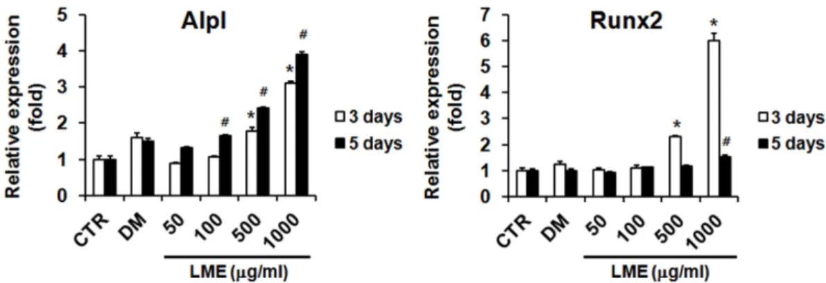
풀무치 추출물을 처리하고 3일 또는 5일 후, MG-63 조골세 포로부터 RNA를 추출하여 골 성장 및 분화와 관련된 유전자 발현의 변화를 확인하고자 하였다(Fig. 3). ALP 활성과 관련된 alkaline phosphatase (Alpl) 유전자의 발현은 CTR군에 비하 여 농도 의존적으로 약 4배까지 증가하였으며 3일 배양했을 때 보다 5일까지 배양한 경우에 유전자 발현량이 보다 높은 것을 확인하였다. 또한 DM군과 유사하거나 높은 Alpl 발현량 은 앞서 ALP 활성 및 염색을 통해 확인한 결과와 유사하였다.
Runt-related transcription factor 2 (Runx2)의 유전자 발현량 은 Alpl과 다르게 3일 배양한 경우 1,000 μg/ml 농도에서 CTR 군에 비해 6배 정도로 매우 높은 증가율을 나타내었으나 5일 배양 후에는 그 발현 양이 감소함을 확인 할 수 있었다. 이러한 결과를 통해 풀무치 추출물에 의해서 Runx2가 먼저 발현이 되고 그 영향으로 Alpl의 발현이 증가한 것으로 사료되며 이는 syringic acid를 사용하여 Alpl과 Runx2 등 유전자 발현을 연 구한 결과에서 Runx2 유전자 발현이 Alpl 유전자 발현에 영향 을 미치며, Alpl의 발현뿐 아니라 분화 초기부터 조골세포의 분화를 조절하는 역할을 한다는 이전의 연구 결과와 유사하다 [1, 30, 34]. 따라서 풀무치 추출물은 Runx2와 Alpl 유전자 발현 에 영향을 주며 이에 따라 조골세포의 분화를 촉진하며 골 형성을 촉진하는 효과가 있을 것으로 판단된다.
MG-63 조골세포의 단백질 발현에 미치는 영향
풀무치 추출물을 처리하고 5일 후에 MG-63 조골세포로부
터 단백질을 추출하여 골 성장 및 분화와 관련된 단백질의
발현을 확인하고자 하였으며 그 결과는 Fig. 4와 같다. ALP
단백질 발현을 확인한 결과 50 μg/ml부터 1,000 μg/ml까지
농도의존적으로 발현량이 증가하는 것을 확인하였다. 이는 앞
서 Alpl 유전자 발현을 확인한 결과와 유사한 것으로 판단된
다. RUNX2 단백질 발현의 경우 유전자 발현 결과와 유사하게
50 μg/ml과 100 μg/ml 농도에서는 증가하지 않았으나 500
μg/ml과 1,000 μg/ml 농도에서는 풀무치 추출물의 농도에
따라 발현량이 증가됨을 확인할 수 있었다. 반면, positive con-
trol인 DM군에서는 RUNX2의 발현량이 증가하지 않았는데
이 결과 또한 앞서 Runx2의 유전자 발현량을 확인한 결과와
유사하였다. 이는 RUNX2의 발현은 ascorbic acid로부터 영향
Fig. 3. Effect of Locusta migratoria extract on osteoblatogenesis related gene expression. MG-63 cells were seeded into 6-well plate (3×10
5cells/well), and then treated with LME (50-1,000 μg/ml) and ascorbic acid with β-glycerophosphate as a differentiation medium (DM). RNA was isolate at 3 days and 5 days after the sample treatment. The levels of Alpl and Runx2 mRNA were determined by quantitative real-time RT-PCR. The data were normalized to Gapdh. Results are shown as the mean
± SD. *p<0.05, compared with the control of 3 days.
#p<0.05, compared with the control of 5 days. CTR, control; DM, 50 μg/ml L-ascorbic acid with 10 mM β-glycerophosphate; LME, Locusta migratoria extract.
Fig. 4. Effect of Locusta migratoria extract on osteoblatogenesis related protein expression. MG-63 cells were seeded into 6-well plate (3×10
5cells/well), and then treated with LME (50-1,000 μg/ml) and ascorbic acid with β-glycerophosphate as a differentiation medium (DM). Protein was isolate at 3 days and 5 days after the sample treatment. The expression levels of ALP and RUNX2 protein were determined by Western blot. The data were normalized to β-actin. Results of densitometric analysis of the Western blot are also shown (right). Results are shown as the mean ± SD. CTR, control; DM, 50 μg/ml L-ascorbic acid with 10 mM β-glycerophosphate; LME, Locusta migratoria extract.
을 받지 않는 이전의 연구에 부합하는 결과인 것으로 판단된 다[35]. 단백질 발현량의 측정 결과에 따라 풀무치 추출물은 MG-63 조골세포에서 ALP 및 RUNX2의 단백질 발현을 증가 시키며 이를 통해 MG-63 조골세포의 분화를 촉진하고 골 형 성 기능을 가지며 결과적으로 골다공증 예방 및 치료에 도움 을 줄 수 있을 것으로 사료된다.
감사의 글
본 연구는 농촌진흥청 어젠다 사업(과제 번호: PJ01311002) 의 지원에 의해 이루어졌으며, 이에 감사드립니다.
References