Differential Cytotoxicity of Penta-O-galloyl-β-D-glucose in Human Cancer and Normal Cell Lines of Various Origins
Hyeon-Jeong Lee1, Min-Gyeong Kim1, Song-Yeong Lee1, Min-Hyock Song1, Yoon-Dong Kim1, Jeong-Sook Ha2, Gie-Joon Jeong2, Gyu-Jin Rho3 and Byeong-Gyun Jeon2,4*
1Gyeongnam Science High School, Jinju 52828, Korea
2Department of Biology Education, College of Education, Gyeongsang National University, Jinju 52828, Korea
3OBS/Theriogenology and Biotechnology, College of Veterinary Medicine, Gyeongsang National University, Jinju 52828, Korea
4Research Institute of Education, Gyeongsang National University, Jinju 52828, Korea Received June 8, 2016 /Revised August 1, 2016 /Accepted September 22, 2016
The present study examined the cytotoxic effects of 1, 2, 3, 4, 6-penta-O-galloyl-β-D-glucose (PGG), known as the pentahydroxy gallic acid ester of glucose, in the various human cancer cell lines (A-549, MDA-MB-231, U87-MG, MCF-7 and PANC-1), normal MRC-5 fetal fibroblasts, and dental papilla tis- sue-derived mesenchymal stem cells (DPSCs). Significantly (p<0.05) lower half maximal inhibitory con- centration (IC50) values were observed in the A-549 and MDA-MB-231 cells showing a high pro- liferation capacity, compared with other cancer and normal cell lines with a relatively low proliferation capacity. The population doubling time (PDT) was significantly (p<0.05) higher in the 10 μM PGG- treated cell lines than those of untreated control cell lines. The present study demonstrated that the IC50 value increases proportionally to the extending PDT. A high cell number with senescence-asso- ciated ß-galactosidase activity was also observed in the 10 μM PGG-treated cells compared with those of untreated control cells. Moreover, the level of telomerase activity was significantly (p<0.05) de- creased with 10 μM PGG treatment, especially in A-549 and MDA-MB-231 cells showing a high pro- liferation capacity. Based on these observations, PGG could serve as a potent agent for cancer chemo- therapy, as its treatment was more effective in cells with a high proliferation capacity.
Key words : Cancer cells, human, PGG, proliferation, telomerase activity
*Corresponding author
*Tel : +82-55-772-2236, Fax : +82-55-772-2239
*E-mail : [email protected]
This is an Open-Access article distributed under the terms of the Creative Commons Attribution Non-Commercial License (http://creativecommons.org/licenses/by-nc/3.0) which permits unrestricted non-commercial use, distribution, and reproduction in any medium, provided the original work is properly cited.
Journal of Life Science 2016 Vol. 26. No. 11. 1320~1329 DOI : http://dx.doi.org/10.5352/JLS.2016.26.11.1320
Introduction
Globally, many people are suffering from various types of cancers. Cancer is generally characterized by an un-con- trolled cell proliferation due to the failure in growth regu- lation and control of metastasis, consequently which invades into other tissues of the body. The untreated cancers lead to death of patients with severe pain. Although, various treatments such as surgery, radiation, chemotherapy, im- munotherapy and others have been successfully applied for cancer therapy, more efficient and economic treatments are required to increase the survival rate of cancer patients.
Several glycoside compounds, including amygdalin (cyano- genic glycoside) from mainly bitter almonds, plant saponins
and ginseng are often used as anti-cancer drugs in chemo- therapy treatment [10, 30, 34]. Recently, 1, 2, 3, 4, 6-Penta-O- galloyl-β-D-glucose (PGG) found in many plant species, a simple hydrolysable tannin, has been considered as a candi- date anti-cancer drug. It has been previously demonstrated that PGG treatment induces cellular cytotoxic effects, includ- ing the induction of cellular apoptotic cell death, elevated expression of activating p53 tumor suppressor, arrest of cell cycle and blocking angiogenesis by inhibiting VEGF ex- pression in several types of human cancer cells [5, 8, 12, 21].
Moreover, different studies have reported a wide variety of biological activities, such as anti-microbial, anti-platelet-co- agulation functions as well as anti-tumor effects in human cancer cells by PGG treatment [4]. Additionally, PGG has been reported to be a strong inhibitor of fatty acid synthase (FAS), a type of lipogenic enzyme, through competition with the active domains of the enzyme subsequently leading to the anti-tumor activity in human cancer cells [35]. Generally, the FAS activity is over-expressed in the cell lines showing high rate of cell proliferation, such as cancer cells [33]. The fatty acid in the cells showing high proliferation rate could
be continuously supplied for new cell membrane, probably presuming that arrest of cell cycle phase can potentially be induced in the cells treated with PGG. However, the cellular cytotoxicity related with cell proliferation rate and pop- ulation doubling time by PGG treatment is not yet inves- tigated. Furthermore, even though PGG possesses the re- markable cellular cytotoxicity against several human cancer cells, the anti-cancer effects in the various human normal and cancer cell lines is not fully understood until recently.
More interestingly, the selective cellular cytotoxicity in the cancer cell lines is an important factor for the process of cancer therapeutic treatment while maintaining less cyto- toxic effects in the normal cell lines.
Cancer cells generally form a cell lumps through un- limited cell division with loss of contact inhibition. The un- limited cell division of cancer cells is tightly regulated with the maintenance of telomeric repeats that consist of repeti- tive DNA sequences (GGTTAGn) at the end of each eukary- otic chromosome, and high telomerase activity is responsible for the maintenance of telomeric repeats [3]. Therefore, the telomerase activity has been considered as a main biomarker in cancer treatment in vitro [18, 28]. Previously, many studies have focused on telomere and telomerase-targeting drug in cancer cell lines [7, 25, 27]. However, the effects on telomer- ase activity are not fully investigated in the various human cancer and normal cell lines treated with PGG.
Herein, we investigated the cytotoxic effects by PGG treat- ment in various types of human cancer and normal cell lines, including A-549 lung adenocarcinoma, MDA-MB-231 breast adenocarcinoma, MCF-7 breast adenocarcinoma, U87-MG brain glioblastoma astrocytoma and PANC-1 pancreatic car- cinoma cancer cells as cancer cell lines, and MRC-5 fetal lung fibroblasts and dental papilla tissue-derived mesenchymal stem cells (DPSCs) of third molar (wisdom tooth) as normal cell lines. The IC50 value by 3-(4, 5-Dimethyl-2-thiazolyl)-2, 5-diphenyltetrazolium bromide (MTT) assay was preferen- tially decided in the each of cell lines treated with PGG.
After PGG treatment at an established concentration, the cy- totoxic effects of PGG were investigated by the changes of population doubling time and senescence-associated-ß-gal- actosidase activity in the cell lines of various. Furthermore, the relative telomerase activity was also examined in the each of cell lines treated PGG.
Materials and Methods Preparation and culture of cells
All chemicals and media used in this study were pur- chased from Sigma (Sigma, St. Louis, MO, USA) and Gibco (Invitrogen, Carlsbad, CA, USA), unless otherwise specified.
The pH and osmolality in media was adjusted to 7.4 and 280 mOsm/kg, respectively. A-549 lung adenocarcinoma, MDA-MB-231 breast Adenocarcinoma, MCF-7 breast Adeno- carcinoma, U87-MG brain glioblastoma astrocytoma, PANC- 1 pancreatic carcinoma cancer cells and MRC-5 normal fetal lung fibroblasts were purchased from the American Type Culture Collection (Manassas, VA, USA). The DPSCs used for this experiments were derived from extracted third molar tooth and the basic characterizations of stem cell in the iso- lated DPSCs were demonstrated in the previous study [15].
All cells were maintained in advanced-Dulbecco's modified eagle medium (A-DMEM) supplemented with 3% fetal bo- vine serum (FBS) and 1.0% penicillin-streptomycin (10,000 IU and 10,000 μg/ml, respectively) at 37.5°C in a humidified atmosphere of 5% CO2 in air and sub-cultured upon con- fluence (70-80%), while culture media were changed twice a week.
Cytotoxicity test by MTT assay
The cellular cytotoxicity of PGG was analyzed by MTT assay and then the half maximal inhibitory concentration (IC50) value was determined in each of the cell lines. Briefly, both cancer and normal cells were seeded at 5×10⁴ per well into a 6-well plate and incubated at 37.5℃ in a humidified atmosphere of 5% CO2 in air. After overnight incubation, media were carefully removed, and replaced with complete A-DMEM media containing 0 (control), 2.5, 5.0, 10, 20 and 50 μM PGG for 48 hr. After washing with Dulbecco's phos- phate-buffered saline (D-PBS) for 3 times, the MTT assay was performed by adding 1 ml of 5 mg/ml MTT stock sol- ution into each well and incubated at 37℃ for 4 hr. After removing MTT stock solution, wells were washed with D-PBS for 3 times. Finally, 200 μl dimethyl sulfoxide (DMSO) solution was added and incubated for 15 min. The DMSO solution was then collected and measured with an ELISA microtiter plate reader (Bio-Tek, Winooski, VT, USA) at 570 nm wavelength and the IC50 value was calculated for each cell lines.
Analysis of PDT and correlation coefficient Both cancer and normal cell lines were seeded at 1×10⁴ cells/well into a 6-well plate and cultured in complete A-DMEM media containing 0 (control) and 10 μM PGG for
7 days at 37.5˚C in a humidified atmosphere of 5% CO2 in air by changing culture medium for every 3 days. After 7 days, cells were harvested with 0.1% (w/v) trypsin and counted using a hemocytometer. The PDT value was calcu- lated using the formula DT = t (log 2)/(log Nt-log No), where t represents the culture time, and No and Nt are the initial and final cell numbers respectively. Furthermore, when IC50 values and PDT were determined, the correlation coefficient analysis was performed between two values us- ing Exel program (Microsoft, Redmond, WA, USA).
Analysis of senescence-associated β-galactosidase activity
The activity of senescence-associated ß-galactosidase was carried out with cell senescence assay kit (Cell Signaling Technology, Danvers, MA, USA) according to the manu- facturer’s protocols. Briefly, the each of cell lines were treat- ed in the media containing with 0 (control) and 10 μM PGG in 6-well plate for 7 days. After being washed with PBS, the cells were fixed with 1 ml fixative solution for 10-15 min at room temperature. The cells were then incubated with 1 ml senescence-associated ß-galactosidase staining solution at 37oC for overnight. The cells stained with blue color were examined under an inverted microscope (Nikon, Tokyo, Japan) equipped with CCD camera and image program.
Analysis of telomerase activity by relative-quantita- tive telomerase repeat amplification protocol (RQ- TRAP)
The conventional ELISA-based TRAP assay was modified for the real time RQ-TRAP using the LightCycler 3.0 (Roche, Mannheim, Germany), and the telomerase activity was quan- tified as previously described [19]. Briefly, 1×10⁵ cells/sam- ple were harvested and lysed in 400 μl of iced 0.5% (v/v) 1·3-[(3-cholamidopropyl) dimethylam-monio] propanesulfonic acid (CHAPS) lysis buffer (pH 7.5) supplemented with 10 mM Tris-HCl, 1 mM MgCl2, 1 mM EGTA, 0.1 mM benzami- dine, 5 mM 2-mercaptoethanol and 10% glycerol for 30 min at 4°C. And the samples were centrifuged for 20 min at 12,000 × g at 4°C. Protein concentration was measured by a spectrophotometer (Mecasys, Daejeon, Korea) and 5 μg of total proteins were used for RQ-TRAP. The RQ-TRAP was performed using the PCR reagent LightCycler FastStart DNA Master SYBR Green 1 (Roche, Germany), containing lysed samples, 2.5 mM MgCl2, 0.02 μg of primer TS (5’-AAT CCG TCG GAG CAG AGT T-3’), 0.04 μg of primer ACX
(5’-GCG CGG CTT ACC CTT ACC CTT ACC CTA ACC-3’), according to the manufacturer’s protocol and final volume was adjusted to 20 μl with RNase-free water. The RQ-TRAP protocol consisted of 30 min incubation at 30°C, followed by a denaturing cycle of 10 min at 94°C, and 40 cycles of PCR (94°C for 30 sec, and 60°C for 90 sec). All samples were quantified using the LightCycler Quantification Software’s (Roche, Mannheim, Germany) second derivative method of crossing point (Cp) determination, and the relative telomer- ase activity of untreated control MRC-5 fibroblasts was con- sidered as 100% for comparison with the treatment groups in five replicates.
Statistical analysis
Differences among the cell groups were analyzed by us- ing one-way analysis of variance (ANOVA). Differences in the IC50 value, population doubling time and relative telo- merase activity were analyzed using a Student’s t-test.
Significance was set at p<0.05.
Results
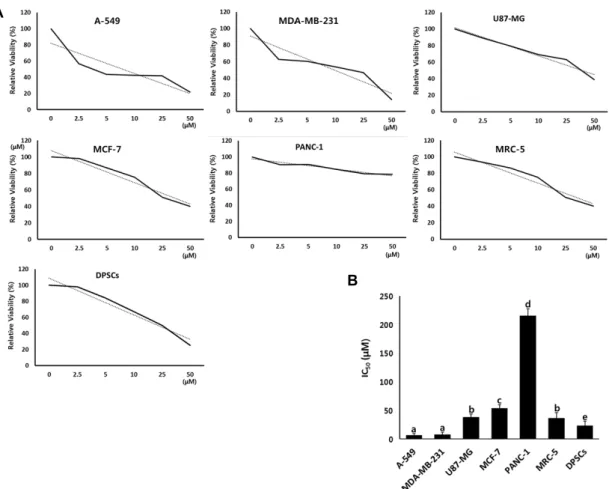
Determination of IC50 value by MTT assay The cytotoxicity test with IC50 values assay was deter- mined by MTT in the A-549, MDA-MB-231, U87-MG, MCF-7 and PANC-1 human cancer cell lines, and DPSCs derived from dental papilla tissues and MRC-5 fetal fibroblasts treat- ed with PGG, as shown in Fig. 1. The IC50 value (mean±SEM) in three replicate was 6.5±3.23, 7.8±4.12, 38.1±5.58, 53.5±6.71, 215.5±12.37, 36.1±10.09 and 23.1±8.35 μM in the A-549, MDA-MB-231, U87-MG, MCF-7, PANC-1, DPSCs and MRC- 5 cell lines, respectively. The IC50 values in the A-549 and MDA-MB-231 cancer cell lines were significantly (p<0.05) lower than other cell lines. Moreover, the IC50 value of PANC-1 cancer cells was interestingly observed at a highest level among all other cell lines.
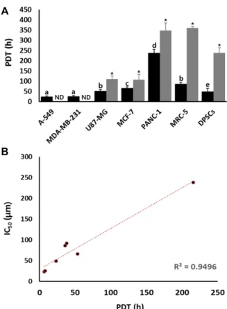
Analysis of population doubling time (PDT) The PDT was analyzed in the untreated control and 10 μM PGG-treated cell lines, and the results were shown in Fig. 2A. The PDT (mean±SEM) in three replicate was 23.2±
3.47, 25.47±3.45, 51.9±6.78, 66.05±6.87, 237.8±17.7, 85.8±7.89 and 49.1±15.23 hours in the untreated control A-549, MDA- MB-231, U87-MG, MCF-7, PANC-1, DPSCs and MRC-5 cell lines, respectively. And the high proliferation rate was ex- hibited in the A-549 and MDA-MB-231 cancer lines. Whereas,
A
B
Fig. 1. Analysis of cytotoxicity and determination of IC50 values by MTT assay in A-549, MDA-MB-231, U87-MG, MCF-7, PANC-1, MRC-5 and DPSCs cell lines treated with PGG. A: Inhibition curves by MTT assay in each cell line. B: Mean±SEM of IC50
values) of each cell line in triplicate. a, b, c, d and e indicate significant (p<.05) difference among each cell lines.
the PDT was 110.3±15.69, 106.2±26.89, 347.5±37.42, 359.5±
8.33 and 237.8±27.22 hours in the U87-MG, MCF-7, PANC-1, DPSCs and MRC-5 cell lines treated with 10 μM PGG, respectively. The PDT was significantly (p<0.05) increased by 10 μM PGG treatment in all of cell lines. Moreover, the cell numbers were exhibited at an extremely low increase in the 10 μM PGG-treated A-549 and MDA-MB-231 cancer cell lines with shorter PDT and higher proliferation rate compared with those of other cell lines, therefore, their PDT could not be determined.
In addition, the correlation coefficient was measured be- tween IC50 values and PDT in each of cell lines, as shown in Fig. 2B. A considerably high correlation coefficient degree (r2=0.9496) was exhibited between IC50 values and PDT, and the IC50 values were proportionally increased along with the increase of PDT in each cell lines.
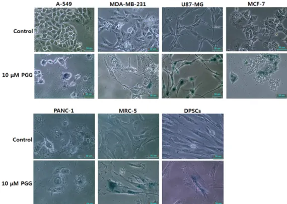
Activity of senescence-associated-β-galactosidase To examine the effect of cellular senescence by PGG treat-
ment, the enzymatic activity of senescence-associated-ß-gal- actosidase was measured in all of cell lines treated with 10 μM PGG, as shown in Fig. 3. The high frequency of cells with senescence-associated-ß-galactosidase activity was ob- served in all the PGG-treated cell lines, compared to those of untreated control cell lines. Furthermore, the cells treated with PGG were gradually changed to enlarged and star- shaped morphology, implying that cellular senescence is proceeding.
Analysis of telomerase activity
To examine the effect of PGG treatment on the telomerase activity, the RQ-TRAP protocol was employed in the un- treated control and 10 μM PGG-treated cancer and normal cell lines. The level of telomerase activity was considered as 100% in the untreated control MRC-5 cells, and the level of telomerase activity in the other cell lines was relatively calculated on the basis of this level. The results are shown in Fig. 4. The level of relative telomere activity was 816±
A
B
Fig. 2. A. Changes of population dubling time (PDT) in A-549, MDA-MB-231, U87-MG, MCF-7, PANC-1, MRC-5 and DPSCs cell lines treated with 10 μM PGG for 7 days.
a, b, c, d and e indicate significant (p<o.05) difference among cell lines. Asterisks (*) indicates significant (p<
o.05) difference between untreated control and 10 μM PGG-treated cell lines, respectively. ND, not determined.
B. Analysis of correlation coefficient between IC50 value and PDT in each cell line. Increased IC50 value against PGG was displayed in cell lines with high PDT than those of relatively low PDT with remarkably high corre- lation coefficient (r2=0.9496).
36.6%, 475±23.3%, 545±32.5%, 1016±72.6%, 698±33.3, 100±15.5
% and 115±26.2 in the untreated control A-549, MDA-MB- 231, U87-MG, MCF-7, PANC-1, MRC-5 and DPSCs cell lines, respectively. As expected, the level of telomerase activity in cancer cell lines was significantly (p<0.05) higher, compared to those of normal MRC-5 and DPSCs cell lines at basal level, reaching the levels over ~8 fold that of MRC-5 fibroblasts.
Whereas the level of relative telomerase activity was 85±20.3, 92±50.1, 292±80.5, 745±80.4, 650±57.1, 88±60.1 and 95±45.1%
in the 10 μM PGG-treated A-549, MDA-MB-231, U87-MG, MCF-7, PANC-1, MRC-5 and DPSCs cell lines treated with 10 μM PGG, respectively. Following PGG treatment, the lev- el of telomerase activity was not significantly (p<0.05) changed in the normal MRC-5 and DPSCs cell lines at the
basal level. However, the level of telomerase activity was significantly (p<0.05) down-regulated in the PGG-treated cancer cell lines. Especially, A-549 and MDA-MB-231 cell lines exhibited highly down-regulated telomerase activity with lower IC50 value, compared to those of other cancer cell lines.
Discussion
In the present study, we evaluated the cytotoxic effects of PGG treatment on the cell proliferation rate, senescence- associated ß-glucosidase activity and telomerase activity in the human cell lines of various origins, including cancer cell lines (A-549, MDA-MB-231, U87-MG, MCF-7 and PANC-1), normal MRC-5 fibroblasts and mesenchymal stem cells de- rived from dental papilla tissues (DPSCs). Our results dem- onstrated that PGG treatment has induced the cellular cyto- toxicity, including inhibition of cell proliferation, high level of senescence-associated ß-glucosidase activity and down- regulation of telomerase activity in the human cell lines of various origins. However, the cytotoxic effects of PGG were different among each cell lines. The higher cytotoxicity was exhibited in cells showing high proliferation capacity than those of low proliferation capacity. Thus, the differential cy- totoxicity of PGG was directly associated with the pro- liferation rate of each cell lines.
PGG is a secondary metabolite with astringent taste, known as the pentahydroxy gallic acid ester of glucose found in the pomegranate, the root of peony, the gallnut of Rhus chinensis Mill and other plant species, and is used as a precursor of gallotannin compound. Several previous studies have demonstrated that PGG treatment is often used for cancer chemotherapy owing to its anti-tumor effects [5, 8, 16].The cells treated with PGG were induced to apoptotic cell death through the mediation of caspase activity in sev- eral human cell lines [14, 29]. In the others studies, PGG treatment induced G0/G1 arrest and DNA replicative S-phase arrest of the cell cycles in human breast cancer cells, MDA-MB-231 and MCF-7 cells. Furthermore, in vivo growth of MDA-MB231 xenograft orally ingested was highly in- hibited without any adverse effect on the host body weight in a dose-dependent manner and growth of DU145 xenograft was also inhibited by activating p53 tumor suppressor path- way and decreasing STAT3 oncogenic signaling [5, 14].
Otherwise, PGG combined to VEGF receptor, and blocked the angiogenesis by inhibiting endothelial cell growth and
Fig. 3. Changes of cell morphology and senescence-associated-β-galactosidase activity in A-549, MDA-MB-231, U87-MG, MCF-7, PANC-1, MRC-5 and DPSCs cell lines treated with 10 μM PGG for 7 days (×200). The high incidences of senescence- associated- β-galactosidase activity (blue) were observed in each of the cell lines treated with 10 μM PGG. Scale bars; 50 μm.
Fig. 4. Changes of telomerase activiy analyzed by RQ-TRAP assay in A-549, MDA-MB-231, U87-MG, MCF-7, PANC- 1, MRC-5 and DPSCs cell lines treated with 10 μM PGG for 7 days. Values indicated the mean telomease activity (mean±SEM) of five replicates and the telomerase activ- ity in untreated control MRC-5 fibroblasts was consid- ered as 100% for comparison with other cell lines. a, b, c and d indicate significant (p<0.05) difference among untreated control cell lines. Asterisks (*) significant (p<
0.05) difference between control and 10 μM PGG-treated cell lines, respectively.
tube formation of new blood vessels via down-regulation of VEGF activity [6, 16, 24]. Our results have also shown
that the cells treated with PGG are induced to the cytotoxic effects, such as inhibition of cell proliferation with prolonged doubling time, induction of cellular senescence by elevated β-galactosidase activity and down-regulation of telomerase activity in various types of human cancer cell lines, and MRC-5 fetal fibroblasts and DPSCs. However, the differ- ential cytotoxicity was exhibited accordingly to the cell lines used in the present study. Moreover, the high cytotoxicity was also exhibited in the normal cell lines (MRC-5 and DPSCs).
The cause of differential cytotoxic effect by PGG treatment in human cell lines of various origins is still unclear. It is well known that the plasma membrane in each cells consists of the phospholipid bilayer having hydrophilic phosphate part and two hydrophobic fatty acids with various proteins.
High proliferating cells require a synthesis of large amount of fatty acids as well as proteins [31]. The synthesis of fatty acids consequently depends on the activity of fatty acid syn- thase (FAS), which is mainly responsible for the synthesis of palmitic acid from acetyl coenzyme A [31]. The activity of FAS enzyme is generally very low or undetectable in nor- mal cells. However, the activity is found to be up-regulated in highly proliferating cells including cancer cells [30, 33].
Moreover, it has been reported that neuronal stem cells or neuronal progenitor cells with high proliferation capacity re- quire a high activity of FAS enzyme-dependent lipogenesis for their proliferation [20], and over-expression 0f FAS activ- ity is tightly associated with an early developmental process of prostate cancer cells and metastasis of cancer cells that spreads to other tissues of the body [32]. Therefore, the defi- ciency of fatty acid for plasma membrane of newly dividing cells may lead to the arrest of cell divisions. Based on this hypothesis, inhibition or down-regulation of FAS activity could be a potential target for cancer chemotherapy or alter- native/adjuvant therapy. Several type of FAS inhibitors, such as C75, cerulenin, orlistat and others have already been tried for cancer treatment [10, 21, 30]. Previously, it has been demonstrated that PGG strongly binds to an active site of FAS, thereby down-regulating FAS activity [7]. However, the potential cytotoxic effects in normal cell lines as well as the anti-tumor effects in various type of cancer cells re- mains to be examined for clinical applications. Our results have demonstrated that PGG induces the cytotoxic effects, including the inhibition of proliferation rate, high sen- escence-associated-ß-galactosidase activity and down-regu- lation of telomerase activity. Especially, the cytotoxic effects was higher in A-549 and MDA-MB-231 cancer cell lines hav- ing high proliferation capacity and short PDT than those of other cell lines, and their IC50 values were also detected at a very low concentrations. As previously reported, the PDT in A-549 and MDA-MB-231 cancer cell lines was approx- imately below 30 hours and exhibited the high proliferation capacity, compared with other cell lines used in the present study [24]. We also observed a markedly low IC50 value in 3T3-L1 mouse embryonic fibroblasts treated with PGG, and these cells also possess rapid cell division capacity (data not shown). Furthermore, even though both MDA-MB-231 and MCF-7 cancer cell lines used in the present study were ad- enocarcinoma tumors derived from female breast epithelial cells, the cellular characterizations based on cancer property were considerably different between two cell lines. MDA- MB-231 cells are generally high malignant tumor cells and possess higher proliferation capacity and shorter PDT than that of MCF-7 cells [1, 12, 27]. However, our results have demonstrated that MDA-MB-231 cells exhibit the lower IC50
value and are more sensitive compared to MCF-7 cells.
Furthermore, accordingly to correlation analysis between the IC50 value and proliferation rate, our data demonstrated that IC50 values against PGG are inversely proportional to the
rate of cell proliferation and directly proportional to the PDT. Whereas the lower IC50 values were observed in MDA- MB-231 and A-549 cell lines with high proliferation capacity and short PDT compared with those of other cell lines with relatively low proliferation capacity and long PDT. A pre- vious study has shown that differential cytotoxic effects and IC50 value are observed in the PGG-treated U251 glio- blastoma tumors and MDA-MB-231 breast adenocarcinoma tumors and U251 cells exhibit lower IC50 value that of MDA-MB-231 cells [7]. It is well known that U251 cells also possess relatively shorter doubling times (~19 hr) than that MDA-MB-231 cells (~28 hr). Therefore, we assumed that the cytotoxic effect of PGG is probably induced by inhibiting synthesis of fatty acid and subsequent process of cell division.
Besides, the PDT was markedly expanded in the cells treated with PGG than those of non-treated control cells, im- plying that the cell proliferation rate is delayed. Other stud- ies have shown that PGG induces arrest of cell cycle at the G1 and S-phase in the various types of human cancer cells treated with PGG by down-regulation of cyclin D1, and in- hibition of DNA polymerase [5, 15]. The mis-regulation of control system for cell cycle induce the unscheduled and un- limited cell proliferation in most of the cancer cells and the arrest of cell cycle at G1 and S phase is a most common status that usually occurs with the inhibition of cell pro- liferation[13]. Furthermore, our results have also demon- strated that the high activity of senescence-associated-ß-gal- actosidase was increased upon PGG-treatment with cellular morphological alternations compared with those of non- treated control cells. The cell size was gradually expanded, and cells were changed into star-shaped and irregular mor- phology in the PGG-treated cells. It has been well demon- strated that the cells at senescent status display an enlarged and irregular cell shape with a large number of cells at G1/S phase of cell cycle [2]. Although, in the present study, the cell cycle analysis was not determined in the PGG-treated cells, we considered that the cells are suspended at G1/S phase of cell cycle. These dramatic alternations were pre- dominantly observed in the PGG-treated A-549 and MDA- MB-231 with high proliferation capacity.
PGG treatment further also induced the down-regulation of telomerase activity resulting in cellular senescence in the human cancer cell lines with the exception for MRC-5 and DPSCs cell lines. Especially, the telomerase activity upon PGG treatment was dramatically decreased in the A-549 and
MDA-MB-231 cell lines, which had high proliferation capacity.
To date, available information on the telomerase activity by PGG treatment is not yet fully reported in the various hu- man cell lines. The telomerase activity and telomeric repeats are also related with cellular senescence [2].The embryonic stem cells and most malignant tumors possess the up-regu- lated telomerase activity that continually maintains or ex- tends telomeric repeats on the 3’ end of the linear eukaryotic DNA strands by adding telomeric repeats [9]. On the other hand, most of the differentiated somatic cells with the down-regulation of telomerase activity are induced to short- ening of their telomeric repeats, and the cells reached at cri- sis status with fully shortened telomeric repeats subsequently leads to cellular senescence [9]. Therefore, it has been re- ported that the level of telomerase activity and length of telomeric repeats are indices of the capacity of limitless cell division and high proliferation. Furthermore, our results have shown that PANC-1, U87-MG and MCF-7 cell lines with low proliferation capacity possess high level of telomer- ase activity, compared with those of A-549 and MDA-MB- 231 cells. Therefore, we have assumed that high level of telo- merase activity is only concerned with unlimited cell pro- liferation capacity, whereas unconcerned with rate of cell proliferation. Even though the shortening of telomeric re- peats by down-regulated telomerase activity are certainly in- duced to cellular senescence and the senescent cells with high level of ß-glucosidase activity were also observed in the present study by PGG treatment, we have presumed that the cellular senescence is probably induced by the cytotox- icity of PGG itself rather than shortening of telomeric repeats. Our previous study has shown that the decreased expression of the genes related with telomerase activity i.e.
telomerase reverse transcriptase (TERT) and telomerase RNA component (TERC) has resulted in down-regulation of telomerase activity [28]. Whereas, the significant changes in the telomerase activity was not observed in the MRC-5 and DPSCs cell lines. We have assumed that this might be due to telomeres activity reached at a critically low level, com- pared with those of cancer cell lines. Although the DPSCs exhibited outstanding characterization of stem cells, such as multi-lineage differentiation capacity, the cells exhibited crit- ically low level of telomeres activity, as MRC-5 normal fibro- blasts [27, 28].
Interestingly, our results have shown that PANC-1 cells with extremely low proliferation capacity exhibited out- standing resistance against PGG treatment and possess very
high IC50 value. As pointed out above, the cell lines with high proliferation capacity require a mass of fatty acid for newly synthesized cell membrane, and the PGG may induce more cytotoxicity effects in the high proliferation capacity, such as A-549 and MDA-MB-231. On the other hand, the present results have shown that PGG treatment also induces the cytotoxicity effects in the normal MRC-5 and DPSCs cell lines. Therefore, PGG treatment or oral administration may induces several side effects, i.e. dyspepsia, hair loss, anemia, weight loss and others by inhibiting the cell division or cell cycle in the normal cell lines. And clinical application of PGG should carefully be considered or examined as a poten- tial toxic compound in various normal somatic cell lines. In the present study, although any intrinsic and cellular in- cidence was not investigated in the PGG-treated cancer cells, PGG against cancer cells have shown a kind of potential chemotherapy compound by inhibiting cell proliferation and down-regulating telomerase activity. Moreover, PGG is more susceptible in the cancer cells with high proliferation capacity.
Acknowledgment
This study was supported by grants from Korean Founda- tion for the Advancement of Science and Creativity.
References
1. Abdullah, A. S., Mohammed, A. S., Abdullah, R., Mirghani, M. E. and Al-Qubaisi, M. 2014. Cytotoxic effects of Mangi- fera indica L. kernel extract on human breast cancer (MCF-7 and MDA-MB-231 cell lines) and bioactive constituents in the crude extract. BMC Complement. Altern. Med. 25, 199.
2. Allsopp, R. C., Vaziri, H., Patterson, C., Goldstein, S., Youn- glai, E. V., Futcher, A. B., Greider, C. W. and Harley, C.
B. 1992. Telomere length predicts replicative capacity of hu- man fibroblasts. Proc. Natl. Acad. Sci. USA 89, 10114-10118.
3. Artandi, S. E. and DePinho, R. A. 2010. Telomeres and telo- merase in cancer. Carcinogenesis 31, 9-18.
4. Cao, Y., Himmeldirk, K. B., Qian, Y., Ren, Y., Malki, A. and Chen, X. 2014. Biological and biomedical functions of Penta-O-galloyl-D-glucose and its derivatives. J. Nat. Med.
68, 465-472.
5. Chai, Y., Lee, H. J., Shaik, A. A., Nkhata, K., Xing, C., Zhang, J., Jeong, S. J., Kim, S. H. and Lu, J. 2010. Penta-O-galloyl-be- ta-D-glucose induces G1 arrest and DNA replicative S-phase arrest independently of cyclin-dependent kinase inhibitor 1A, cyclin-dependent kinase inhibitor 1B and P53 in human breast cancer cells and is orally active against triple negative xenograft growth. Breast Cancer Res. 12, R67.
6. Cryan, L. M., Bazinet, L., Habeshian, K. A., Cao, S., Clardy, J., Christensen, K. A. and Rogers, M. S. 2013. 1,2,3,4,6- Penta-O-galloyl-β-D-glucopyranose inhibits angiogenesis via inhibition of capillary morphogenesis gene 2. J. Med.
Chem. 56, 1940-1945.
7. Dikmen, Z. G., Gellert, G. C., Jackson, S., Gryaznov, S., Tressler, R., Dogan, P., Wright, W. E. and Shay, J. W. 2005.
In vivo inhibition of lung cancer by GRN163L: a novel hu- man telomerase inhibitor. Cancer Res. 65, 7866-7873.
8. Dong, Y., Yin, S., Jiang, C., Luo, X., Guo, X., Zhao, C., Fan, L., Meng, Y., Lu, J., Song, X., Zhang, X., Chen, N. and Hu, H. 2014. Involvement of autophagy induction in penta- 1,2,3,4,6-O-galloyl-β-D-glucose-induced senescence-like growth arrest in human cancer cells. Autophagy 10, 296-310.
9. Falandry, C., Bonnefoy, M., Freyer, G. and Gilson, E. 2014.
Biology of cancer and aging: a complex association with cel- lular senescence. J. Clin. Oncol. 32, 2604-2610.
10. Flavin, R., Peluso, S., Nguyen, P. L. and Loda, M. 2010. Fatty acid synthase as a potential therapeutic target in cancer.
Future Oncol. 6, 551-562.
11. Funayama, R. and Ishikawa, F. 2007. Cellular senescence and chromatin structure. Chromosoma 116, 431-440.
12. Ghate, N. B., Chaudhuri, D., Sarkar, R., Sajem, A. L., Panja, S., Rout, J. and Mandal, N. 2013. An antioxidant extract of tropical lichen, Parmotrema reticulatum, induces cell cycle arrest and apoptosis in breast carcinoma cell line MCF-7.
PLoS One 8, e82293.
13. Hartwell, L. H. and Kastan, M. B. 1994. Cell cycle control and cancer. Science 266, 1821-1828.
14. Hu, H., Lee, H. J., Jiang, C., Zhang, J., Wang, L., Zhao, Y., Xiang, Q., Lee, E. O., Kim, S. H. and Lü, J. 2008. Penta- 1,2,3,4,6-O-galloyl-beta-D-glucose induces p53 and inhibits STAT3 in prostate cancer cells in vitro and suppresses pros- tate xenograft tumor growth in vivo. Mol. Cancer Ther. 7, 2681-2691.
15. Hu, H., Zhang, J., Lee, H. J., Kim, S. H. and Lü, J. 2009.
Penta-O-galloyl-beta-D-glucose induces S- and G(1)-cell cy- cle arrests in prostate cancer cells targeting DNA replication and cyclin D1. Carcinogenesis 30, 818-823.
16. Huh, J. E., Lee, E. O., Kim, M. S., Kang, K. S., Kim, C. H., Cha, B. C., Surh, Y. J. and Kim, S. H. 2005. Penta-O-gallo- yl-beta-D-glucose suppresses tumor growth via inhibition of angiogenesis and stimulation of apoptosis: roles of cyclo- oxygenase-2 and mitogen-activated protein kinase pathways.
Carcinogenesis 26, 1436-1445.
17. Jeon, B. G., Jang, S. J., Park, J. S., Subbarao, R. B., Jeong, G. J., Park, B. W. and Rho, G. J. 2015. Differentiation poten- tial of mesenchymal stem cells isolated from human dental tissues into non-mesodermal lineage. Anim. Cells Syst. 19, 321-331.
18. Jeon, B. G., Kumar, B. M., Kang, E. J., Maeng, G. H., Lee, Y. M., Hah, Y. S., Ock, S. A., Kwack, D. O., Park, B. W.
and Rho, G. J. 2011. Differential cytotoxic effects of sodium meta-arsenite on human cancer cells, dental papilla stem cells and somatic cells correlate with telomeric properties and gene expression. Anticancer Res. 31, 4315-4328.
19. Jeon, B. G., Kwack, D. O. and Rho, G. J. 2011. Variation of telomerase activity and morphology in porcine mesen- chymal stem cells and fibroblasts during prolonged in vitro culture. Anim. Biotechnol. 22, 197-210.
20. Knobloch, M., Braun, S. M., Zurkirchen, L., von Schoultz, C., Zamboni, N., Araúzo-Bravo, M. J., Kovacs, W. J., Karalay, O., Suter, U., Machado, R. A., Roccio, M., Lutolf, M. P., Semenkovich, C. F. and Jessberger, S. 2013. Metabolic con- trol of adult neural stem cell activity by Fasn-dependent lipogenesis. Nature 493, 226-230.
21. Kuhajda, F. P., Pizer, E. S., Li, J. N., Mani, N. S., Frehywot, G. L. and Townsend, C. A. 2000. Synthesis and antitumor activity of an inhibitor of fatty acid synthase. Proc. Natl.
Acad. Sci. USA 97, 3450-3454.
22. Kwon, T. R., Jeong, S. J., Lee, H. J., Lee, H. J., Sohn, E. J., Jung, J. H., Kim, J. H., Jung, D. B., Lu, J. and Kim, S. H.
2012. Reactive oxygen species-mediated activation of JNK and down-regulation of DAXX are critically involved in penta-O-galloyl-beta-d-glucose-induced apoptosis in chron- ic myeloid leukemia K562 cells. Biochem. Biophys. Res. Com- mun. 424, 530-537.
23. Lee, S. J., Lee, H. M., Ji, S. T., Lee, S. R., Mar, W., Kim, J.
H., Jung, D. B., Lu, J. and Kim, S. H. 2012. 1,2,3,4,6-Penta- O-galloyl-beta-D-glucose blocks endothelial cell growth and tube formation through inhibition of VEGF binding to VEGF receptor. Cancer Lett. 208, 89-94.
24. Limame, R., Wouters, A., Pauwels, B., Fransen, E., Peeters, M., Lardon, F., De Wever, O. and Pauwels, P. 2012.
Comparative analysis of dynamic cell viability, migration and invasion assessments by novel real-time technology and classic endpoint assays. PLoS One 7, e46536.
25. Maida, Y. and Masutomi, K. 2015. Telomerase reverse tran- scriptase moonlights: Therapeutic targets beyond telomer- ase. Cancer Sci. 106, 1486-1492.
26. Makarević, J., Rutz, J., Juengel, E., Kaulfuss, S., Tsaur, I., Nelson, K., Pfitzenmaier, J., Haferkamp, A. and Blaheta, R.
A. 2014. Amygdalin influences bladder cancer cell adhesion and invasion in vitro. PLoS One 9, e110244.
27. Mender, I., Gryaznov, S. and Shay, J. W. 2015. A novel telo- merase substrate precursor rapidly induces telomere dys- function in telomerase positive cancer cells but not telomer- ase silent normal cells. Oncoscience 22, 693-695.
28. Moon, J. Y., Kim, S. W., Yun, G. M., Lee, H. S., Kim, Y.
D., Jeong, G. J., Ullah, I., Rho, G. J. and Jeon, J. B. 2015.
Inhibition of cell growth and down-regulation of telomerase activity by amygdalin in human cancer cell lines. Anim. Cells Syst. 19, 295-304.
29. Pan, M. H., Lin, J. H., Lin-Shiau, S. Y. and Lin, J. K. 1999.
Induction of apoptosis by penta-O-galloyl-beta-D-glucose through activation of caspase-3 in human leukemia HL-60 cells. Eur. J. Pharmacol. 381, 171-183.
30. Pandey, P. R., Liu, W., Xing, F., Fukuda, K. and Watabe, K. 2013. Anti-cancer drugs targeting fatty acid synthase (FAS). Recent Pat. Anticancer Drug Discov. 7, 185-197.
31. Simons, K. and Ikonen, E. 1997. Functional rafts in cell membranes. Nature 387, 569-572.
초록:사람의 다양한 조직에서 기원하는 암세포 및 정상세포에 대한 penta-O-galloyl-β-D-glucose의 세포독성 효과
이현정1․김민경1․이송영1․송민혁1․김윤동1․하정숙2․정계준2․노규진3․전병균2,4*
(1경남과학고등학교, 2경상대학교 사범대학 생물교육과, 3경상대학교 수의과대학 수의학과, 4경상대학교 교육연구원)
본 연구는 다당체의 한 종류인 penta-O-galloyl-β-D-glucose (PGG)가 사람의 여러 조직에서 기원하는 여러 암 세포주(A-549, MDA-MB-231, U87-MG, MCF-7 및 PANC-1), 정상 MRC-5 태아 섬유아세포 그리고 사랑니에서 유
래한 간엽줄기세포(DPSCs)에 미치는 세포독성 효과를 조사하였다. IC50값은 다른 세포주에 비해 높은 증식률을
나타내는 A-549 및 MDA-MB-231 암세포주에서 유의적으로 낮게 관찰되었다. 10 uM의 PGG가 포함된 배양액에
서 세포를 7일 동안 배양한 결과, 세포배가시간은 모든 세포주에서 유의적으로 늘어났고, 세포배가시간과 IC50값
의 관계를 조사한 결과, 세포배가시간이 늘어남에 따라 IC50값은 비례적으로 증가됨을 증명하였다. 또한, 10 uM의
PGG로 처리된 세포주들은 노화와 관련된 ß-galactosidase의 활성도가 높게 관찰되었다. 특히, telomerase 활성도 는 A-549 및 MDA-MB-231 암세포주에서 다른 세포주에 비하여 현저히 감소하는 것을 관찰하였다. 이러한 결과를 바탕으로 PGG는 높은 증식률을 보이는 암세포주에서 높은 세포독성효과를 나타내어 잠재적인 항암물질임을 증 명하였다.
32. Swinnen, J. V., Roskams, T., Joniau, S., Van Poppel, H., Oyen, R., Baert. L., Heyns, W. and Verhoeven, G. 2002.
Overexpression of fatty acid synthase is an early and com- mon event in the development of prostate cancer. Int. J.
Cancer 98, 19-22.
33. Veigel, D., Wagner, R., Stübiger, G., Wuczkowski, M., Filipits, M., Horvat, R., Benhamú, B., López-Rodríguez, M. L., Leisser, A., Valent, P., Grusch, M., Hegardt, F. G., García, J., Serra, D., Auersperg, N., Colomer, R. and Grunt, T. W.
2015. Fatty acid synthase is a metabolic marker of cell pro-
liferation rather than malignancy in ovarian cancer and its precursor cells. Int. J. Cancer 136, 2078-2090.
34. Wong, A. S., Che, C. M. and Leung, K. W. 2015. Recent advances in ginseng as cancer therapeutics: a functional and mechanistic overview. Nat. Prod. Rep. 32, 256-272.
35. Zhao, W., Wang, Y., Hao, W., Zhao, M. and Peng, S. 2015.
In vitro inhibition of fatty acid synthase by 1,2,3,4,6-pen- ta-O-galloyl-β-D-glucose plays a vital role in anti-tumour activity. Biochem. Biophys. Res. Commun. 445, 346-351.