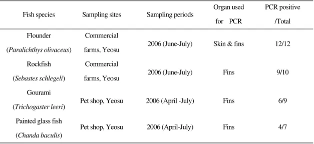
173 Lymphocystis disease virus (LCDV) is the causative agent of lymphocystis disease developing a tumor like cluster composed of hypertrophied fibroblast cells with cytoplasmic inclusions (Wolf, 1998). The tumors are usually found from the skin, fins and oral regions both in the marine and fresh- water fishes worldwide. Lymphocystis disease affects more than 125 different wild and cultured fish species, causing important economic losses globally. In Korea, lymphocystis disease is the com- mon fish disease, especially in aquaculture of olive flounder Paralichthys olivaceus, rockfish Sebastes schlegeli and sea bass Lateolabrax sp. (Kitamura et al., 2006a, 2006b). Additionally, in aquarist shops, the disease has been observed in ornamental fish species.
From the first report of the electron microscopic observation (Walker, 1962), great advances have been made in LCDV studies such as morphology,
detection method and genome (Zwillenberg and wolf, 1968; Darai et al., 1983; Tidona and Darai, 1997; Tidona et al., 1998; Webby and Kalmakoff, 1998; Zhang et al., 2004; Cano et al., 2006; Kita- mura et al., 2006a, 2006b). However, few researchers have reported on the protein profiles of LCDV (Flugel et al., 1982; Schnitzler and Darai, 1993; Garcia-Rosado et al., 2004). In the present study, LCDV from marine fish (olive flounder and rockfish) and freshwater ornamental fish (gourami, Trichogaster leeri, and painted glassfish, Chanda baculis) was detected, and proteins of the viruses were compared.
Materials and Methods
Fish sampling
The lymphocystis disease-affected freshwater ornamental fish, gourami and painted glass fish
Comparison of lymphocystis disease virus proteins between marine and freshwater fish
Mosharrof Hossain, Sung-Ju Jung, Wi-Sik Kim, Seok-Ryel Kim
�and Myung-Joo Oh
�Division of Food Science and Aqualife Medicine, Chonnam National University, Yeosu-550-749, Korea
�
Pathology division, National Fisheries Research and Development Institute, Busan 619-902, Korea
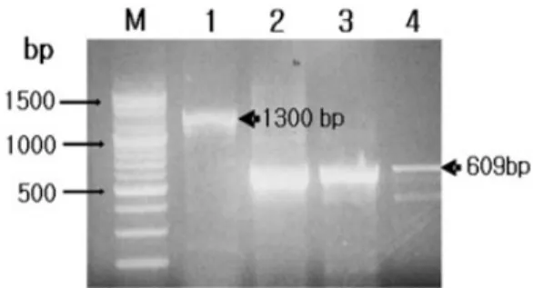
Lymphocystis disease virus (LCDV) was detected from olive flounder Paralichthys olivaceus, painted glass fish Chanda baculis, gourami Trichogaster leeri and rockfish Sebastes schlegeli, and proteins of the viruses were compared. The major capsid protein (MCP) gene-specific primer sets successfully amplified approximately 1300 bp nucleotides from the olive flounder and 600 bp nucleotides from painted glass fish, gourami and rockfish isolates, respectively. In western blotting analysis using anti-LCDV mouse polyclonal serum, major antigenic proteins had 21, 26, 45, 50, 80, 110 and 120 kDa in olive flounder, 26, 47 and 80 kDa in painted glass fish, 26, 46, 80 and 92 kDa in gourami, 26, 44, 49, 80 and 105 in rockfish, respectively. All the marine and freshwater isolates showed only common antigens of approximately 26 kDa and 80 kDa.
These results suggest that antigenic protein profiles of LCDVs may vary depending upon fish species.
Key words: Lymphocystis disease virus, Anti-LCDV serum, Viral protein, Western blot
�