Copyright ⓒ 2018 by Korean Society for Surgery of the Hand, Korean Society for Microsurgery, and Korean Society for Surgery of the Peripheral Nerve. All Rights reserved.
This is an Open Access article distributed under the terms of the Creative Commons Attribution Non-Commercial License (http://creativecommons.org/licenses/by-nc/4.0/) which permits unrestricted non-commercial use, distribution, and reproduction in any medium, provided the original work is properly cited.
Vulvar cancer is rare and usually requires tumor abla- tion surgery, which results in a need for functional and aesthetic reconstruction1,2. When the defect is small, re- construction can be done with primary closure, local flap, and skin graft. When the defect is large, reconstruction can be done using the pedicled deep inferior epigastric artery perforator (DIEP) flap, the gracillis musculocu- taneous flap, and free flaps1. As the lotus pedal flap and the perforator flap are increasingly recognized, excellent
functional and aesthetic outcomes are reported by per- forming reconstruction with a thin perforator flap around the vulva3.
The pedal flap based on the internal pudendal artery perforator, various types of internal pudendal artery per- forator flaps, or other options can be used2. The internal pudendal artery perforator flap is designed based on the vessel running along the vulva margin, which is safe and reliable, but if the tumor is large and extensive excision Hand and
Microsurgery
두 종류의 천공지 피판을 이용한 양측 외음부 재건: 증례 보고
기세휘
1,2ㆍ심승현
2ㆍ이상환
21인하대학교 의과대학 성형외과학교실, 2인하대병원 성형외과
Bilateral Vulvar Reconstruction Using Two Different Types of Perforator Flap: A Case Report
Sae Hwi Ki
1,2, Seung Hyun Sim
2, Sang Hwan Lee
21Department of Plastic Surgery, Inha University School of Medicine, Incheon, Korea
2Department of Plastic Surgery, Inha University Hospital, Incheon, Korea
We reconstructed an extensive vulvar defect with the internal pudendal artery and the anterior obturator artery perforator flap and compared the two types of flaps. A 57-year-old female patient had a 15×13 cm2 defect on both sides around the vagina after vulvectomy. Due to the injury in the internal pudendal artery, we used the internal pudendal artery perforator on the right side, and the anterior obturator artery perforator flap on the left side. The internal pudendal artery perforator flap could be rotated easily 120 degrees toward the vulvar defect. However, as the anterior obturator artery perforator flap was difficult to rotate, reconstruction was performed by rotation and advancement. The anterior obturator artery perfora- tor flap is a viable alternative method that can be used when it is difficult to use the internal pudendal artery perforator flap during vulvar reconstruction.
Key Words: Vulvar neoplasms, Perforator flap, Rotation
Received September 18, 2017, Revised [1] October 16, 2017, [2] November 6, 2017, Accepted November 7, 2017 Corresponding author: Sae Hwi Ki
Department of Plastic Surgery, Inha University Hospital, Inha University School of Medicine, 27 Inhang-ro, Jung-gu, Incheon 22332, Korea TEL: +82-32-890-3870, FAX: +82-32-890-2918, E-mail: [email protected], ORCID: http://orcid.org/0000-0001-9194-9681
This study was supported by the Inha University Research Grant.
Case Report
needs to be done, another surgical method must be select- ed because of artery injury2. In this case, it was difficult to use the internal pudendal artery perforator, and recon- struction was carried out using the anterior obturator ar- tery perforator at the more distal site of the medial thigh.
Therefore, the authors compare the two different types of flaps with our experience.
CASE REPORT
A 57-year-old female patient had a 0.8×0.5 cm2 mass at the lower 1/3 of vulva labia majora. In the biopsy and pelvis computed tomography, she was diagnosed with stage IIIB squamous cell carcinoma of the vulva. In the Department of Gynecology at this hospital, the tumor was diagnosed as malignant on the frozen biopsy performed during operation. While vulvectomy, bilateral inguino- femoral lymph node dissection, pelvic and para-aortic lymph node dissection were underway, the excision range of the vulva became larger, and primary closure or local flap coverage became difficult. Thus, an emergency operation was performed for reconstruction. After wide vulvectomy, a 15×13 cm2 defect was observed on both sides around the vagina and urethra opening, while the anus was intact (Fig. 1).
Because of the emergency operation, we could not evaluate the vascular condition around the vulva. For reconstruction of the vulva defect, a fasciocutaneous flap based on the internal pudendal artery perforator was de- signed, and if the use of perforator flap proved difficult, the obturator artery based gracillis musculocutaneous V-Y advancement flap was prepared (Fig. 1). The inter- nal pudendal arteries on both sides were checked using a hand-held doppler, but due to the vessel injury caused by
the wide tumor excision, the internal pudendal artery was not found on the left side. Reconstruction was planned using the internal pudendal artery perforator for the right side and the anterior obturator artery perforator for the left side.
We made the skin incision around the right posterior medial thigh, and dissected the skin flap via supra-fascial plane while we tried to find the perforator from the proxi- mal side to the distal side of the medial thigh. The skin perforator was present in the inside of the lower margin of the vascular triangle from the ischial tuberosity to apex of the coccyx and came from the internal pudendal artery.
We elevated the 15×7 cm2 sized perforator flap for the defect of the right side vulva. The flap thickness around the internal pudendal artery perforator was 1.9 cm, the elevated flap was rotated clockwise by 120 degrees, and tension-free closure was conducted for the vulvar defect (Fig. 2A). The donor site could be closed primarily.
For the left vulva defect, suprafascial dissection was done from the proximal thigh to the distal over the gracil- lis muscle, and a proper size of perforator was found, specifically the anterior obturator artery perforator. The
Fig. 1. Both sides of the vulvar defect; preparation of the medial thigh V-Y advancement flap.
Fig. 2. (A) Internal pudendal ar- tery perforator flap, right side (ro- tation state). (B) Anterior obturator artery perforator flap, left side (before advancement, rotation).
A B
subcutaneous fat tissue thickness over the gracillis muscle was 3.5 cm. We elevated the 18×8 cm2 sized perforator flap for the left vulva defect. The position of the perfora- tor was very deep and more distal than the position of the internal pudendal artery perforator (Fig. 2B). The elevat- ed flap was advanced and rotated counterclockwise by 90 degrees. Tension-free primary closure could be done for the donor site (Fig. 3).
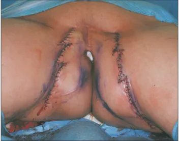
Two weeks later, there was partial necrosis on the right side flap, but it healed spontaneously. There were no other complications. Five months later, volume reduction of the both flaps multiple Z-plasty were performed due to flap bulkiness and scar contracture (Fig. 4).
DISCUSSION
The vulvar region is an organ that represents female identity, and when reconstructing the defect occurring af- ter wide surgical resection of vulva cancer, the functional and aesthetic aspects of the vagina and urethra, as well as the coverage of the soft tissue defect, must be considered.
The wide range of reconstructive options for vulvar re- construction includes skin grafts, skin flaps, fasciocutane- ous flaps and myocutaneous flaps1,2,4.
In reconstruction for extensive vulvar defects, medial thigh V-Y advancement flap, the gracillis musculocu- taneous flap, the pedicled DIEP flap, and free flap are often selected, but these methods pose the disadvantage of low aesthetic and functional satisfaction due to thick
flap thickness after reconstruction3. The lotus pedal flap and the internal pudendal artery perforator flap centered on the gluteal fold are excellent alternatives in the aes- thetic and functional aspects, because thin covering is possible, allow a better anatomical reconstruction, respect inguinal–crural crease and leave minimal scars in folds5,6. Hashimoto et al.2 reported that these flaps are quick, safe, sensitive and more aesthetically effective. As an alterna- tive flap, O`Dey et al.7 presented the anterior obturator ar- tery perforator flap. They said that the anterior obturator artery perforator pierces the gracilis muscle proximally on the level of its thin aponeurosis as an indirect or mus- culocutaneous perforator near to the inferior pubic ramus.
They suggest that the anterior obturator artery perforator flap could be thinner because both the septocutaneous and musculocutaneous anterior obturator perforators ex- tend nearly perpendicularly to the subdermal plexus. In our case, because of the injury on the internal pudendal artery, it was difficult to use the internal pudendal artery perforator flap. Based on the assessment that a reliable flap had to be selected, the obturator artery perforator flap was used first, having the medial thigh V-Y advancement flap, the anterior gracillis musculocutaneous flap and free flaps as the second choice8-10.
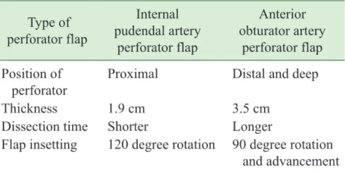
Since two different types of perforator flaps were performed on either side, advantages and disadvantages could be compared (Table 1). The position of the ante-
Fig. 3. Immediate post-surgery.
Fig. 4. Five months post-surgery. After debulking of the flap and multiple Z-plasty.
rior obturator artery perforator was more distal than that of the internal pudendal artery perforator, and the skin thickness over the gracillis was thicker than that of the internal pudendal artery perforator. In addition, due to the thickness, dissection time of the anterior obturator artery perforator was longer without initial flap thinning pro- cedure on both sides. The position of the perforator was very deep and more distal than the position of the internal pudendal artery perforator. The arc of rotation of the flap was limited due to the thick flap, deep placed perforator, insecurity of the vascular pedicle torsion or kinking8. Be- cause propeller pattern rotation is difficult, advancement and rotation is considered a safer method rather than rota- tion alone.
The internal pudendal artery perforator flap was thinner and rotation was easier than the anterior obturator artery perforator flap. However, the reconstruction method with the anterior obturator perforator flap was safe, and can be a good alternative choice for vulvar reconstruction when it is difficult to use the internal pudendal artery perforator flap.
CONFLICTS OF INTEREST
The authors have nothing to disclose.
REFERENCES
1. Höckel M, Dornhöfer N. Vulvovaginal reconstruction for neoplastic disease. Lancet Oncol. 2008;9:559-68.
2. Hashimoto I, Abe Y, Nakanishi H. The internal pudendal artery perforator flap: free-style pedicle perforator flaps for vulva, vagina, and buttock reconstruction. Plast Re- constr Surg. 2014;133:924-33.
3. Bodin F, Weitbruch D, Seigle-Murandi F, Volkmar P, Bruant-Rodier C, Rodier JF. Vulvar reconstruction by a
“supra-fascial” lotus petal flap after surgery for malignan- cies. Gynecol Oncol. 2012;125:610-3.
4. Al-Benna S, Tzakas E. Postablative reconstruction of vul- var defects with local fasciocutaneous flaps and superficial fascial system repair. Arch Gynecol Obstet. 2012;286:443- 8.
5. Sawada M, Kimata Y, Kasamatsu T, et al. Versatile lotus petal flap for vulvoperineal reconstruction after gyneco- logical ablative surgery. Gynecol Oncol. 2004;95:330-5.
6. Tan BK, Kang GC, Tay EH, Por YC. Subunit principle of vulvar reconstruction: algorithm and outcomes. Arch Plast Surg. 2014;41:379-86.
7. O’Dey DM, Bozkurt A, Pallua N. The anterior Obturator Artery Perforator (aOAP) flap: surgical anatomy and ap- plication of a method for vulvar reconstruction. Gynecol Oncol. 2010;119:526-30.
8. Lazzaro L, Guarneri GF, Rampino et al. Vulvar recon- struction using a “V-Y” fascio-cutaneous gluteal flap: a valid reconstructive alternative in post-oncological loss of substance. Arch Gynecol Obstet. 2010;282:521-7.
9. Chen YC, Scaglioni MF, Kuo YR. Profunda artery per- forator based V-Y rotation advancement flap for total vulvectomy defect reconstruction--A case report and lit- erature review. Microsurgery. 2015;35:668-71.
10. Lee JH, Shin JW, Kim SW, et al. Modified gluteal fold V-Y advancement flap for vulvovaginal reconstruction.
Ann Plast Surg. 2013;71:571-4.
Table 1. Comparsion of internal pudendal artery perforator flap and anterior obturator artery perforator flap
Type of perforator flap
Internal pudendal artery
perforator flap
Anterior obturator artery
perforator flap Position of
perforator Proximal Distal and deep
Thickness 1.9 cm 3.5 cm
Dissection time Shorter Longer
Flap insetting 120 degree rotation 90 degree rotation and advancement
두 종류의 천공지 피판을 이용한 양측 외음부 재건: 증례 보고
기세휘
1,2ㆍ심승현
2ㆍ이상환
21인하대학교 의과대학 성형외과학교실, 2인하대병원 성형외과
57세 여자 환자로 외음부 편평세포암으로 외음부 절제술을 시행하며 생긴 15×13 cm2의 결손 부위에 서로 다른
천공지 피판을 동시에 사용하여 재건하고 두 종류 피판을 비교하여 보았다. 환자의 우측 부위 결손은 내음부동맥 천공지 피판을 이용하고 좌측 부위의 결손은 내음부동맥이 수술 시 손상되어 앞폐쇄동맥 천공지 피판을 이용하여 재건하였다. 내음부동맥 천공지 피판은 120도로 회전하고 앞폐쇄동맥 천공지 피판은 회전과 전진을 동시에 진행하 여 재건을 시행하여 좋은 결과를 얻었다. 내음부동맥 천공지 피판을 사용할 수 없을 때 앞폐쇄동맥 천공지 피판은 좋은 선택이 될 수 있을 것으로 생각한다.
색인단어: 외음부암, 천공지 피판, 회전
접수일 2017년 9월 18일 수정일 1차: 2017년 10월 16일, 2차: 2017년 11월 6일 게재확정일 2017년 11월 7일
교신저자 기세휘
22332, 인천시 중구 인항로 27, 인하대학교 의과대학 인하대병원 성형외과학교실 TEL 032-890-3870 FAX 032-890-2918 E-mail [email protected] ORCID: http://orcid.org/0000-0001-9194-9681