29
책임저자: 김지훈, 강원도 강릉시 사천면 방동리 415
210-711, 울산대학교 강릉아산병원 외과 Tel: 033-610-3220, Fax: 033-610-4960 E-mail: [email protected]
접수일:2010년 10월 28일, 게재승인일:2010년 12월 8일 본 논문은 2009년도 강릉아산병원(임상의학연구소 학술연구비)의 지원을 받아 연구되었음.
장막침범이 없는 진행성 위암의 2년 이내 재발과 연관된 E-cadherin, β-catenin, Cdx2, MMP7 발현양상
울산대학교 의과대학 강릉아산병원 외과학교실, 1병리학교실
김지훈ㆍ엄대운
1ㆍ김찬욱ㆍ최남규ㆍ곽진호ㆍ최건무ㆍ장혁재ㆍ한명식
Expression of E-cadherin, β-catenin, Cdx2 and MMP7 in pT2 and N1/N2 Gastric Cancer: Relationship with Tumor Recurrence within 2-Year Period
Ji Hoon Kim, M.D., Dae Yoon Eom, M.D.1, Chan Wook Kim, M.D., Nam Kyu Choi, M.D., Jin Ho Kwak, M.D., Gun Moo Choi, M.D., Hyuck Jae Jang, M.D., Myung Sik Han, M.D.
Departments of Surgery and 1Pathology, College of Medicine, University of Ulsan, Gangneung Asan Hospital, Gangneung, Korea
Purpose: The aim of this study was to examine the expression of E-cadherin, β-catenin, Cdx2, MMP7 in gastric cancer and to evaluate the clinical significance of these molecules in tumor recurrence within 2 years of pT2 and N1/N2 gastric cancer.
Methods: In 122 patients who underwent radical resection of gastric cancer, we investigated the association between the expression of these molecules and clinicopathologic factors by immunohistochemistry. The included criteria were pT2 and N1 or N2 (6th AJCC TNM).
Results: The expression of MMP7 was significantly associated with N stage (N1 vs. N2) (P=0.011). The negative expression of β-catenin was strongly correlated with tumor recurrence within a 2-year period. However, the expression of these molecules was not related with recurrent sites. Multivariate analysis demonstrated that negative expression of β-catenin was an independent predictor for tumor recurrence within 2 years (OR 2.366; 95%CI 1.056∼5.297; P=0.036).
Conclusion: Negative expression of β-catenin may serve as a significant indicator for predicting tumor recurrence within a 2-year period in pT2 and N1/N2 gastric cancer. (J Korean Surg Soc 2011;80:29-35)
Key Words: E-cadherin, β-catenin, Cdx2, MMP7, Gastric cancer 중심 단어: E-cadherin, β-catenin, Cdx2, MMP7, 위암
서 론
암으로 대표되는 악성질환의 가장 중요한 특징 중의 하
나인 원격전이가 되기 위해서는 종양세포의 세포-세포간 유착, 세포-기질간 유착과 같은 조직안정성이 무너지고, 기 저막이 파괴되며, 신생혈관형성과정과 혈관 및 림프관을 따라 이동하며, 원발 부위가 아닌 이소 장기에 군집화를 이 루는 것이 필요하다. 이러한 전이과정에 관여하는 여러 생 물표지인자들이 많이 연구되어있으며, 위암과 관련된 여러 인자들이 알려져 있다.(1) 대표적인 세포접착분자로 알려진 E-cadhrein, β-catenin의 발현소실 및 이상발현이 위암의 나 쁜 예후 인자이며,(2) 여러 종류의 기질 금속단백분해효소 (matrix metalloproteinase, MMP) 중에서 MMP7의 양성 발현
보이는 인자라는 보고가 있다.(4,5)
현재 근치적 수술을 받는 위암환자들 중에서 조기위암의 비율이 50% 이상이며 수술 후 5년 생존율이 90%를 상회하 고 있다. 이들은 보조적 항암치료의 대상이 되지 않는다. 하 지만 진행성 위암으로 진단된 환자들의 생존율은 병기에 따라 차이가 있지만 불량한 예후를 보인다. 그리고 근치적 위암수술을 시행 받은 진행성 위암환자들은 수술 후 첫 2년 이내 대부분 재발된다고 보고하고 있다.(6)
저자들은 수술 후 진행성 위암으로 진단된 환자들 중에 서 장막침범이 없으며(pT2), 림프절전이(N1, N2)가 있는 진 행성 위암환자들을 연구 대상으로 선택하였으며, 이들은 대부분 수술 후 보조적 항암치료를 시행하며, 짧은 주기로 추적관찰을 하고 있다. 이렇게 비교적 동질의 집단에서 재 발유무의 차이를 예측할 수 없는 것이 현실이다. 이번 연구 에서 비교적 동질의 진행성위암 환자들의 재발유무의 차이 가, 위에 언급한 여러 생물표지인자들과 어떤 관련성이 있 는지를 알고자 하였다.
방 법
1) 연구 대상
2000년 1월부터 2008년 12월까지 외과에서 위암으로 근 치적 위 절제술을 시행한 환자 중 제6판 AJCC분류에 기준 하여 pT2, N1 혹은 N2병기로 확인된 122명을 대상으로 하 였다. 장막침범이 있는(pT3, pT4) 진행성 위암에서는 복막 전이가 가장 흔한 재발양상이므로 면역염색 결과의 의미를 분석하는데 혼동 가능성이 있어 이번 연구 대상에 제외하 였다. 후향적으로 의무기록을 검토하여 성별, 연령, 종양의 크기(≤5 cm, >5 cm), 종양의 깊이(pT2a, pT2b), 림프절 전 이상태(N1, N2), 조직학적 분화도(WHO,1997)를 조사하였 다. 재발유무는 복부골반컴퓨터 단층촬영, 복부초음파, 양 전자방출단층촬영 등을 통하여 확인하였다. 평균 추적관찰 기간은 43.29개월(범위 3.50∼107.20개월)이었다.
2) 면역조직화학 염색
검체는 포르말린으로 고정된 조직의 파라핀 포매 블록을 이용하였으며, 2 mm 직경의 조직 실린더를 이용하여 각 블 록에서 병변 부위를 채취하여 재배열하였다. 면역조직화학
E-cadherin 항체 (Zymed Inc., SanFrancisco., CA, USA, 1:
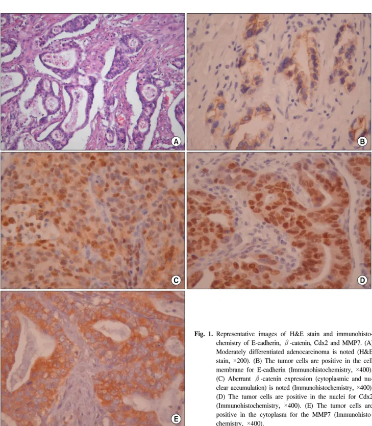
300희석), β-catenin 항체 (Leica Biosystem, Newcastle, UK, 1:300희석), Cdx2 항체 (Cell Marque Corporation, Rocklin, CA, USA, 1:500희석), MMP7 항체 (Abcam, Cambridge, UK, 1:500희석)가 사용되었으며, 상온에서 2시간 반응시 켰다. 3-Amino-9-ethylcarbazole이 색소원(chromogen)으로 사 용되었으며, Meyer’s hematoxylin으로 대조 염색하였다. 면 역조직화학 염색에 있어서 E-cadherin은 막성표현(membranous expression) 패턴이며, 종양세포의 25% 이하가 염색이 안된 경우를 발현 소실(음성)으로 판정하였으며, β-catenin 면역 염색 역시 E-cadherin 기준과 동일하였으며, 반면에 종양세 포의 핵염색이 발현되었을 경우 이상 발현으로 판정하였 다. MMP7 면역염색은 세포질(cytoplasm) 혹은 세포막 패턴 으로 염색되며 종양세포의 5%이상일 때 양성으로 판정하 였다. 그리고 Cdx2의 면역염색은 세포질염색 보다는 핵염 색 패턴이 많으며 종양세포의 5% 이상 염색되었을 경우 양 성으로 판정하였다(Fig. 1).
3) 통계분석
E-cadherin, β-catenin, Cdx2, MMP7의 면역염색 표현과 임상병리요인간의 분석은 카이제곱 검정으로 이용하였으 며, 생존율 분석은 Kaplan-Meier 방법 및 Cox회귀모형을 이 용하였다. 유의 수준은 P<0.05로 정의하였다.
결 과
1) 임상병리학적 특성과 면역염색 발현과의 관계
E-cadherin의 발현양상에서 모든 정상 위 점막에서 막성 발현이 뚜렷이 관찰되었으며, 위암세포에서의 발현은 122 예 중 33 (27%)예에서 발현되지 않았으며, 34 (28%)예는 면 역 염색 면적이 25% 미만인 경우로 관찰되었다. 25% 이상 의 염색을 보인 예는 55 (45%)로 이 중 21예는 25∼50%, 32예는 50∼75%, 2예에서 75∼100%의 염색을 보였다. 65세 이상에서 음성이 더 많이 관찰되었다(P=0.015). 그리고 조 직학적 차이는 P값이 0.072로 통계적 유의성이 없었다.
β-catenin의 발현양상은 정상적인 위 점막에서 모두 뚜 렷한 막성 발현이 관찰되었으며, 핵 염색은 관찰되지 않았 다. 위암세포에서의 염색을 보면, 34 (28%)예에서 발현되지
Fig. 1. Representative images of H&E stain and immunohisto- chemistry of E-cadherin, β-catenin, Cdx2 and MMP7. (A) Moderately differentiated adenocarcinoma is noted (H&E stain, ×200). (B) The tumor cells are positive in the cell membrane for E-cadherin (Immunohistochemistry, ×400).
(C) Aberrant β-catenin expression (cytoplasmic and nu- clear accumulation) is noted (Immunohistochemistry, ×400).
(D) The tumor cells are positive in the nuclei for Cdx2 (Immunohistochemistry, ×400). (E) The tumor cells are positive in the cytoplasm for the MMP7 (Immunohisto- chemistry, ×400).
않았으며, 13 (11%)예에서 25% 미만의 염색을 보였다. 8 (7%)예에서 핵염색이 관찰되었다. 핵염색이 관찰된 8예의 경우는 이상발현으로, 통계적 편의를 위해 25% 이하 음성 판정에 포함하여 통계처리를 하였다. β-catenin는 여성에서 양성 판정이 더 많음을 관찰하였다(P=0.022).
Cdx2의 발현양상을 보면, 종양세포에서 발현되었으며, 정 상적인 상피세포에서는 관찰할 수 없었으며, 양성으로 판 정된 경우가 80 (66%)예에서 관찰되었다. 조직학적 분화도 가 높은 군에서 양성판정의 예가 더 많이 관찰되었으나 통 계적으로 유의성은 없었다(P=0.088).
Table 1. Relationship between the results of immunohistochemistry and clinicopathological features
E-cadherin β-catenin Cdx2 MMP7
Negative N=67
Positive N=55
Negative N=55
Positive N=67
Negative N=42
Positive N=80
Negative N=47
Positive N=75 Age
≤65 19 28 20 27 18 29 22 25
>65 48 27 35 40 24 51 25 50
P=0.015 P=0.711 P=0.558 P=0.181
Gender
Male 54 38 47 45 34 58 24 58
Female 13 17 8 22 8 22 13 17
P=0.205 P=0.022 P=0.379 P=0.666
Histological
Differentiated 38 22 26 34 16 44 18 42
Undifferentiated 29 33 29 33 26 36 29 33
P=0.072 P=0.720 P=0.088 P=0.065
Depth
T2a 12 17 14 15 12 17 9 20
T2b 55 38 41 52 30 63 38 55
P=0.134 P=0.831 P=0.379 P=0.389
N stage
N1 42 36 32 46 25 53 37 41
N2 25 19 23 21 17 27 10 34
P=0.850 P=0.259 P=0.552 P=0.011
Size
≤5 cm 35 26 29 32 21 40 25 36
>5 cm 32 29 26 35 21 40 22 39
P=0.716 P=0.716 P=1.000 P=0.710
Table 2. Relationship between the results of immunohistochemistry and 2-year disease free survival rate (2YDFSR)
E-cadherin β-catenin Cdx2 MMP7
Negative N=63
Positive N=51
Negative N=50
Positive N=64
Negative N=36
Positive N=78
Negative N=41
Positive N=73
Recurrence within 2-Year 24 12 21 15 11 25 9 27
2YDFSR (%) 61.9 76.5 58.0 76.6 69.4 67.9 78.0 63.0
P-value 0.172 0.044 0.763 0.103
병기에서 더 높은 양성발현을 보였다(P=0.011). 그리고 조 직학적 분화도가 높은 군에서 양성발현이 많이 관찰되었지 만 통계적 유의성은 없었다(P=0.065)(Table 1).
2) 면역염색 발현여부와 2년 무병생존율 및 재발부위와 의 연관성
122명의 환자들의 평균 추적관찰 기간은 평균 43.29개월
예, β-catenin 음성인 경우는 21 (42%)예, Cdx2 양성인 경우 는 25 (32%)예, MMP7 양성의 경우는 27 (37%)예에서 관찰 되었다. 2년 무병생존율은 β-catenin 음성의 경우가 58.0%
로 양성의 경우(76.6%)보다 통계적으로 유의하게 낮게 관 찰되었다(P=0.044)(Table 2). 재발부위와의 연관성에서는 의 미 있는 결과를 관찰하지 못하였다(Table 3).
각 면역염색의 결과와 임상병리학적 요인들의 2년 무병생
Table 3. Relationship between the results of immunohistochemistry and recurrent sites
E-cadherin β-catenin Cdx2 MMP7
Negative N=63
Positive N=51
Negative N=50
Positive N=64
Negative N=36
Positive N=78
Negative N=41
Positive N=73
Peritoneum 3 9 5 7 4 8 3 9
P=0.221 P=0.361 P=1.000 P=0.532
Liver 6 9 6 9 4 11 4 11
P=0.785 P=0.264 P=0.772 P=0.567
Lymph node 2 5 3 4 2 5 2 5
P=0.457 P=0.697 P=1.000 P=1.000
Lung 0 1 0 1 0 1 0 1
P=1.000 P=0.439 P=1.000 P=1.000
Bone 1 0 1 0 1 0 0 1
P=0.447 P=1.000 P=0.316 P=1.000
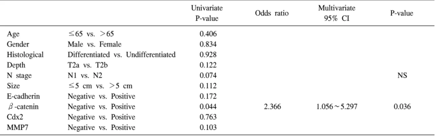
Table 4. Univariate and multivariate analysis with respect to 2-year disease free survival rate Univariate
P-value Odds ratio Multivariate
95% CI P-value
Age ≤65 vs. >65 0.406
Gender Male vs. Female 0.834
Histological Differentiated vs. Undifferentiated 0.928
Depth T2a vs. T2b 0.122
N stage N1 vs. N2 0.074 NS
Size ≤5 cm vs. >5 cm 0.112
E-cadherin Negative vs. Positive 0.172
β-catenin Negative vs. Positive 0.044 2.366 1.056∼5.297 0.036
Cdx2 Negative vs. Positive 0.763
MMP7 Negative vs. Positive 0.103
존율과의 단변량 분석에서 β-catenin 면역염색 결과가 의 미 있는 요인으로 분석되었으며(P=0.044), N병기(N1, N2)는 P값이 0.074로 나타났다. 이러한 단변량 분석결과를 토대로 Cox회귀모델을 이용한 다변량 분석에서는 β-catenin 면역 염색 결과가 독립적으로 2년 무병생존율과 관련 있는 것으 로 파악되었다(P=0.036, OR=2.366)(Table 4).
고 찰
대한위암학회에서 시행한 ‘2009전국위암등록사업’자료 를 보면 조기위암의 비율이 해마다 증가하여 2009년에는 조기위암이 57.7%, 진행성위암이 42.3%로 조사되었다. 이 러한 조기위암의 증가와 치료 기술의 발전으로 전체 위암 의 생존율이 향상되고 있으나, 진행성 위암은 복막전이, 간 전이, 림프절전이 등 원격 전이로 인하여 그 예후가 불량하 다. 원발 병소의 악성세포가 원격 전이가 되기 위해서는 우
선 세포와 세포 사이의 접착이 떨어져야 하며, 이후 국소 침습, 이동, 신생혈관생성, 원격 장기 내에서의 성장 등의 여러 단계를 거쳐야 한다.(1) 세포 사이의 구조적 안정화를 유지하는데 필요한 세포접착분자들은 세포-세포 결합, 세 포-세포 외 기질 결합, 세포간 상호작용에 관여하는 것으로 알려져 있다. 여러 세포접착분자들 중에서 상피세포의 세 포간 유착에 매우 중요한 역할을 하는 E-cadherin는 동종친 화성 상호작용을 하는 칼슘 의존성인 막성 당단백질로, 세 포질내 위치한 부위는 고인산화된 곳으로 catenin과 복합체 를 형성한 후 세포내 골격단백질인 actin과 결합하여 정상 적인 세포막 형성과 골격을 유지하는데 큰 역할을 담당하 고 있다.(7) E-cadherin-catenin 복합체의 변성은 세포분화의 소실과 고 침윤성 및 원격전이와 연관성이 있다.(8) 이번 연 구결과에서 E-cadherin, β-catenin의 발현소실이 조직학적 분화도, 종양의 침범 깊이, 림프절 전이상태등과의 관련성 을 찾지 못하였다.
상적인 위 점막에서는 발견되지 않으나, 장 상피화생의 위 점막에서는 대부분 발견되며,(10) 위암에서 Cdx2의 발현은 좀더 나은 예후 및 림프절 전이와 음의 상관관계를 보인다 고 보고하고 있다.(11) 하지만 이번 연구에서는 림프절 전 이상태와 연관성은 없었으며, 분화도가 좋은 경우에서 양 성 발현 경향이 높았으나 통계학적으로 의미는 없었다 (P=0.088). 본 연구에서 림프절 전이가 있는 경우(N1, N2)를 대상으로 하였기 때문에 림프절 전이상태와 연관성이 나타 나지 않은 것으로 보인다. 아마도 림프절 전이가 없는 경우 와 비교하였다면 의미 있는 결과가 나올 수 있었을 것으로 생각된다.
세포 외 기질(extracellular matrix, ECM)은 세포-세포, 혹은 세포-ECM 상호작용을 매개함으로써 구조적 골격형성을 지지하는데 도움을 주며, ECM 구성성분의 분해는 기질 금 속단백분해효소(matrix metalloproteinase, MMP)에 의해 대 부분 조절된다. MMP는 아연의존성 펩티드내부분해효소 (zinc dependent endopeptidase)로 최소 25종류로 구성되어있 다.(12) 그 중에서 MMP7은 단백당(proteoglycans), fi- bronectin, laminin, 4형 콜라겐과 같은 광범위한 기질들에 대 한 단백 분해능을 가진다.(13) 이러한 MMP7는 암 조직에서 과 발현되고, 세포성장, 세포자가사멸, 침습성, 원격전이, 혈관생성 등의 암 진행과 연관되어 있다.(14) 특히 위암에 서 림프절 전이와 같은 침습성과 관련이 있다.(15) 이번 연 구의 결과를 보면, N1인 경우 MMP7의 양성 발현율이 52.6%, N2의 경우는 77.3%로 림프절전이가 높을 때 양성 발현율이 높게 나타났다(P=0.011).
Gabbert 등(16)은 E-cadherin의 이상 발현이 위암의 생존 율과 관련이 있다고 하였으며, Zhou 등(17)과 Jawhari 등(18) 에 의하면 E-cadherin보다 β-catenin의 이상발현이 나쁜 생 존예후를 보인다고 하였다. Chen 등(19)은 MMP7 양성 발현 의 환자들에서 복막전이로 인한 사망이 더 많으며, 의미 있 게 나쁜 예후를 보인다고 하였다. 저자들은 단변량 분석에 서 β-catenin 발현소실 및 이상 발현이 2년 생존율과 통계 적으로 의미 있는 관련성을 나타내었다. 하지만 재발유형 과 관련해서는 E-cadherin, β-catenin, Cdx2, MMP7 면역염 색 모두 특별한 연관성을 발견하지 못하였다. 저자들은 연 구대상으로 진행성위암을 선정하였음에도 장막침범이 있 는 경우를 제외한 것은 그 재발양상이 대부분 복막재발이
현소실 및 이상 발현이 의미 있게 나쁜 2년 생존율을 보이 는 독립인자로 관찰되었다.
결 론
β-catenin의 발현 소실 및 이상 발현은 재발 부위와는 관 련성이 없었으나, 2년 이내 재발을 예측할 수 있는 의미 있 는 인자로서 그 역할을 할 수 있을 것으로 생각된다.
REFERENCES
1) John M, Peter MH, Mark AI, Joe WG, Craig BT. The Molecular Basis of Cancer. 3rd ed. Philadelphia: Saunders;
2008.
2) Joo YE, Rew JS, Kim HS, Choi SH, Park CS, Kim SJ.
Changes in the E-cadherin-catenin complex expression in early and advanced gastric cancers. Digestion 2001;64:111-9.
3) Yonemura Y, Endou Y, Fujita H, Fushida S, Bandou E, Taniguchi K, et al. Role of MMP-7 in the formation of peri- toneal dissemination in gastric cancer. Gastric Cancer 2000;3:
63-70.
4) Mizoshita T, Tsukamoto T, Nakanishi H, Inada K, Ogasawara N, Joh T, et al. Expression of Cdx2 and the phenotype of ad- vanced gastric cancers: relationship with prognosis. J Cancer Res Clin Oncol 2003;129:727-34.
5) Seno H, Oshima M, Taniguchi MA, Usami K, Ishikawa TO, Chiba T, et al. CDX2 expression in the stomach with intestinal metaplasia and intestinal-type cancer: Prognostic implications.
Int J Oncol 2002;21:769-74.
6) Yoo CH, Noh SH, Shin DW, Choi SH, Min JS. Recurrence following curative resection for gastric carcinoma. Br J Surg 2000;87:236-42.
7) Takeichi M. Cadherin cell adhesion receptors as a morphoge- netic regulator. Science 1991;251:1451-5.
8) Shiozaki H, Oka H, Inoue M, Tamura S, Monden M. E-cad- herin mediated adhesion system in cancer cells. Cancer 1996;77(8 Suppl):1605-13.
9) Suh E, Traber PG. An intestine-specific homeobox gene regu- lates proliferation and differentiation. Mol Cell Biol 1996;16:
619-25.
10) Eda A, Osawa H, Yanaka I, Satoh K, Mutoh H, Kihira K, et al. Expression of homeobox gene CDX2 precedes that of CDX1 during the progression of intestinal metaplasia. J Gastroenterol 2002;37:94-100.
11) Fan Z, Li J, Dong B, Huang X. Expression of Cdx2 and hep-
atocyte antigen in gastric carcinoma: correlation with histo- logic type and implications for prognosis. Clin Cancer Res 2005;11:6162-70.
12) Nelson AR, Fingleton B, Rothenberg ML, Matrisian LM.
Matrix metalloproteinases: biologic activity and clinical impli- cations. J Clin Oncol 2000;18:1135-49.
13) Shiomi T, Okada Y. MT1-MMP and MMP-7 in invasion and metastasis of human cancers. Cancer Metastasis Rev 2003;22:
145-52.
14) Adachi Y, Yamamoto H, Itoh F, Hinoda Y, Okada Y, Imai K. Contribution of matrilysin (MMP-7) to the metastatic path- way of human colorectal cancers. Gut 1999;45:252-8.
15) Ajisaka H, Yonemura Y, Miwa K. Correlation of lymph node metastases and expression of matrix metalloproteinase-7 in pa- tients with gastric cancer. Hepatogastroenterology 2004;51:
900-5.
16) Gabbert HE, Mueller W, Schneiders A, Meier S, Moll R, Birchmeier W, et al. Prognostic value of E-cadherin expression
in 413 gastric carcinomas. Int J Cancer 1996;69:184-9.
17) Zhou YN, Xu CP, Han B, Li M, Qiao L, Fang DC, et al.
Expression of E-cadherin and beta-catenin in gastric carcinoma and its correlation with the clinicopathological features and pa- tient survival. World J Gastroenterol 2002;8:987-93.
18) Jawhari A, Jordan S, Poole S, Browne P, Pignatelli M, Farthing MJ. Abnormal immunoreactivity of the E-cadherin- catenin complex in gastric carcinoma: relationship with patient survival. Gastroenterology 1997;112:46-54.
19) Chen JQ, Zhan WH, He YL, Peng JS, Wang JP, Cai SR, et al. Expression of heparanase gene, CD44v6, MMP-7 and nm23 protein and their relationship with the invasion and metastasis of gastric carcinomas. World J Gastroenterol 2004;
10:776-82.
20) Kuramoto M, Shimada S, Ikeshima S, Matsuo A, Yagi Y, Matsuda M, et al. Extensive intraoperative peritoneal lavage as a standard prophylactic strategy for peritoneal recurrence in patients with gastric carcinoma. Ann Surg 2009;250:242-6.