Ann Hepatobiliary Pancreat Surg 2018;22:75-78
https://doi.org/10.14701/ahbps.2018.22.1.75
Case Report
Right adrenal gland pseudocyst masquerading as a large symptomatic hepatic cyst: Single incision laparoscopic (SILS)
resection and a review of current literature
Koy Min Chue1, Giap Hean Goh2, and Alfred Wei Chieh Kow3
1Department of Surgery, University Surgical Cluster, National University Hospital, 2Department of Pathology, National University Hospital, 3Division of Hepatobiliary and Pancreatic Surgery and Liver Transplantation,
Department of Surgery, University Surgical Cluster, National University Hospital, Singapore
Adrenal pseudocysts are rare entities, which are usually asymptomatic. Large symptomatic adrenal pseudocysts may cause compressive symptoms. The etiology of these cysts is unknown, although the cyst wall is all lined by fibrous tissue, without any epithelial or endothelial lining. We report a case of a 26-year-old lady who presented with a sympto- matic right adrenal pseudocyst measuring 7.6 cm in size. Magnetic resonance imaging confirmed the presence of a right retroperitoneal cystic lesion which was hyperintense on T2 sequencing. An attempted single incision transumbilical laparoscopic surgery (SILS) was performed to excise the right adrenal pseudocyst. However, due to the retro-hepatic nature of the lesion and as the medial wall of the cyst was adherent to the inferior vena cava, an additional 5 mm port was inserted to facilitate retraction of the liver. The post-operative period was uneventful. She was successfully discharged from the hospital as a day surgery patient. The final pathology showed an adrenal pseudocyst. (Ann Hepatobiliary Pancreat Surg 2018;22:75-78)
Key Words: Adrenalectomy; Adrenal gland; Laparoscopy; Pseudocyst
Received: June 4, 2017; Revised: July 5, 2017; Accepted: July 7, 2017 Corresponding author: Alfred Wei Chieh Kow
Division of Hepatobiliary and Pancreatic Surgery and Liver Transplantation, Department of Surgery, University Surgical Cluster, National University Hospital, 1E, Kent Ridge Road, NUHS Tower Block, Level 8, Singapore 119228
Tel: +65-6779-5555, Fax: +65-6777-8427, E-mail: [email protected]
Copyright Ⓒ 2018 by The Korean Association of Hepato-Biliary-Pancreatic Surgery
This is an Open Access article distributed under the terms of the Creative Commons Attribution Non-Commercial License (http://creativecommons.org/
licenses/by-nc/4.0) which permits unrestricted non-commercial use, distribution, and reproduction in any medium, provided the original work is properly cited.
Annals of Hepato-Biliary-Pancreatic Surgery ∙ pISSN: 2508-5778ㆍeISSN: 2508-5859
INTRODUCTION
Adrenal pseudocysts are rare entities consisting of a cystic lesion surrounded by a wall of fibrous connective tissue.1 Although the etiology of adrenal pseudocyst for- mation is unknown, it is postulated to occur due to malfor- mation and hemorrhage from adrenal veins into the adre- nal gland, cystic degeneration of a primary adrenal neo- plasm or degeneration of a vascular neoplasm.2-4 Most adrenal pseudocysts are asymptomatic and require no treatment,5 and they are diagnosed incidentally with the increasing use of imaging modalities. For symptomatic adrenal pseudocysts, treatment options include either per- cutaneous drainage6 or surgical excision.1,2,5,7 However, as these cysts are filled with old, hemorrhagic fluid, and it may be difficult to differentiate them from necrotic, malignant neoplasms, surgical excision is preferred.7 Furthermore, with
the advent of laparoscopic surgery, most of the sympto- matic adrenal pseudocysts can be excised safely via the laparoscopic approach.2,5 We report a case of a patient with a 7.6 cm-sized right adrenal pseudocyst that was ex- cised laparoscopically.
CASE
A 26-year-old lady, presented to our department with a three-month history of abdominal bloating associated with constant, non-radiating right flank discomfort. This was associated with symptoms such as early satiety, al- though there was no vomiting. She had no other sig- nificant medical conditions, and the systematic review was unremarkable. There was no history of trauma to the abdomen. There was also no evidence of any hormonal disorder. She underwent magnetic resonance imaging
76 Ann Hepatobiliary Pancreat Surg Vol. 22, No. 1, February 2018
Fig. 1. (A) MRI T2-weighted image showing a 7.6×5.7 cm-sized right adrenal cyst. (B) MRI T2-weighted image showing a right adrenal cyst measuring 6.5 cm in width.
Fig. 2. (A) A self-constructed glove port for single-incision laparoscopic surgery. (B) Wound protector with a glove port stretching over it.
(MRI) of her abdomen in her home country, which re- vealed a 7.6×5.7 cm cystic lesion in the right lobe of the liver that was hyperintense on T2 sequencing (Fig. 1A and B). She had previously undergone percutaneous cyst fluid aspiration in her home country prior to seeking treatment in Singapore, but her symptoms had recurred within 2 weeks after the procedure. The cyst fluid cytology during the initial aspiration was benign. Considering the previous cytology findings, with no radiological suspicion of ma- lignancy on the MRI scan, she was offered single incision laparoscopic surgical cyst resection.
A transumbilical approach was employed to gain access into the abdomen using the open Hasson technique.8 A self-made glove port was created to attempt SILS surgery (Fig. 2A). The glove port was made using medium Alexis O wound Protector/ Retractor (Applied Medical, Rancho
Santa Margarita, CA 92688) with a latex glove modified to accommodate 5 laparoscopic ports including a camera port (Fig. 2B). The camera port was fitted onto the index finger of the glove, and the other four 5mm ports were attached to the other digits. All the working ports were 5 mm in size. A diagnostic laparoscopy was performed, which showed that the cyst was not of hepatic origin.
Instead, there was a smooth cystic mass in a retro- peritoneal location, posterior to the liver, situated adjacent to the inferior vena cava and attached inferiorly to the right adrenal gland (Fig. 3). Midway through the SILS ex- cision of the cyst, an additional 5 mm port was inserted in the right hypochondriac region to facilitate liver retraction. The cyst was separated and excised using a combination of blunt dissection and LigaSure (Valley Lab, Boulder, CO) without any difficulty. Part of the right
Koy Min Chue, et al. Adrenal pseudocyst masquerading as hepatic cyst: Single incision laparoscopic resection 77
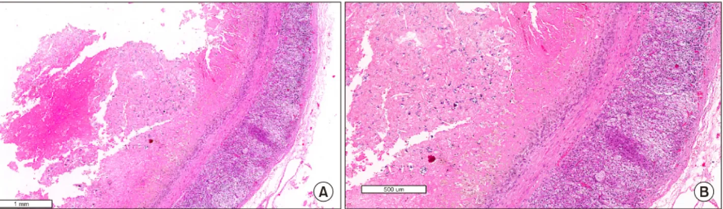
Fig. 4. (A) Adrenal Pseudocyst. The cyst wall was devoid of an epithelial or endothelial lining and it was composed of fibrous tissue with haemorrhagic fibrinous material, cholesterol clefts and calcifications. Adrenal parenchymal tissue was identified (Hematoxylin and eosin ×40). (B) Adrenal Pseudocyst (Hematoxylin and eosin ×100).
Fig. 3. Adrenal Pseudocyst situated posterior to the liver.
adrenal gland was excised together with the cyst as they were adherent. The cyst fluid was found to be brownish, likely indicating previous hemorrhage into the cyst. The cyst fluid was sent for cytology, while the cyst wall was sent for pathological examination. Postoperatively, the pa- tient made an unremarkable recovery and was discharged on the same day. The patient had no postoperative com- plications and was reassessed 2 months after surgery. The previous abdominal bloating and early satiety had re- solved completely at the latest clinical review.
Histologic examination of the cyst wall revealed adre- nal cortical tissue enveloping a cyst comprising of a thick rim of dense collagenous fibrous tissue, with no dis- cernible epithelial or endothelial lining (Fig. 4A and B).
The cyst wall itself showed scattered calcifications, with degenerative eosinophilic material, cholesterol clefts and
hemosiderophages. No granulomata or malignancy was identified. The cytology of the cyst fluid revealed blood and rare inflammatory cells, consistent with previous hemorrhage.
DISCUSSION
Adrenal pseudocysts are rare. To date, one of the larg- est recorded adrenal pseudocyst was reported by Davenport et al.9 in a woman measuring up to 22 cm.
These pseudocysts are picked up incidentally on imaging or autopsy, or less commonly, they may present with compressive symptoms.6,7 They are seen more commonly in females.7,9 In our patient, given the findings suggestive of previous hemorrhage, we postulate that she had experi- enced previous adrenal hemorrhage, likely to be sponta- neous, as there was no history of trauma or infection.1,2,6,7 The formation of the pseudocyst was likely to be the re- sult of the hematoma enclosed by a fibrotic cavity.6,7
This case highlights the importance of proper evaluation of a symptomatic cystic lesion in the posterior right hypo- chondriac region. These cystic masses must be dis- tinguished from other benign or malignant lesions that can originate from the surrounding structures.2,10 In this pa- tient, the differential diagnoses included a right posterior sector liver cyst, a right renal cyst, a retroperitoneal cyst, or rarely, a bowel duplication cyst in the subhepatic region. As in all adrenal lesions >6 cm, the risk of malig- nancy increases.2,11 A single-incision laparoscopic surgery was offered as the cyst did not exhibit any suspicious fea- tures on preoperative imaging. However, in order not to
78 Ann Hepatobiliary Pancreat Surg Vol. 22, No. 1, February 2018
compromise any potential oncologic outcomes should the cyst appear suspicious intraoperatively, an anterior surgi- cal approach was undertaken with a low threshold for con- version to conventional laparoscopic or even open surgery.
Since the first laparoscopic adrenalectomy by Gagner et al.12 in 1992, in well selected patients, laparoscopic sur- gery was shown to reduce blood loss, decrease length of stay with quicker commencement of oral intake and return of bowel function.13-15 Single-incision laparoscopic sur- gery for the adrenal gland, performed either via a retro- peritoneoscopic or transabdominal approach, has been gaining popularity,16-20 with similar outcomes as those of conventional laparoscopic surgery, in addition to im- proved cosmesis.16 Ishida et al.20 demonstrated that despite the feasibility of single-incision transumbilical laparo- scopic adrenal surgery, there were obvious technical diffi- culties compared to conventional laparoscopy. Hora et al.17 performed single incision laparoscopic adrenalec- tomies strictly on small tumors with a non-malignant eti- ology, in their series of 18 cases. In our patient, despite adequate positioning, she had a large floppy liver which was partially obscuring the surgical field. Furthermore, the cyst wall was thin and fragile, and there were no bent in- struments, which limited the amount of manipulation pos- sible with the single incision technique. One additional ports was inserted to retract the liver superiorly, in order to achieve optimal exposure of the retrohepatic region to successfully resect the right adrenal pseudocyst. To the best of our knowledge, our case describes the first at- tempted single-incision laparoscopic surgery of an adrenal pseudocyst and the lessons learned from this experience.
A single incision approach might be feasible in a smaller cystic lesion, with or without bent instruments to help to achieve proper triangulation.
In conclusion, adrenal pseudocysts are rare. With ad- vanced imaging techniques, accurate diagnosis is possible and symptomatic adrenal pseudocysts can be safely re- sected via minimally invasive surgery.
REFERENCES
1. Tagge DU, Baron PL. Giant adrenal cyst: management and re- view of the literature. Am Surg 1997;63:744-746.
2. Kim BS, Joo SH, Choi SI, Song JY. Laparoscopic resection of
an adrenal pseudocyst mimicking a retroperitoneal mucinous cystic neoplasm. World J Gastroenterol 2009;15:2923-2926.
3. Gaffey MJ, Mills SE, Fechner RE, Bertholf MF, Allen MS Jr.
Vascular adrenal cysts. A clinicopathologic and immunohistochemical study of endothelial and hemorrhagic (pseudocystic) variants. Am J Surg Pathol 1989;13:740-747.
4. Medeiros LJ, Lewandrowski KB, Vickery AL Jr. Adrenal pseu- docyst: a clinical and pathologic study of eight cases. Hum Pathol 1989;20:660-665.
5. AsalZare M, Shakiba B, Asadpour AA, Ghoreifi A. Laparoscopic management of symptomatic giant adrenal pseudocyst: a case report. Urol J 2014;11:1517-1520.
6. Habra MA, Feig BW, Waguespack SG. Image in endocrinology:
adrenal pseudocyst. J Clin Endocrinol Metab 2005;90:3067-3068.
7. Khilnani GC, Kumar A, Bammigatti C, Sharma R, Gupta SD.
Hemorrhagic pseudocyst of the adrenal gland causing acute ab- dominal pain. J Assoc Physicians India 2008;56:379-380.
8. Hasson HM. A modified instrument and method for laparoscopy.
Am J Obstet Gynecol 1971;110:886-887.
9. Davenport M, Pollard K, Smith SE, MacMahon MJ. Adrenal cysts--report, review and classification. Postgrad Med J 1988;64:
71-73.
10. Ghandur-Mnaymneh L, Slim M, Muakassa K. Adrenal cysts:
pathogenesis and histological identification with a report of 6 cases. J Urol 1979;122:87-91.
11. Khoda J, Hertzanu Y, Sebbag G, Lantsberg L, Barky Y. Adrenal cysts: diagnosis and therapeutic approach. Int Surg 1993;78:
239-242.
12. Gagner M, Lacroix A, Bolté E. Laparoscopic adrenalectomy in Cushing's syndrome and pheochromocytoma. N Engl J Med 1992;327:1033.
13. Haveran LA, Novitsky YW, Czerniach DR, Kaban GK, Kelly JJ, Litwin DE. Benefits of laparoscopic adrenalectomy: a 10-year single institution experience. Surg Laparosc Endosc Percutan Tech 2006;16:217-221.
14. Wang HS, Li CC, Chou YH, Wang CJ, Wu WJ, Huang CH.
Comparison of laparoscopic adrenalectomy with open surgery for adrenal tumors. Kaohsiung J Med Sci 2009;25:438-444.
15. Maccabee DL, Jones A, Domreis J, Deveney CW, Sheppard BC.
Transition from open to laparoscopic adrenalectomy: the need for advanced training. Surg Endosc 2003;17:1566-1569.
16. Shi TP, Zhang X, Ma X, Li HZ, Zhu J, Wang BJ, et al.
Laparoendoscopic single-site retroperitoneoscopic adrenalec- tomy: a matched-pair comparison with the gold standard. Surg Endosc 2011;25:2117-2124.
17. Hora M, Ürge T, Stránský P, Trávníček I, Pitra T, Kalusová K, et al. Laparoendoscopic single-site surgery adrenalectomy - own experience and matched case-control study with standard laparo- scopic adrenalectomy. Wideochir Inne Tech Maloinwazyjne 2014;9:596-602.
18. Hirano D, Hasegawa R, Igarashi T, Satoh K, Mochida J, Takahashi S, et al. Laparoscopic adrenalectomy for adrenal tu- mors: a 21-year single-institution experience. Asian J Surg 2015;38:79-84.
19. Kwak HN, Kim JH, Yun JS, Son BH, Chung WY, Park YL, et al. Conventional laparoscopic adrenalectomy versus laparo- scopic adrenalectomy through mono port. Surg Laparosc Endosc Percutan Tech 2011;21:439-442.
20. Ishida M, Miyajima A, Takeda T, Hasegawa M, Kikuchi E, Oya M. Technical difficulties of transumbilical laparoendoscopic sin- gle-site adrenalectomy: comparison with conventional laparo- scopic adrenalectomy. World J Urol 2013;31:199-203.