http://dx.doi.org/10.4174/astr.2015.89.5.254 Annals of Surgical Treatment and Research
Effect of extramucin pools in gastric cancer patients
Ki-Hyun Kim*, Si-Hak Lee*, Cheol-Woong Choi1, Su-Jin Kim1, Chang-In Choi2, Dae-Hwan Kim2, Tae-Yong Jeon2, Dong-Heon Kim2, Sun-Hwi Hwang
Departments of Surgery and 1Internal Medicine, Pusan National University Yangsan Hospital, Yangsan, 2Department of Surgery, Pusan National University School of Medicine, Busan, Korea
INTRODUCTION
Mucinous gastric adenocarcinoma (MGC) is a rare type of cancer that is defined by the World Health Organization (WHO) as a gastric adenocarcinoma with >50% extracellular mucin pools within the tumors [1]. MGC comprises approximately 2%–
5% of all gastric carcinomas [2-5]. The clinicopathologic charac- teristics of MGC remain controversial. MGC has shown a poorer prognosis than nonmucinous gastric adenocarcinoma (NMGC) [6-8]. In contrast, another study suggested no differ ence in the survival rate of MGC and NMGC.
Clinically, it is possible to find cases with extracellular mucin
pools lower than a half of the tumor area (Fig. 1). However, the exact prevalence rate is unknown. It could be missing from the pathologic report because gastric cancer with lower than 50%
tumor volume of extracellular mucin pool adenocarcinoma (LEMPC) is not included in the diagnostic criteria of WHO classi- fication. Moreover, if a pathologist is not aware of LEMPC, ex tra - cellular mucin pools may be overlooked. According to the WHO classification, those cases are classified as NMGC, not MGC.
Treatment of LEMPC is similar to that of NMGC, even though LEMPC tends to be more progressive [9].
The clinicopathologic feature of LEMPC patients is not clear.
To clarify the effect of extracellular mucin pools, we compared Purpose: Mucinous gastric adenocarcinoma (MGC) is defined by the World Health Organization as a gastric adenocarcinoma with >50% extracellular mucin pools within the tumors. In this study, we attempted to analyze the clinicopathologic features of patients pathologically diagnosed as gastric cancer with lower than 50% tumor volume of extracellular mucin pool adenocarcinoma (LEMPC). We compared MGC versus nonmucinous gastric adenocarcinoma (NMGC). We were used in abbreviations LEMPC for NMGC including extracellular mucin pool.
Methods: Files of 995 patients with gastric cancer NMGC (n = 935), MGC (n = 20), LEMPC (n = 40) who underwent curative resection at Pusan National University Yangsan Hospital from December 2008 to December 2013 were retrospectively analyzed. All pathologic reports after curative resection and evaluated clinicopathologic features were reviewed to identify the effect of extracellular mucin pools in gastric cancer.
Results: Compared with the NMGC patients, the clinicopathological features of MGC patients were as follows: more frequent open surgery, larger tumor size, more advanced T stage and N stage, more positive lymph node metastasis, and perineural invasion. LEMPC patients showed similar features compared with NMGC patients. MGC and LEMPC patients showed similar clinicopathological features, except T stage and lymph node metastasis.
Conclusion: LEMPC can be thought of as a previous step of MGC. It is reasonable to consider LEMPC patients in the diagnostic criteria of MGC, and to adequately treat.
[Ann Surg Treat Res 2015;89(5):254-260]
Key Words: Mucinous adenocarcinoma, Stomach, Neoplasms
Reviewed January February March April May June July August September October November
*Ki-Hyun Kim and Si-Hak Lee contributed equally to this study as co-first authors.
Copyright ⓒ 2015, the Korean Surgical Society
cc Annals of Surgical Treatment and Research is an Open Access Journal. All articles are distributed under the terms of the Creative Commons Attribution Non- Commercial License (http://creativecommons.org/licenses/by-nc/4.0/) which permits unrestricted non-commercial use, distribution, and reproduction in any Received June 22, 2015, Revised July 26, 2015, Accepted August 17, 2015
Corresponding Author: Sun-Hwi Hwang
Department of Surgery, Pusan National University Yangsan Hospital, 20 Geumo-ro, Mulgeum-eup, Yangsan 50612, Korea
Tel: +82-55-360-2124, Fax: +82-55-360-2154 E-mail: [email protected]
the clinicopathologic features of NMGC patients with MGC and LEMPC patients to determine the effect of extracellular mucin pools. We also evaluated the independent prognostic factors from the point of recurrence.
METHODS
Subjects
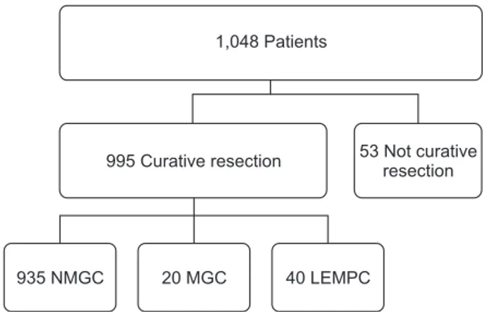
A retrospective review was done of 1,048 gastric cancer patients treated at Pusan National University Yangsan Hospital between December 2008 and December, 2013. Curative resection was performed in 995 pa tients (Fig. 2). MGC defined as extracellular mucin pools oc cupying over a half the tumor area was observed [1]. NMGC is classified as no extracellular mucin pool in tumor. LEMPC is defined as NMGC with including extracellular mucin pool.
All the diagnosis followed by the last report of pathologists.
Clinicopathologic variables including gender, age, body mass index (BMI), tumor location, surgical methods, tumor size, depth of invasion, number of lymph node metastasis, number of resected lymph node, lymph node metastasis, Lauren classifica tion, lymphatic invasion, vascular invasion, and perineural invasion were compared between MGC and NMGC patients, and between MGC and LEMPC patients. We investi- gated the variables in accordance with an established guideline [10]. Tumor staging was evaluated by using the TNM system of the International Union Against Cancer [10].
Prognostic factors associated with the recurrence of gastric cancers were also investigated. A disease-free survival (DFS) curve was prepared to identify the most significant prognostic factors for gastric cancer in our hospital based on recurrence.
The diagnosis of recurrence was confirmed by radiological findings, computed tomography or positron emission tomo- graphy, and endoscopic biopsy with histopathological findings.
Statistical analyses
Data was statistically analyzed using the chi-square test and Student t-test. Odds ratios (ORs) were estimated with 95%
confidence intervals (CIs). DFS curves were plotted using the Kaplan-Meier method and the log-rank test was used for univa- riate risk factors for recurrence. In multivariate analysis, the Cox proportional-hazards regression F model was used for inde- pendent risk factors of recurrence. All statistics were conducted using IBM SPSS Statistics ver. 21.0 (IBM Co., Armonk, NY, USA).
Statistical significance was defined as P < 0.05.
RESULTS
All 995 patients underwent curative resection. MGC and LEMPC patients comprise 2% and 6%, respectively, of all patients.
A B
Fig. 1. Extracellular mucin pools are lower than one half of the tumor area (A, Hematoxylin and eosin stain x20) and mucinous gastric adenocarcinoma (B, Hematoxylin and eosin stain).
20 MGC
935 NMGC 40 LEMPC
53 Not curative resection 995 Curative resection
1,048 Patients
Fig. 2. Flow chart of the study. NMGC, nonmucinous gastric adenocarcinoma; MGC, mucinous gastric adenocarcinoma;
LEMPC, gastric cancer with <50% tumor volume of extra- cellular mucin pool carcinoma.
Clinicopathological comparison between NMGC and other groups
Mean ages of MGC, NMGC, and LEMPC patients were similar. Gender, BMI, and tumor location did not significantly differ in the three groups. MGC and LEMPC patients received more open gastrectomies with extended lymph node dissection than NMGC patients (both P < 0.001). MGC tumor size of was
larger than NMGC tumors (P < 0.001). T stage, N stage, and node metastasis were higher in MGC patients than in NMGC patients (P < 0.001). LEMPC showed more advanced T stage (P
< 0.001) and node metastasis than LMGC (P < 0.001). However, the N stage of LEMPC was not significantly different from that of NMGC (P < 0.0109). More positive perineural invasion was apparent in LEMPC than in NMGC (P < 0.010) (Table 1).
Table 1. Clinicopathological comparison between NMGC and other groups
Variable LEMPC (n = 40) P-valuea) NMGC (n = 935) P-valueb) MGC (n = 20)
Sex 0.112 0.369
Male 31 (77.5) 611 (65.3) 15 (75.0)
Female 9 (22.5) 324 (34.7) 5 (25.0)
Age (yr) 61.53 ± 8.75 0.366 60.22 ± 10.97 0.068 64.75 ± 10.96
Body mass index (kg/m2) 24.33 ± 3.18 0.146 23.52 ± 3.42 0.739 23.27 ± 3.05
Location 0.541 0.476
Upper body 9 (22.5) 149 (15.9) 4 (20.0)
Mid bod 8 (20.0) 196 (21.0) 2 (10.0)
Lower body 23 (57.5) 590 (63.1) 14 (70)
Surgery 0.008 <0.001
Open 28 (70.0) 455 (48.7) 14 (70.0)
Laparoscopic 12 (30.0) 480 (51.3) 6 (30.0)
Size (cm) 4.66 ± 2.41 0.477 4.32 ± 3.03 <0.011 6.06 ± 2.99
Depth <0.001 <0.001
T1 13 (32.5) 567 (60.6) 4 (20.0)
T2 9 (22.5) 97 (10.4) 2 (10.0)
T3 12 (30.0) 133 (14.2) 3 (15.0)
T4 6 (15.0) 138 (14.8) 11 (55.0)
N stage 0.109 <0.001
N0 21 (52.5) 656 (60.6) 5 (25)
N1 8 (20.0) 101 (10.4) 5 (25)
N2 5 (12.5) 78 (14.2) 4 (20)
N3 6 (50) 100 (14.8) 6 (30)
Resected LN 37.18 ± 13.77 0.785 36.55 ± 14.15 0.913 36.20 ± 14.65
Node dissection
Below D2 8 (20.0) <0.001 432 (46.2) 0.006 3 (15.0)
Above D2 32 (80.0) <0.001 503 (53.8) 0.006 17 (85.0)
Node metastasis 0.018 <0.001
+ 19 (47.5) 279 (29.8) 15 (75.0)
– 21 (52.5) 656 (70.2) 5 (25.0)
Lauren classification 0.130 0.641
Intestinal 25 (62.5) 470 (50.3) 9 (45.0)
Diffuse 15 (37.5) 465 (49.7) 11 (55.0)
Lymphatic invasion 0.120 0.168
+ 13 (32.5) 206 (22.0) 7 (35.0)
– 27 (67.5) 729 (78.0) 13 (65.0)
Vascular invasion 0.826 0.355
+ 4 (10.0) 84 (9.0) 3 (15.0)
– 36 (90.0) 851 (91.0) 17 (85.0)
Perineural invasion <0.010 0.390
+ 18 (45.0) 247 (26.4) 7 (35.0)
– 22 (55.0) 688 (73.6) 13 (65.0)
Values are presented as number (%) or mean ± standard deviation.
NMGC, nonmucinous gastric adenocarcinoma; LEMPC, gastric cancer with <50% tumor volume of extracellular mucin pool adenocarcinoma; MGC, mucinous gastric adenocarcinoma; LN, lymph node.
Table 2. Clinicopathological comparison between NMGC and all extra celluarmucin pool gastric adenocarcinomas
Variable Nonextracellular mucin pool
(n = 935)
All extracellular mucin poola)
(n = 60) P-value
Sex 0.730
Male 611 (65.3) 46 (76.7)
Female 324 (34.7) 14 (23.3)
Age (yr) 60.22 ± 10.97 62.60 ± 9.57 0.101 Body mass index
(kg/m2) 23.52 ± 3.42 23.97 ± 3.15 0.322
Location 0.436
Upper body 149 (15.9) 13 (21.7)
Mid bod 196 (21.0) 10 (16.7)
Lower body 590 (63.1) 37 (61.7)
Surgery <0.001
Open 455 (48.7) 42 (70.0)
Laparoscopic 480 (51.3) 18 (30.0)
Size (cm) 4.32 ± 3.03 5.13 ± 2.68 0.043
Depth <0.001
T1 567 (60.6) 17 (28.3)
T2 97 (10.4) 11 (18.3)
T3 133 (14.2) 15 (25.0)
T4 138 (14.8) 17 (28.3)
N stage <0.001
N0 656 (60.6) 26 (43.3)
N1 101 (10.4) 13 (21.7)
N2 78 (14.2) 9 (15.0)
N3 100 (14.8) 12 (20.0)
Resected LN 36.55 ± 14.15 36.85 ± 13.95 0.874
Node dissection <0.001
Below D2 432 (46.2) 11 (18.3)
Above D2 503 (53.8) 49 (81.7)
Node metastasis <0.001
+ 279 (29.8) 34 (56.7)
– 656 (70.2) 26 (43.3)
Lauren classification 0.337
Intestinal 470 (50.3) 34 (56.7)
Diffuse 465 (49.7) 26 (43.3)
Lymphatic invasion 0.043
+ 206 (22.0) 20 (33.3)
– 729 (78.0) 40 (66.7)
Vascular invasion 0.485
+ 84 (9.0) 7 (11.7)
– 851 (91.0) 53 (88.3)
Perineural invasion 0.010
+ 247 (26.4) 25 (41.7)
– 688 (73.6) 35 (58.3)
Values are presented as number (%) or mean ± standard deviation.
NMGC, nonmucinous gastric adenocarcinoma; LEMPC, gastric cancer with <50% tumor volume of extracellular mucin pool adenocarcinoma; MGC, mucinous gastric adenocarcinoma; LN, lymph node.
a)All extracellular mucin pool: MGC with LEMPC.
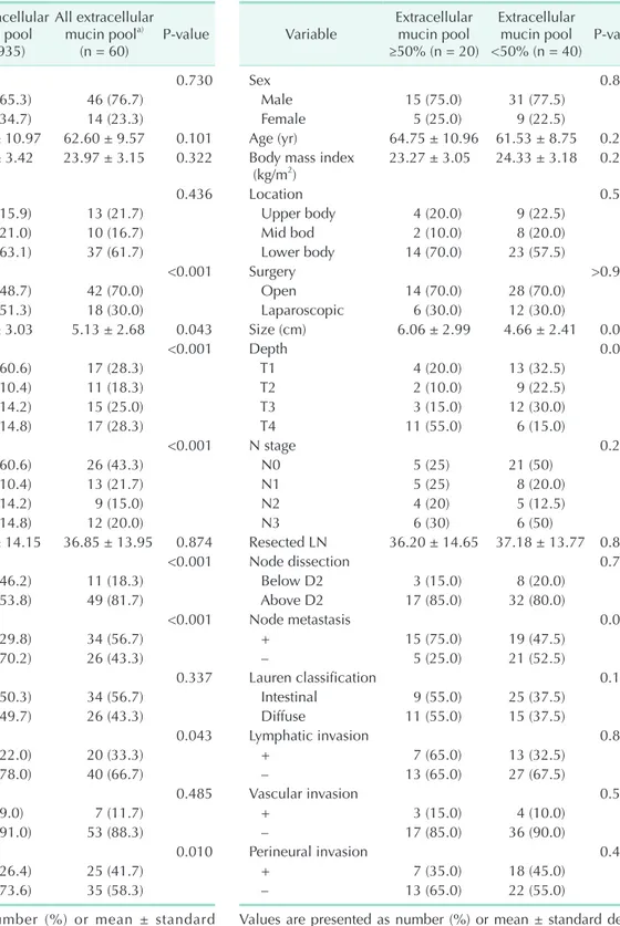
Table 3. Clinicopathological comparisons between LEMPC and MGC
Variable Extracellular mucin pool
≥50% (n = 20)
Extracellular mucin pool
<50% (n = 40) P-value
Sex 0.829
Male 15 (75.0) 31 (77.5)
Female 5 (25.0) 9 (22.5)
Age (yr) 64.75 ± 10.96 61.53 ± 8.75 0.222 Body mass index
(kg/m2) 23.27 ± 3.05 24.33 ± 3.18 0.223
Location 0.555
Upper body 4 (20.0) 9 (22.5)
Mid bod 2 (10.0) 8 (20.0)
Lower body 14 (70.0) 23 (57.5)
Surgery >0.999
Open 14 (70.0) 28 (70.0)
Laparoscopic 6 (30.0) 12 (30.0)
Size (cm) 6.06 ± 2.99 4.66 ± 2.41 0.056
Depth 0.014
T1 4 (20.0) 13 (32.5)
T2 2 (10.0) 9 (22.5)
T3 3 (15.0) 12 (30.0)
T4 11 (55.0) 6 (15.0)
N stage 0.214
N0 5 (25) 21 (50)
N1 5 (25) 8 (20.0)
N2 4 (20) 5 (12.5)
N3 6 (30) 6 (50)
Resected LN 36.20 ± 14.65 37.18 ± 13.77 0.801
Node dissection 0.736
Below D2 3 (15.0) 8 (20.0)
Above D2 17 (85.0) 32 (80.0)
Node metastasis 0.043
+ 15 (75.0) 19 (47.5)
– 5 (25.0) 21 (52.5)
Lauren classification 0.197
Intestinal 9 (55.0) 25 (37.5)
Diffuse 11 (55.0) 15 (37.5)
Lymphatic invasion 0.846
+ 7 (65.0) 13 (32.5)
– 13 (65.0) 27 (67.5)
Vascular invasion 0.570
+ 3 (15.0) 4 (10.0)
– 17 (85.0) 36 (90.0)
Perineural invasion 0.459
+ 7 (35.0) 18 (45.0)
– 13 (65.0) 22 (55.0)
Values are presented as number (%) or mean ± standard devia- tion.
LEMPC, gastric cancer with <50% tumor volume of extracellular mucin pool adenocarcinoma; MGC, mucinous gastric adenocar- cinoma; LN, lymph node.
Clinicopathological comparison between NMGC and all extracellular mucin pool gastric adeno- carcinomas
NMGC was compared with all extracellular mucin pool gastric adenocarcinomas. All extracellular mucin pool gastric adeno carcinomas involved more open gastrectomies (P < 0.001) with extended lymph node dissection (P < 0.001) than NMGC.
All extracellular mucin pool gastric adenocarcinomas featured a larger tumor size and more positive node metastasis (P <
0.001), lymphatic invasion (P < 0.043), and perineural invasion (P < 0.001) than NMGC. All extracellular mucin pool gastric adenocarcinomas showed more advanced T stage (P < 0.001) and N stage (P < 0.001) (Table 2).
Clinicopathological comparison between LEMPC and MGC
MGC featured higher T stage (P < 0.014) and more positive lymph node metastasis (P < 0.043) than LEMPC. Other clinico- pathologic characteristics were not significantly different (Table 3).
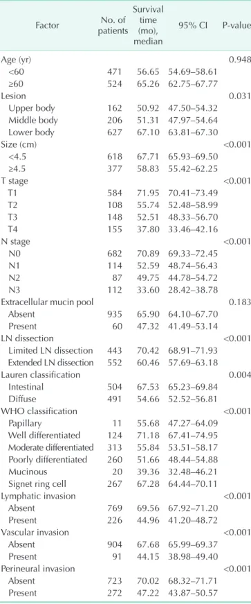
Prognostic factors for DFS in patients with curative gastric cancer
In univariate analysis, prognostic factors of all patients were tumor lesion (P = 0.031), tumor size (P < 0.001), T stage (P <
0.001), N stage (P < 0.001), extent of lymph node dissection (P <
0.001), WHO classification (P < 0.001), lymphatic invasion (P <
0.001), vascular invasion (P < 0.001), and perineural invasion (P <
0.001). Extracellular mucin pool was not a prognostic factor of re- currence (Table 4).
Comparison of Recurrence rates with NMGC, MGC and LEMPC revealed no differences in DFS curves (P < 0.170) (Fig. 3).
Table 4. Prognostic factors for disease-free survival in patients with curative gastric cancer evaluated by univariate analysis using the Kaplan-Meier method and log-rank test
Factor No. of
patients
Survival time (mo), median
95% CI P-value
Age (yr) 0.948
<60 471 56.65 54.69–58.61
≥60 524 65.26 62.75–67.77
Lesion 0.031
Upper body 162 50.92 47.50–54.32 Middle body 206 51.31 47.97–54.64 Lower body 627 67.10 63.81–67.30
Size (cm) <0.001
<4.5 618 67.71 65.93–69.50
≥4.5 377 58.83 55.42–62.25
T stage <0.001
T1 584 71.95 70.41–73.49
T2 108 55.74 52.48–58.99
T3 148 52.51 48.33–56.70
T4 155 37.80 33.46–42.16
N stage <0.001
N0 682 70.89 69.33–72.45
N1 114 52.59 48.74–56.43
N2 87 49.75 44.78–54.72
N3 112 33.60 28.42–38.78
Extracellular mucin pool 0.183
Absent 935 65.90 64.10–67.70
Present 60 47.32 41.49–53.14
LN dissection <0.001
Limited LN dissection 443 70.42 68.91–71.93 Extended LN dissection 552 60.46 57.69–63.18
Lauren classification 0.004
Intestinal 504 67.53 65.23–69.84
Diffuse 491 54.66 52.52–56.81
WHO classification <0.001
Papillary 11 55.68 47.27–64.09
Well differentiated 124 71.18 67.41–74.95 Moderate differentiated 313 55.84 53.51–58.17 Poorly differentiated 260 51.66 48.44–54.88
Mucinous 20 39.36 32.48–46.21
Signet ring cell 267 67.28 64.44–70.11
Lymphatic invasion <0.001
Absent 769 69.56 67.92–71.20
Present 226 44.96 41.20–48.72
Vascular invasion <0.001
Absent 904 67.68 65.99–69.37
Present 91 44.15 38.98–49.40
Perineural invasion <0.001
Absent 723 70.02 68.32–71.71
Present 272 47.22 43.87–50.57
CI, confidence interval; LN, lymph node; WHO, World Health Organization.
0
Time after operation (mo) 1.0
0.8
0.6
0.4
0.2
0
Disease-freesurvival
80 P = 0.170
60 40
20
NMGC LEMPC MGC
Fig. 3. Disease-free survival curves using the Kaplan- Meier method for gastric cancers diagnosed NMGC and all extracelluarmucin pool gastric carcinoma. NMGC, nonmu- cinous gastric adenocarcinoma; LEMPC, gastric cancer with
<50% tumor volume of extracellular mucin pool adeno-
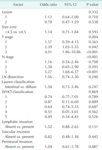
In multivariate analysis, prognostic factors for recurrence rate were T stage and N stage (Table 5).
DISCUSSION
Our results show that approximately 2% of the gastric cancer patients were MGC patients. The total gastric cancer patient’s rate increased to 6% when all extracellular mucin pool gastric adenocarcinoma patients were included. These results did not show the difference that has been previously reported [9].
However, the prevalence of LEMPC is not well known. This is because LEMPC is not a WHO diagnostic criteria and it can be overlooked by endoscopists, clinicians, and even pathologists. If pathologists do not diagnosed with MGC, can be omit reported extracellular mucin pool composition.
The clinicopathologic features of MGC include large size [6,11,12], deeper invasion [13,14], and higher N stage than that of NMGC. In this study, MGC differed from NMGC in tumor
size, T stage, N stage, and node metastasis. When the MGC is diagnosed, it is usually in a more advanced stage than NMGC.
More extracelluar mucin pools within a tumor indicate a more pro gressive state of disease. Overexpressed transmembrane mucins are considered to be a signal of cell growth and survival.
Extra cellular mucin pools derived from overexpressed trans- membrane mucin are relevant to the progression of carcinoma [15]. In this study, MGC featured more advanced T and N stages than NMGC.
LEMPC also showed similar results with MGC, having more advanced T stage and more positive node metastasis than NMGC. LEMPC and MGC patients received more radical surgery than NMGC patients. LEMPC and MGC showed similar cilinico- pathologic features. Just, MGC had a larger tumor size, more advanced T stage and more positive node metastasis than LEMPC. This result suggests that LEMPC is an earlier state of MGC irrespective of other WHO classification. For this reason, it has been suggested to consider extracellular mucin pools carcinoma of more than 30% as MGC [9].
Additionally, all extracellular mucin pool carcinomas featured larger tumor size, more advanced T stage and N stage, more aggressive lymph node dissection with open surgery, more positive node metastasis, lymphatic invasion, and perineural invasion than NMGC. All extracellular mucin pool gastric adenocarcinomas were in a more advanced state than NMGC, regardless of Lauren classification. Therefore, we can consider all extracellular mucin pool gastric adenocarcinomas included with the diagnostic criteria of MGC. In this study, we analyzed the effect on the prognosis in terms of recurrence of gastric cancer. Univariate analysis showed that tumor size, T stage, N stage, range of lymph node dissection, Lauren classification, WHO classification, lymphatic invasion, vascular invasion, and perineural invasion were significantly correlated with gastric cancer recurrence in all patients treated by curative gastrectomy at our hospital. However, only T stage and N stage were identified as independent prognostic factors for gastric cancer recurrence in the multivariate analysis. It is well known that prognostic factors for gastric cancers are influenced by tumor invasion depth, lymph node metastasis, and complete tumor removal [16-18]. Therefore, we assume extracellular mucin pools affect T stage and N stage, then the prognosis of gastric cancer recurrence
Limitations of the study include its retrospective design, and short follow-up. Specimens were not reassessed by pathologists again for the study. If all the specimens were reassessed by pathologists again for the study, then we can get high quality data. Moreover, we can calculate cutoff value of a linear/log relationship between mucin percent and clinical endpoints.
Despite these limitations, the results clearly show that the extracellular mucin pool gastric adenocarcinomas have more aggressive characteristics in T stage and N stage than NMGC.
Table 5. Prognostic factors for recurrence rate in patients with curative gastric cancer evaluated by multivariate analysis using the Cox proportional hazard model
Factor Odds ratio 95% CI P-value
Lesion 0.332
1 1.12 0.64–2.00 0.710
2 0.78 0.47–1.29 0.338
Size (cm)
<4.5 vs. ≥4.5 1.14 0.71–1.84 0.593
T stage 0.004
1 1.57 0.59–4.15 0.363
2 2.39 1.03–5.55 0.042
3 4.51 1.86–10.86 <0.001
N stage <0.001
1 1.16 0.54–2.46 0.708
2 1.38 0.65–2.90 0.395
3 3.27 1.68–6.37 <0.001
LN dissection 1.56 0.74–3.30 0.240
Lauren classification
Intestinal vs. diffuse 1.58 0.73–3.46 0.247
WHO classification 0.869
1 0.74 0.77–7.01 0.789
2 0.87 0.11–6.60 0.889
3 0.64 0.74–5.55 0.687
4 0.51 0.05–5.01 0.562
5 0.49 0.54–4.43 0.526
Lymphatic invasion
Absent vs. present 1.52 0.88–2.63 0.131 Vascular invasion
Absent vs. present 0.82 0.48–1.38 0.442 Perineural invasion
Absent vs. present 1.04 0.61–1.78 0.887 CI, confidence interval; LN, lymph node; WHO, world health organization.
This suggests that in the treatment of these cancers, aggressive treatment is needed. More rigorous treatment is expected at the time of the operation rather than additional surgery if extracellular mucin pool gastric adenocarcinoma is predicted before surgery.
The amount of extracellular mucin pool content is not an independent prognostic factor for the recurrence rate. However, it needs to be seriously considered when diagnosed. When a preoperative biopsy result includes extracellular mucin pool carcinoma, we should be more careful in implementing the treatment, such as protection from mucin spillage while operating and more extensive lymph node dissection. Cautious consi- deration about treatment of extracellular mucin pool gastric adenocarcinoma patients may improve prognosis.
In conclusion, the clinicopathological characteristics of LEMPC
patients are similar to MGC patients, except for T stage and positive lymph node metastasis. Patients with MGC had more progressed T stage and node metastasis than patients with LEMPC. We suggest that LEMPC can be thought of as a previous step of MGC. It is reasonable to consider LEMPC patients in the diagnostic criteria of MGC. To improve prognosis, we need more rigorous treatment for extracellular mucin pool gastric carcinoma. The results and conclusions of this paper are helpful for pathologists and surgeons when they make accurate diagnosis and adequate treatment.
CONFLICTS OF INTEREST
No potential conflict of interest relevant to this article was reported.
1. Bosman FT; World Health Organization;
International Agency for Research on Cancer. WHO classification of tumours of the digestive system. 4th ed. Lyon: Inter - national Agency for Research on Cancer;
2010. (World Health Organization classifi- cation of tumours; v. 3).
2. Hoerr SO, Hazard JB, Bailey D. Prognosis in carcinoma of the stomach in relation to the microscopic type. Surg Gynecol Ob- stet 1966;122:485-94.
3. Adachi Y, Mori M, Kido A, Shimono R, Maehara Y, Sugimachi K. A clinicopath- ologic study of mucinous gastric carci- noma. Cancer 1992;69:866-71.
4. Brander WL, Needham PR, Morgan AD.
Indolent mucoid carcinoma of stomach. J Clin Pathol 1974;27:536-41.
5. Epstein J, Lieberman PH. Mucinous ade- nocarcinoma of the prostate gland. Am J Surg Pathol 1985;9:299-308.
6. Kawamura H, Kondo Y, Osawa S, Nisida Y, Okada K, Isizu H, et al. A clinicopathologic study of mucinous adenocarcinoma of the stomach. Gastric Cancer 2001;4:83-6.
7. Lim SW, Kim DY, Kim YJ, Kim SK. Clinic- opathologic features of mucinous gastric
carcinoma. Dig Surg 2002;19:286-90.
8. Kunisaki C, Akiyama H, Nomura M, Matsuda G, Otsuka Y, Ono HA, et al. Cli- nicopathologic characteristics and surgical outcomes of mucinous gastric car cinoma.
Ann Surg Oncol 2006;13:836-42.
9. Hyung WJ, Noh SH, Shin DW, Yoo CH, Kim CB, Min JS, et al. Clinicopathologic charac teristics of mucinous gastric adeno- carcinoma. Yonsei Med J 1999;40:99-106.
10. Sano T, Aiko T. New Japanese classifica- tions and treatment guidelines for gastric cancer: revision concepts and major re- vised points. Gastric Cancer 2011;14:97- 100.
11. Sassa H, Kino I. A comparative study on mucinous carcinoma of the stomach and large intestine. II. Mucin histochemical study (author's transl). Nihon Shokakibyo Gakkai Zasshi 1979;76:861-70.
12. Hidaka S, Tanaka K, Takeshita H, Sumida Y, Fukuoka H, Abo T, et al. Clinicopath- ology and prognosis of mu cinous gastric carcinoma. Hepatogastroenterology 2008;
55:791-4.
13. Zhang M, Zhu GY, Zhang HF, Gao HY, Han
XF, Xue YW. Clinicopathologic characteris- tics and prognosis of mucinous gastric carcinoma. J Surg Oncol 2010;102: 64-7.
14. Koufuji K, Takeda J, Toyonaga A, Kodama I, Aoyagi K, Yano S, et al. Mucinous adenocarcinoma of the stomach-clinic- opathological studies. Kurume Med J 1996;43:289-94.
15. Kufe DW. Mucins in cancer: function, prognosis and therapy. Nat Rev Cancer 2009;9:874-85.
16. Kim DH, Kim SM, Hyun JK, Choi MG, Noh JH, Sohn TS, et al. Changes in post- operative recurrence and prognostic risk factors for patients with gastric cancer who underwent curative gastric resection during different time periods. Ann Surg Oncol 2013;20:2317-27.
17. Lo SS, Wu CW, Chen JH, Li AF, Hsieh MC, Shen KH, et al. Surgical results of early gastric cancer and proposing a treatment strategy. Ann Surg Oncol 2007;14:340-7.
18. Shiraishi N, Sato K, Yasuda K, Inomata M, Kitano S. Multivariate prognostic study on large gastric cancer. J Surg Oncol 2007;96:
14-8.