Gallbladder perforation: a single center experience of 32 cases
Gopalakrishnan Gunasekaran, Debasis Naik, Ashwani Gupta, Vimal Bhandari, Manigandan Kuppusamy, Gaind Kumar, and Niuto S Chishi
Department of Surgery, Vardhman Mahavir Medical College and Safdarjung Hospital, New Delhi, India
Backgrounds/Aims: Gallbladder perforation is a rare but potentially fatal disease. We herein present our clinical experi- ence in diagnosis and management of 32 cases of gallbladder perforation. Methods: This retrospective study was con- ducted with inclusion of all cases of gallbladder perforation that presented to our hospital from January 2012 to November 2014. Cases of traumatic gallbladder perforation and patients younger than 12 years of age were excluded from this study. Results: This study included 32 patients (13 males and 19 females). The mean age of patients was 55.9 years. Gallbladder perforation was most common in the 5th and 6th decade of life. The mean age of patients with type I, II, and III gallbladder perforation was 57.0 years, 57.6 years, and 49.8 years, respectively. The most com- mon site of perforation was the fundus, followed by the body and Hartmann’s pouch (24 : 5 : 2). Most of the type I gallbladder perforations were diagnosed intraoperatively, type II gallbladder perforations were diagnosed by enhanced abdominal computed tomography, and type III gallbladder perforations were diagnosed during laparoscopic chol- ecystectomy converted to open cholecystectomy for cholelithiasis. Mortality was highest in patients with type I gall- bladder perforation. The mean hospital stay was 10.1 days, 6.4 days, and 9.2 days in patients with type I, II, and III gallbladder perforation, respectively. The histopathologic analysis in 28 patients who were operated on showed acute cholecystitis in 19 cases, acute-on-chronic cholecystitis in 4 cases, chronic cholecystitis in 4 cases, and mucinous adenocarcinoma of the gallbladder in a single case. Conclusions: Gallbladder perforation represents a special diag- nostic and surgical challenge. Appropriate classification and management are essential. (Korean J Hepatobiliary Pancreat Surg 2015;19:6-10)
Key Words: Gallbladder perforation; Acute cholecystitis; Cholecysto-enteric fistula; Hole sign; Percutaneous catheter drainage
Received: December 19, 2014; Revised: January 10, 2015; Accepted: February 17, 2015 Corresponding author: Debasis Naik
Department of Surgery, Vardhman Mahavir Medical College and Safdarjung Hospital, New Delhi 110029, India Tel: +91-11-26707225, Fax: +91-9968845306, E-mail: [email protected]
Copyright Ⓒ 2015 by The Korean Association of Hepato-Biliary-Pancreatic Surgery
This is an Open Access article distributed under the terms of the Creative Commons Attribution Non-Commercial License (http://creativecommons.org/
licenses/by-nc/3.0) which permits unrestricted non-commercial use, distribution, and reproduction in any medium, provided the original work is properly cited.
Korean Journal of Hepato-Biliary-Pancreatic Surgery ∙ pISSN: 1738-6349ㆍeISSN: 2288-9213
INTRODUCTION
Gallbladder perforation is a rare but potentially fatal disease; its presentation can vary and therefore is a di- lemma for early diagnosis. It is usually a complication of acute cholecystitis with or without gallstones.1 Most per- forations are subacute, causing a pericholecystic abscess.
Acute free perforation with bile peritonitis and chronic perforation with an internal biliary fistula are rare. The fundus of the gallbladder is the most common site of per- foration because of its poor blood supply.2 Because of the infrequent and rare occurrence of gallbladder perforation, data regarding its incidence and management are lacking.
We herein present our experience of 32 cases of gall- bladder perforation that presented to our hospital from
January 2012 to November 2014.
MATERIALS AND METHODS
The present retrospective study was conducted in the Department of Surgery, Safdarjung Hospital, New Delhi, India by including all cases of gallbladder perforation that presented to our hospital from January 2012 to November 2014. Cases of traumatic gallbladder perforation and pa- tients younger than 12 years of age were excluded from this study.
Patient data was collected from the Medical Records Department and the Department of Pathology, Safdarjung Hospital, New Delhi. The patients included were classi- fied as per the Niemeier’s classification of gallbladder
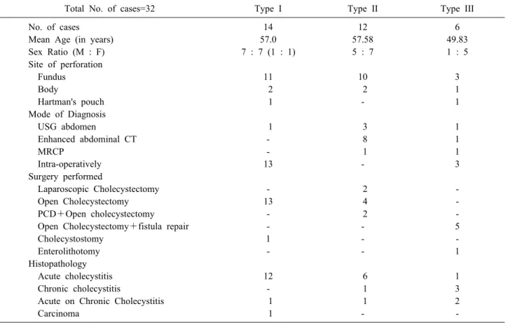
Table 1. Table showing data collected from gallbladder perforation patients
Total No. of cases=32 Type I Type II Type III
No. of cases 14 12 6
Mean Age (in years) 57.0 57.58 49.83
Sex Ratio (M : F) 7 : 7 (1 : 1) 5 : 7 1 : 5
Site of perforation
Fundus 11 10 3
Body 2 2 1
Hartman's pouch 1 - 1
Mode of Diagnosis
USG abdomen 1 3 1
Enhanced abdominal CT - 8 1
MRCP - 1 1
Intra-operatively 13 - 3
Surgery performed
Laparoscopic Cholecystectomy - 2 -
Open Cholecystectomy 13 4 -
PCD+Open cholecystectomy - 2 -
Open Cholecystectomy+fistula repair - - 5
Cholecystostomy 1 - -
Enterolithotomy - - 1
Histopathology
Acute cholecystitis 12 6 1
Chronic cholecystitis - 1 3
Acute on Chronic Cholecystitis 1 1 2
Carcinoma 1 - -
perforation,3 and the following data were analysed: age, sex, mode of diagnosis, site of perforation, procedure per- formed, outcome, and histopathology (Table 1).
RESULTS
After our extensive search, 32 patients (13 males and 19 females) were included in the study. The mean age of patients was 55.9 years (male patients, 54.6 years and fe- male patients, 56.7 years). Gallbladder perforation was most common in the 5th and 6th decade of life (range, 14-75 years). Patients were further categorised as having type I, type II, and type III gallbladder perforation as per the Niemeier’s classification (Table 1).3
There were 14 patients with type I gallbladder perfo- ration (7 males and 7 females), 12 patients with type II gallbladder perforation (5 males and 7 females), and 6 pa- tients with type III gallbladder perforation (1 male and 5 females). The mean age of patients with type I, II, and III gallbladder perforation was 57.0 years, 57.6 years, and 49.8 years, respectively.
Comorbid disease was present in 18 patients, 12 pa-
tients were known diabetics, 4 patients were known cases of ischaemic heart disease, and 2 patients had both dia- betes and hypertension as comorbidity.
The most common site of perforation was the fundus, followed by the body and Hartmann’s pouch (24 : 5 : 2).
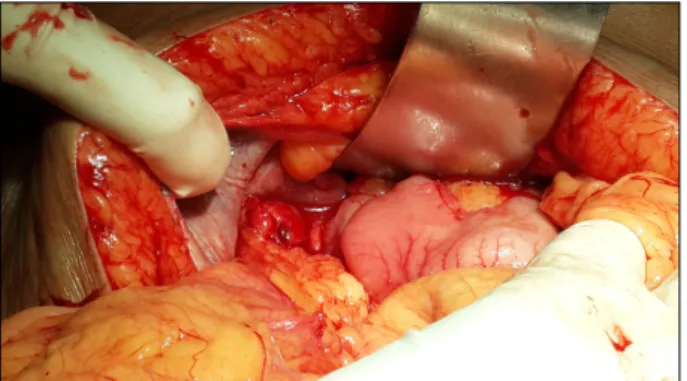
The site of perforation could not be identified in a single case with type III gallbladder perforation due to dense ad- hesions, and this case was of gallstone ileus (Fig. 1). A proximal enterolithotomy (Fig. 2) was performed and a 2-stage procedure was planned for the patient.
Most of the spontaneous gallbladder perforations were diagnosed intraoperatively (9 gallbladder perforations were diagnosed during exploratory laparotomy for perforation peritonitis and 7 gallbladder perforations were diagnosed while the patients were undergoing laparoscopic chol- ecystectomy for cholelithiasis). Most of the type I gall- bladder perforations were diagnosed intraoperatively (Figs. 3, 4), type II gallbladder perforations were diag- nosed by enhanced abdominal computed tomography (CT), and type III gallbladder perforations were diagnosed during laparoscopic cholecystectomy converted to open cholecystectomy for cholelithiasis. Abdominal ultrasono-
Fig. 1. Intraoperative photograph showing dense adhesions around the gallbladder in a patient with gallstone ileus (type III gallbladder perforation).
Fig. 5. Magnetic resonance cholangiopancreatography show- ing type II gallbladder perforation and a huge localised collection.
Fig. 2. Proximal longitudinal enterolithotomy performed for gallstone ileus.
Fig. 3. Intraoperative photograph showing the perforation site in a patient with type I gallbladder perforation.
Fig. 4. Cholecystectomy specimen showing perforation in the body of the gallbladder.
graphy was useful in diagnosing 5 cases of gallbladder perforation and magnetic resonance cholangiopancreatog- raphy (MRCP) was useful in diagnosing 2 cases of gall- bladder perforation (Fig. 5).
In the type III gallbladder perforations, cholecystocolic
fistula was the most common (3 cases) and there was 1 case of cholecystoduodenal and cholecystogastric fistula each.
Among the 12 patients with type II gallbladder perfo- ration, 10 were managed conservatively and were asked to follow-up in the outpatient department. Patients with type II gallbladder perforation had localised signs with no systemic toxic features and were managed conservatively with antibiotics, intravenous fluids, and analgesics. All of these patients had responded to the treatment by the time we performed further abdominal imaging for arriving at the diagnosis, and hence they were discharged with advice to follow up for elective cholecystectomy as a routine pro-
tocol of our hospital. Of these 10 patients, 4 were treated by open cholecystectomy, 2 were treated by laparoscopic cholecystectomy, and the remaining 4 were lost to fol- low-up. Two cases of type II gallbladder perforation were managed with percutaneous catheter drainage followed by open cholecystectomy after a period of approximately 2 months. In contrast, all cases of type I gallbladder perfo- ration underwent exploratory laparotomy.
Mortality was highest (4 of 14 cases, 28.5%) in patients with type I gallbladder perforation (in none of the patients with type II gallbladder perforation and in one patient with type III gallbladder perforation). Mortality was high in patients with type I gallbladder perforation because of their poor general condition at the time of presentation, delayed presentation to our hospital, and associated co-morbidities. The mean hospital stay was 10.1 days in patients with type I gallbladder perforation, 6.4 days in patients with type II gallbladder perforation, and 9.2 days in patients with type III gallbladder perforation.
The histopathologic analysis in 28 patients who were operated on showed acute cholecystitis in 19 patients, acute-on-chronic cholecystitis in 4 patients, chronic chol- ecystitis in 4 patients, and mucinous adenocarcinoma of the gallbladder in one patient. The youngest patient with gallbladder perforation was a male aged 14 years who had an enteric etiology proven by tissue culture and positive Widal test.
DISCUSSION
Acute uncomplicated cholecystitis is more commonly seen in females, but spontaneous gallbladder perforation is more common in males.4 However, in this study a high- er incidence of gallbladder perforation was observed in fe- males (males : females=13 : 19).
Gallbladder perforation results due to persistent occlu- sion of the cystic duct by an impacted calculus, causing a rise in intracholecystic pressure, epithelial injury, release of phospholipases, degradation of cell membranes, and in- tense inflammatory reaction.5
Infections, malignancy, trauma, drugs (e.g. cortico- steroids), and systemic diseases such as diabetes mellitus and atherosclerotic heart disease are the predisposing factors.6 In our retrospective study, comorbid disease was present in 18 patients, 12 patients were known diabetics,
4 patients were known cases of ischaemic heart disease, and 2 patients had both diabetes and hypertension as comorbidity.
Niemeier, in 1934, classified free gallbladder perfo- rations into 3 types. Type I (acute) is associated with gen- eralised biliary peritonitis, type II (subacute) consists of localised collection of fluid at the site of perforation, peri- cholecystic abscess and localised peritonitis, while type III (chronic) comprises formation of internal or external fistulae.3 The number of type I : type II : type III gall- bladder perforation in our study was 14 : 12 : 6.
Fundus is the most distal part with regards to blood supply and therefore this makes it the most common site for perforation which can occur as early as 2 weeks or several weeks after the onset of cholecystitis.7 This was also observed in our study with the most common site of perforation being the fundus, followed by the body and Hartmann’s pouch.
The ultrasonographic appearances of gallbladder perfo- ration are diverse and non-specific. They include wall thick- ening (>3 mm), distension (largest diameter >3.5-4.0 cm), gallstones, coarse intracholecystic echogenic debris and bile duct dilatation. Distension of the gallbladder and ede- ma of its wall may be the earliest detectable signs of im- minent perforation. The ‘hole sign’ (a defect in the gall- bladder wall) is the most specific finding.7
Although standard abdominal CT plays an important role in diagnosing gallbladder perforation, upper abdomi- nal CT for acute cholecystitis, in which pericholecystic fluid is found by ultrasonography, may increase the rate of preoperative diagnosis of gallbladder perforation.8 The advantage of MRCP is its superb ability to detect stones in the bile ducts, biliary dilatation, and the relationship of a pericholecystic fluid collection to the abdominal wall and gallbladder. This information can aid in surgical planning.9
Most of the type I gallbladder perforations were diag- nosed intraoperatively, type II gallbladder perforations were diagnosed by enhanced abdominal CT, and type III gallbladder perforations were diagnosed during laparo- scopic cholecystectomy converted to open cholecystec- tomy for cholelithiasis. Type I perforation, which presents with acute abdomen and features of peritonitis, was diag- nosed in most of the patients intraoperatively after open- ing the abdomen, this might be due to the non-availability
of enhanced abdominal CT as an emergency investigation tool in our hospital. However, type II perforations were diagnosed by enhanced abdominal CT, which could be made available for the diagnosis due to the subacute na- ture of the disease allowing us time to investigate the patients.
Cholecystectomy, drainage of abscess if it is present, and abdominal lavage are usually sufficient to treat gall- bladder perforation. Cholecystectomy may be difficult in type III gallbladder perforations. If a cholecystectomy is performed, additional surgical procedures such as repair of the fistula may be required. Cholecystectomy can be performed after the infection is relieved by ultra- sonography-guided percutaneous drainage in type II gall- bladder perforations.8
Laparoscopic cholecystectomy can be performed for acute, gangrenous, and/or perforated cholecystitis, but a conversion may be necessary in case of difficulty like an unclear anatomy.8
In conclusion, gallbladder perforation represents a spe- cial diagnostic and surgical challenge. Early classification and appropriate management are crucial. We think that this study might be helpful in management of patients
with gallbladder perforation.
REFERENCES
1. Khan SA, Gulfam, Anwer AW, Arshad Z, Hameed K, Shoaib M. Gallbladder perforation: a rare complication of acute chole- cystitis. J Pak Med Assoc 2010;60:228-229.
2. Teefey SA, Wechter DG. Sonographic evaluation of peri- cholecystic abscess with intrahepatic extension. J Ultrasound Med 1987;6:659-662.
3. Niemeier OW. Acute free perforation of the gall-bladder. Ann Surg 1934;99:922-924.
4. Simmons TC, Miller C, Weaver R. Spontaneous gallbladder perforation. Am Surg 1989;55:311-313.
5. Aljiffry M, Walsh M, Peltekian K, Molinari M. Type II gall bladder perforation with abdominal wall abscess in a cirrhotic patient: case report and review of the literature. J Surg Educ 2008;65:367-371.
6. Alvi AR, Ajmal S, Saleem T. Acute free perforation of gall blad- der encountered at initial presentation in a 51 years old man:
A case report. Cases J. 2009;2:166
7. Sood BP, Kalra N, Gupta S, Sidhu R, Gulati M, Khandelwal N, et al. Role of sonography in the diagnosis of gallbladder perforation. J Clin Ultrasound 2002;30:270-274.
8. Derici H, Kara C, Bozdag AD, Nazli O, Tansug T, Akca E.
Diagnosis and treatment of gallbladder perforation. World J Gastroenterol 2006;12:7832-7836.
9. Karcaaltincaba M, Hohenwalter MD, Erickson SJ, Taylor AJ.
MRCP findings of gallbladder perforation and pericholecystic abscess. CMIG Extra: Cases 2004;28:59-61.