DNA Methylation Changes Following 5-azacitidine Treatment in Patients with Myelodysplastic Syndrome
DNA methyltransferase inhibitor, 5-azacitidine (AC) is effective in myelodysplastic syndromes (MDS) and can induce re-expression in cancer. We analyzed the methylation of 25 tumor suppressor genes in AC-treated MDS. Hypermethylation of CDKN2B, FHIT, ESR1, and IGSF4 gene was detected in 9/44 patients. In concordance with the clinical response, a lack of or decreased methylation in 4 patients with hematologic improvements and persistent methylation in 4 others with no response was observed. The mRNA expression of CDKN2B, IGSF4, and ESR1 was significantly reduced in MDS. Our results suggest that methylation changes contribute to disease pathogenesis and may serve as marker to monitor the efficacy of treatments.
Key Words: Demethylation; DNA Methylation; Myelodysplastic Syndromes; DNA Methyltransferase Inhibitors; Azacitidine; MS-MLPA
Huong Thi Thanh Tran1, Hee Nam Kim1, Il-Kwon Lee1, Yeo-Kyeoung Kim2, Jae-Sook Ahn2, Deok-Hwan Yang2, Je-Jung Lee2, and Hyeoung-Joon Kim1,2
1Genome Research Center for Hematopoietic Diseases, Chonnam National University Hwasun Hospital, Hwasun; 2Department of Hematology- Oncology, Chonnam National University Medical School, Gwangju, Korea
Received: 14 July 2010 Accepted: 9 November 2010 Address for Correspondence:
Hyeoung-Joon Kim, MD
Department of Hematology-Oncology, Chonnam National University Hwasun Hospital, 160 Ilsim-ri, Hwasun 519-809, Korea
Tel: +82.61-379-7637, Fax: +82.61-379-7736 Email: [email protected]
The study was supported by a grant from the Korea Health 21 R&D Project, Ministry of Health and Welfare, Republic of Korea (A01-0385-A70604-07M7-00000A).
DOI: 10.3346/jkms.2011.26.2.207 • J Korean Med Sci 2011; 26: 207-213 Oncology & Hematology
INTRODUCTION
Myelodysplastic syndrome (MDS) is a clonal stem cell disorder characterized by ineffective hematopoiesis in one or more of the lineages in the bone marrow and an increased tendency to- wards acute leukemia transformation (1). MDS patients were treated with supportive care measures only; high-dose chemo- therapy programs modeled after acute myelogenous leukemia (AML) therapy or allogenic stem cell transplantation (1, 2). Re- cently, 5-azacitidine (AC) and 5-aza-2-deoxycitidine (DAC) have been used as hypomethylating agents for the treatment of MDS.
The aberrant methylation of CpG islands or CpG sites, in or near the promoter region of many genes was found to be asso- ciated with the transcriptional inactivation of important tumor suppressor genes (TSG) (3-5). These TSG are also often silenced in hematopoietic malignancies by DNA hypermethylation (6).
Many of these genes plays roles in multiple fundamental path- ways that include cell cycle control (p16INK4a, p15INK4a, Rb1, p27Kip1, p14ARF, and p73), DNA repair (BRCA1 and MGMT), apoptosis inhibition (DAPK, TMS1, and caspase-8), tumor in- vasion/metastasis (E-cadherin, APC, TIMP3, and VHL), and growth factor responses (ER and EphA3) (7). The DNA methyl- transferase (DNMT) inhibitors AC and DAC can induce the func-
tional re-expression of genes silenced by cancer that cause growth arrest and apoptosis in tumor cells (8). The p15 (CDKN2B) of hypermethylation genes may play a causal role in hematologic malignancies such as MDS and AML (3). The methylation of p15 occurs in patients with these disorders and is associated with an increased frequency (or density) and advanced disease (9, 10).
In response to the treatment with AC, one study showed remark- able differential demethylation in several myeloid leukemia cell lines and a significant but transient demethylation in MDS pa- tients. Genomic DNA methylation patterns have revealed wide- spread demethylation in array-based analysis (11).
Recently, AC and DAC are clinically active in MDS (12-14) and other hematologic malignancies. Therapeutic strategies have been developed to effectively inhibit methylation in these and other malignancies such as CDKN2B (15) that can be reactivat- ed after treatment with methylation inhibitors. The aberrant methylation of CpG islands represents an ideal candidate for diagnostic and prognostic cancer markers (16).
We analyzed expression and methylation changes of 25 TSG identified as potential epigenetic markers after Vidaza® (azaciti- dine; AC) treatment in MDS that further the correlation to clinical response. Based on a complete cell blood count (CBC) and bone marrow findings, our data indicates that methylation changes
after AC treatment were correlated with the clinical responses of patients. Our study also confirmed that methylation was as- sociated with a reduced mRNA expression in patients with MDS.
Thus, DNA methylation changes can be used as a biomarker in predicting the response to a particular chemotherapeutic agent.
MATERIALS AND METHODS Patient samples
A total of 44 MDS patients (30 men and 14 women; age range, 15-82 yr), including 13 with refractory anemia (RA), one case of RA with ringed sideroblasts (RARS), 9 cases of refractory cyto- penia with multilineage dysplasia (RCMD), one case of refrac- tory cytopenia with multilineage dysplasia and ringed sidero- blasts (RCMD-RS), 9 cases of RA with excess blasts-1 (RAEB-1), 9 cases of RA with excess blasts-2 (RAEB-2) and 2 cases of MDS- unclassified (MDS-U) (WHO classification), were examined before (as part of the initial diagnostic process) and during treat- ment with AC. The AC dose for all patients was 75 mg/m2/d for 7 days and repeated on a 28-day cycle. The response was as- sessed prior to each cycle of treatment. Therapy was continued for three cycles after complete remission (CR) or until progres- sive disease or toxicity in patients with partial remission (PR) or hematologic improvement (HI). Blood was obtained before or at the beginning of the first cycle (as before treatment) and at the beginning of the 4th, 5th, and 6th cycles (in turn as after 3, 4, and 5 cycle’s treatment). The characteristics of the patients are summarized in Table 1. Samples were obtained from patients who were admitted between 2004 and 2008 at Chonnam Nation- al University Hwasun Hospital (Jeonnam, Korea). Peripheral blood (PB)-MNCs from 3 healthy donors served as non-malig- nant controls.
DNA methylation and fragment analyses
Genomic DNA was extracted from PB using a QIAamp DNA
Blood Mini kit (Qiagen, Valencia, CA, USA) according to the manufacturer’s protocol. An ME001 probe mix kit was used for methylation-specific multiplex ligation-dependent probe am- plification (MS-MLPA), according to the manufacturer’s instruc- tions (MRC-Holland, Amsterdam, Netherlands). The kit contains 25 sequences corresponding to TSG that are frequently silenced by methylation in different tumors but are unmethylated in the blood-derived DNA of healthy individuals. MS-MLPA starts with sample DNA denaturation and the hybridization of the MLPA probes their specific DNA targets. Methylated hybrids of the sample DNA are prevented from being digested by HhaI, and are then amplified by PCR to generate a signal on an ABI 3100 sequencer (Applied Biosystems, Foster City, CA, USA). The data shown are the mean of triplicate reactions.
Data normalization and analysis were performed using the built-in MLPA application in GeneMarker ver. 1.5 (Soft-Genet- ics). To determine the methylation status, the normalized height- ratio data for each ligated sample were compared with the height- ratio data for the same sample after digestion with HhaI. Aber- rant methylation was scored when the calculated methylation was > 10% (hypermethylation). Percentages below this level were regarded as background.
RNA isolation and quantitative real-time RT-PCR
RNA was extracted using the Trizol reagent (Invitrogen, Carls- bad, CA, USA) according to the manufacturer’s instructions.
First-strand cDNA was synthesized from RNA using random primers and a Moloney murine leukemia virus (MMLV) reverse transcriptase, according to the manufacturer’s protocol. For the quantification of CDKN2B, FHIT, IGSF4, and ESR1 transcripts, real-time PCR was performed using a 72-well Rotor-Gene RG- 3000 (Corbett Research, Sydney, Australia) with a 10 μL final re- action mixture containing 250 nM in each primer, 1 × SYBR pre- mix Ex Taq (Takara, Tokyo, Japan), and 2 μL of cDNA. The prim- er sequences and annealing temperatures used to amplify each of the genes tested are listed in Table 2. The reaction mixture was preheated to 95°C for 10 sec, followed by 45 cycles of 95°C for 10 sec, 60°C for 20 sec, and 72°C for 20 sec. As a reference transcript for each expression level, the housekeeping gene β2- microglobin was amplified. All reactions included negative con- trols where the reverse transcriptase was omitted.
Table 1. Characteristics of the study group (44 patients)
Characteristic No. (%)
≥ 60 yr old 31 (70.5)
Number of females 14 (31.8)
WHO
Low-risk (RA, RARS, RCMD, and unclassified )
High-risk (RAEB-1 and RAEB-2 ) 26 (59)
18 (41) IPSS
Low Intermediate 1 Intermediate 2 High
3 (6.8) 27 (61.4) 11 (25)
3 (6.8)
Blast ≤ 5% 26 (59)
Response after three or four cycles 22 (50)
Methylation of at least one gene 9 (20.5)
WHO, World Health Organization; RA, refractory anemia; RARS, RA with ringed sidero- blasts; RCMD, refractory cytopenia with multilineage dysplasia; RAEB, RA with excess blasts; IPSS, International Prognostic Scoring System.
Table 2. Primers used for quantitative real-time PCR Target Product
size (bp) GenBank Primer sequence (5´-3´) CDKN2B 129 NM_078487.2 F: TGATTAGCACTTGGGTGACG R: CCTCCTCCACTTTGTCCTCA FHIT 140 NM_002012.1 F: AAACATTTCCATGGGACCTCTCT
R: GAGCTCCTCATAGATGCTGTCATTC
IGSF4 129 NM_014333 F: CTGGTCCCACCACGTAATCT
R: GTTCCCTTTGAACCACCTGA
ESR1 108 NM_004451.3 F: CCTATCTCAGGGAGGGAAGG
R: TGCTCTCCAAGTCCCACTCT
Statistical analysis
For the mRNA data, 95% confidence intervals (CIs) were calcu- lated to assess the significance of the 2-∆∆CT (17). Differences in continuous variables were analyzed by the Mann-Whitney U- test for distributions between two groups. All statistical analyses were performed using SPSS software (ver.12.0). For all analyses, the P values were two-tailed; a P values < 0.05 was considered statistically significant.
Ethics statement
This study was approved by the institutional review board of the Chonnam National University Hwasun Hospital in Hwasun, Korea (IRB number: CNUHHIRB 2009-22). At the time of PB col- lection, all case and control subjects provided informed consent to participate in this study.
RESULTS
Methylation status in MDS patients and methylation reversal after 3-5 courses of AC treatment corresponded to the clinical response
Alterations in DNA methylation are common in a variety of tu- mors, as well as during development (18). All MDS patients at the time of diagnosis were screened for gene methylation; only those samples containing at least one methylated gene, were analyzed further by MS-MLPA following 3-5 cycles of AC. The PB in this study is similar to that taken previously (10, 11, 20, 24).
Hypermethylation of at least one gene was detected in 9/44 cases (20.5%) that included CDKN2B, FHIT, IGSF4, and ESR1 in MDS (Table 3), but not in any of the control samples. Our results in- dicate increased methylation among high-risk patients (5/18, 27.8%) compared with low-risk patients (4/26, 15.4%) (P < 0.05).
Either CDKN2B or ESR1 was methylated in 5 patients (5/44, 11.4%), whereas FHIT and IGSF4 was methylated in 3 patients (3/44, 6.8%) and 2 patients (2/44, 4.5%) respectively.
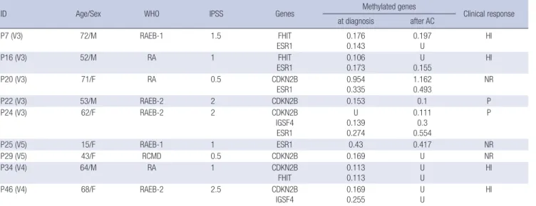
We examined the methylation status of 25 TSG after 3-5 cour- ses of AC treatment in 9 MDS patients with methylation at the time of diagnosis, and assessed the subsequent change in meth- ylation in association with the correlative clinical response. At the time that blood was obtained after treatment, the clinical re- sponse was evaluated respectively. Increased or reduced meth- ylation level of at least 10% is considered reinforcement or reduc- tion respectively. Our data revealed an agreement between de- methylation and the clinical response in 8 of 9 cases (Table 4).
Patients (P20, P25, P22, and P24) had the persistent methylation that correlated to either no clinical response or tends to leuke- mia. Specially, P24 (RAEB-2 subtype) progressed to leukemia, showed increased methylation level at ESR1 and IGSF4 as well as de novo methylation of CDKN2B. The pretreated methylated genes were reduced or loss for 3 patients (P16, P34, and P46) who exhibited an increase in hematologic improvement (HI). In pa- tients P34 and P46, no methylation was observed after treatment.
Patient P16 exhibited ESR1 and FHIT methylation and then re- mained only ESR1. In the case of patient P7, the results indicat-
Table 3. Methylation frequencies of tumor suppressor genes in 44 AC-treated MDS patients
Gene Function Location Methylated
No. %
TIMP3 Apoptosis regulation, Tyrosine kinase signalling 22q12.3 0 0
APC Wnt signalling, Cell adhesion 5q21-q22 0 0
CDKN2A/p14-ARF Cell cycle control, Apoptosis regulation 9p21 0 0
MLH1 Cell cycle control, Mismatch repair 3p22.1 0 0
ATM DNA repair, Apoptosis regulation, Cell cycle control, TLR signalling 11q23 0 0
RARβ Transcription regulation, Signal transduction 3p24 0 0
CDKN2B/p15 Cell cycle control 9p21 5 11
HIC1 Cell cycle control, Transcription regulation 17p13.3 0 0
CHFR Cell cycle control, Ubiquitination 12q24.33 0 0
PAH L-phenylalanine catabolism, Amino acid biosynthesis 12q23 0 0
CASP8 Apoptosis regulation 2q33.2 0 0
CDKN1B/p27 Cell cycle control 12p13.2 0 0
PTEN Cell adhesion, Apoptosis regulation, Cell cycle control 10q23.3 0 0
BRCA2 DNA repair, Cell cycle control 13q12.3 0 0
CD44 Cell adhesion 11p212 0 0
DAPK1 Apoptosis regulation 9q34.1 0 0
TP73 Cell cycle control 1p36 0 0
VHL Transcription factor binding Apoptosis regulation, Cell cycle control 3p25.3 0 0
ESR1 Transcription regulation, Signal transduction, Cell growth regulation 6q25.1 5 11
RASSF1 Cell cycle control, Ras signalling 3p21.3 0 0
FHIT Cell cycle control, Nucleotide metabolism 3p14.2 3 7
IGSF4 Cell adhesion 11q23 2 4.5
CDH13 Cell adhesion 16q24.2 0 0
ed a loss of ESR1 and remained FHIT methylation with no ac- companying clinical response after 3 courses (the patient re- fused the fourth course of AC); however, hematologic improve- ment was observed in this patient after follow-up. It is possible that the therapeutic effect may be delayed with a response oc- curring several weeks after treatment (19). Although patient P29 (RCMD subtype) had no clinical response, the methylation at CDKN2B disappeared after 5 courses of AC.
Methylation status of CDKN2B and FHIT using pyrosequenc- ing in MDS patients with methylation at the time of diagnosis was confirmed in the association shown between DNA methyla- tion reversal and a clinical response (data not shown). The meth- ylation status changed following 3-5 courses of AC, a demethyl-
ation agent, (correlated with the clinical response observed), based on recommended criteria.
Reduced gene expression in the MDS patients versus controls
The transcriptional silencing of TSG associated with DNA meth- ylation is a common epigenetic event in hematologic malignan- cies. To verify the contribution of DNA methylation to the inac- tivation of transcription, we compared mRNA expression levels in the MDS and control group. The mean level of mRNA expres- sion for CDKN2B, FHIT, IGSF4, and ESR1 was higher in the con- trol group (16 PB samples) than in the MDS group (30 PB samples) (Fig. 1). The difference for CDKN2B, IGSF4, and ESR1 mRNA Table 4. Methylation reversal in AC-treated MDS patients and their clinical responses
ID Age/Sex WHO IPSS Genes Methylated genes
Clinical response at diagnosis after AC
P7 (V3) 72/M RAEB-1 1.5 FHIT
ESR1
0.176 0.143
0.197 U
HI
P16 (V3) 52/M RA 1 FHIT
ESR1 0.106
0.173 U
0.155 HI
P20 (V3) 71/F RA 0.5 CDKN2B
ESR1 0.954
0.335 1.162
0.493 NR
P22 (V3) 53/M RAEB-2 2 CDKN2B 0.153 0.1 P
P24 (V3) 62/F RAEB-2 2 CDKN2B
IGSF4 ESR1
U 0.139 0.274
0.111 0.3 0.554
P
P25 (V5) 15/F RAEB-1 1 ESR1 0.43 0.417 NR
P29 (V5) 43/F RCMD 0.5 CDKN2B 0.169 U NR
P34 (V4) 64/M RA 1 CDKN2B
FHIT 0.113
0.113 U
U HI
P46 (V4) 68/F RAEB-2 2.5 CDKN2B
IGSF4 0.169
0.255 U
U HI
V (No.), number of cycles of AC treatment; U, Unmethylated; HI, Hematologic improvement; NR, no response; P, progression to AML.
Fig. 1. Quantitative analysis of mRNA expression of CDKN2B, FHIT, IGSF4, and ESR1 in 30 MDS patients and 16 normal controls were analyzed at diagnosis. MDS group was significantly lower level in CDKN2B, IGSF4, and ESR1 (P < 0.05). The horizontal bars indicate the median value for each sample group.
Rel. mRNA expressionRel. mRNA expression Rel. mRNA expressionRel. mRNA expression
CDKN2B
IGSF4
FHIT
ESR1 Normal
Normal
Normal
Normal MDS
MDS
MDS
MDS P < 0.001
P < 0.001
P = 0.343
P < 0.001 60
40
20
0
40 30 20 10 0
4 3 2 1 0
80 60 40 20 0
expression was statistically significant (P < 0.05) whereas the difference for FHIT was not (P = 0.343). Additionally, most of the individuals for whom CDKN2B, FHIT, IGSF4, and ESR1 were initial methylated showed reduced mRNA compared with the median value. Despite the lack of a clinical response in the 9 sam- ples (in terms of their mRNA levels after 3 5 courses of AC), we also found evidence for the reactivation of gene expression in response to the demethylation agent (Fig. 2).
DISCUSSION
Many genes important for most of the carcinogenic pathways are hypermethylated in MDS (7). DNMT inhibitors, that inhibit DNA methylation in various studies (15, 20, 21), represent a ma- jor advance in treatment (13, 14); however, it is still unclear wheth- er methylation reversal corresponds to a clinical response. The present study examined genome-specific methylation changes in MDS patients who received conventional AC treatment using MS-MLPA. Our results indicate a higher degree of methylation among high-risk patients (5/18, 27.8%) versus low-risk patients (4/26, 15.4%; P < 0.05), similar to previous reports (9, 22). Inter- estingly, a correlation was detected between clinical responses based on conventional hematologic improvement and quanti- tative gene methylation dynamics that followed 3-5 courses of AC treatment.
Many studies have shown that the demethylation and reacti- vation of some genes correlate with drug responses to DAC and AC in primary cells such as MDS patients (15, 21), and AML (20).
In addition, the large-scale demethylating activity of AC was shown in a subset of MDS patients (11). These results are simi-
lar to the result that responding patients with a partial recovery (or complete recovery) showed a reduction in or the absence of methylated genes and that patients with an AML transformation (or no response) showed the maintenance to reinforcement of methylation status after treatment. However, Raj et al. (23) showed an AC reduced CDKN2B methylation uncorrelated with the re- sponse. Currently, no association between the clinical response and baseline methylation or methylation reversal in the bone marrow or purified CD34+ cells was reported (24).
The authors focused on molecular changes during cycle 1 to ensure the study of proximal changes in the diseased cells. How- ever, our present findings indicate that important epigenetic al- terations may occur in later cycles. The poor responses of the patients with strong methylation (such as P20, P24, and P25) sug- gest the need for tailored therapy (alternative doses and combi- nation strategies) to optimize their responses. It was previously shown that methylation reversal during cycle 1 predicted the clinical response in MDS/AML patients treated with AC and phenylbutyrate (25). A limited response in cytopenia was de- tected in patient P29; however, a lack of CDKN2B hypermethyl- ation and reactivation of mRNA expression was detected earlier during AC treatment. This indicated that the demethylating agent became activated and that other unknown mechanisms may have prevented hematologic improvements (i.e., a demethyl- ation-independent response). In their review, Silverman et al.
(26) presented a mechanism for a hematologic response, with an AC blocking leukemia inhibitory factor, oncostatin M, inter- leukin (IL)-6, and IL-11; alternatively; AC may act as a biologi- cal response modifier to restore normal hematopoiesis. It is pos- sible that the methylation/demethylation of genes other than Fig. 2. Demethylation due to AC treatment in the methylated samples was associated with mRNA re-expression in the MDS group after 3-5 treatment cycles. Data shown are representative of three independent experiments. P means patient number.
Rel. mRNA expression Rel. mRNA expression
CDKN2B FHIT
150 60 20 10 0
10 8 6 4 2 0 Before
AC treatment
Before AC treatment After
AC treatment
P20
P34 P24
P34 P22 P7 P29 P46
P16
After AC treatment
Rel. mRNA expression Rel. mRNA expression
IGSF4 ESR1
10 8 6 4 2 0
120 100 80 60 40 20 0 Before
AC treatment Before
AC treatment
P20 P24 P25 P24
P7
P46
P16
After
AC treatment After
AC treatment
CDKN2B is critical to this response (7). Taken together, these data indicate that methylation reversal can be used as a molec- ular marker following AC treatment. Consistent with this, gene methylation increased during disease progression with little to no reversal occurring after AC treatments in conjunction with the lack of a clinical response or leukemic transformation may be indicative of a poor prognosis.
The most widespread epigenetic abnormality in tumorigen- esis is the silencing of a gene transcription associated with a rise in DNA methylation in normally unmethylated gene promoter regions. In this study, we found that the mRNA expression of several methylated genes, including CDKN2B, IGSF4, and ESR1, in the MDS group was reduced at the time of the initial diagno- sis of the patient. In contrast, the mRNA expression of FHIT in MDS was not significantly altered when compared with the con- trols. The results indicate that CDKN2B, IGSF4, and ESR1 expres- sion is involved in the pathogenesis of MDS at an early stage while FHIT expression appears to be important at a later phase in the development of MDS (27). Additionally, because the ex- pression level decreased in association with methylation at the time of diagnosis (while the expression level increased in response to treatment with AC) the reduction in transcription caused by methylation might be involved in the pathogenesis and disease progression of MDS.
When compared with the incidences reported by others (29), e.g. for p15 (15), and for FHIT (27), low methylation incidences in this study were observed. This may be due to the underesti- mation for the methylation of TSG such as a limited number of CpGs in MS-MLPA (28), detection in non-purification of malig- nant cells and low frequency (41%) patients with a blast more than 5%. We also omitted clinical responders without baseline methylation that seemed to mask potential associations between the reversal of methylation and a clinical response. Thus, other means of controlling the response of patients (such as histone code or apoptosis) should be considered.
In conclusion, changes in methylation caused by AC treatment may contribute to the clinical response of patients, as shown by the improvement of specific hematologic factors in our study.
Demethylation agent-induced treatment is associated with hy- pomethylation and the reestablishment of a gene-specific ex- pression. The sustained response observed in patients with AC requires validation in a larger cohort. Additional studies that in- clude the identification of relevant TSG for drug-induced de- methylation are needed to confirm the results.
REFERENCES
1. Corey SJ, Minden MD, Barber DL, Kantarjian H, Wang JC, Schimmer AD. Myelodysplastic syndromes: the complexity of stem-cell diseases. Nat Rev Cancer 2007; 7: 118-29.
2. Atallah E, Garcia-Manero G. Treatment strategies in myelodysplastic
syndromes. Cancer Invest 2008; 26: 208-16.
3. Baylin SB. DNA methylation and gene silencing in cancer. Nat Clin Pract Oncol 2005; 2 Suppl 1: S4-11.
4. Hong SJ, Kang MI, Oh JH, Jung YC, Kim YH, Kim SJ, Choi SH, Seo EJ, Choi SW, Rhyu MG. DNA methylation and expression patterns of key tissue-specific genes in adult stem cells and stomach tissues. J Korean Med Sci 2009; 24: 918-29.
5. Park SH, Jung KC, Ro JY, Kang GH, Khang SK. 5’ CpG island methyla- tion of p16 is associated with absence of p16 expression in glioblastomas.
J Korean Med Sci 2000; 15: 555-9.
6. Krug U, Ganser A, Koeffler HP. Tumor suppressor genes in normal and malignant hematopoiesis. Oncogene 2002; 21: 3475-95.
7. Galm O, Herman JG, Baylin SB. The fundamental role of epigenetics in hematopoietic malignancies. Blood Rev 2006; 20: 1-13.
8. Berg T, Guo Y, Abdelkarim M, Fliegauf M, Lübbert M. Reversal of p15/
INK4b hypermethylation in AML1/ETO-positive and -negative myeloid leukemia cell lines. Leuk Res 2007; 31: 497-506.
9. Tien HF, Tang JH, Tsay W, Liu MC, Lee FY, Wang CH, Chen YC, Shen MC. Methylation of the p15(INK4B) gene in myelodysplastic syndrome:
it can be detected early at diagnosis or during disease progression and is highly associated with leukaemic transformation. Br J Haematol 2001;
112: 148-54.
10. Uchida T, Kinoshita T, Nagai H, Nakahara Y, Saito H, Hotta T, Murate T.
Hypermethylation of the p15INK4B gene in myelodysplastic syndromes.
Blood 1997; 90: 1403-9.
11. Stresemann C, Bokelmann I, Mahlknecht U, Lyko F. Azacytidine causes complex DNA methylation responses in myeloid leukemia. Mol Cancer Ther 2008; 7: 2998-3005.
12. Silverman LR, McKenzie DR, Peterson BL, Holland JF, Backstrom JT, Beach CL, Larson RA; Cancer and Leukemia Group B. Further analysis of trials with azacitidine in patients with myelodysplastic syndrome: stud- ies 8421, 8921, and 9221 by the Cancer and Leukemia Group B. J Clin Oncol 2006; 24: 3895-903.
13. Silverman LR, Demakos EP, Peterson BL, Kornblith AB, Holland JC, Odchimar-Reissig R, Stone RM, Nelson D, Powell BL, DeCastro CM, El- lerton J, Larson RA, Schiffer CA, Holland JF. Randomized controlled tri- al of azacitidine in patients with the myelodysplastic syndrome: a study of the cancer and leukemia group B. J Clin Oncol 2002; 20: 2429-40.
14. Kantarjian H, Issa JP, Rosenfeld CS, Bennett JM, Albitar M, DiPersio J, Klimek V, Slack J, de Castro C, Ravandi F, Helmer R 3rd, Shen L, Nimer SD, Leavitt R, Raza A, Saba H. Decitabine improves patient outcomes in myelodysplastic syndromes: results of a phase III randomized study. Can- cer 2006; 106: 1794-803.
15. Daskalakis M, Nguyen TT, Nguyen C, Guldberg P, Köhler G, Wijermans P, Jones PA, Lübbert M. Demethylation of a hypermethylated P15/INK4B gene in patients with myelodysplastic syndrome by 5-Aza-2’-deoxycyti- dine (decitabine) treatment. Blood 2002; 100: 2957-64.
16. Baylin SB, Herman JG. DNA hypermethylation in tumorigenesis: epi- genetics joins genetics. Trends Genet 2000; 16: 168-74.
17. Livak KJ, Schmittgen TD. Analysis of relative gene expression data using real-time quantitative PCR and the 2(-Delta Delta C(T)) Method. Meth- ods 2001; 25: 402-8.
18. Das PM, Singal R. DNA methylation and cancer. J Clin Oncol 2004; 22:
4632-42.
19. Issa JP, Garcia-Manero G, Giles FJ, Mannari R, Thomas D, Faderl S, Bayar
E, Lyons J, Rosenfeld CS, Cortes J, Kantarjian HM. Phase 1 study of low- dose prolonged exposure schedules of the hypomethylating agent 5-aza- 2’-deoxycytidine (decitabine) in hematopoietic malignancies. Blood 2004;
103: 1635-40.
20. Yang AS, Doshi KD, Choi SW, Mason JB, Mannari RK, Gharybian V, Luna R, Rashid A, Shen L, Estecio MR, Kantarjian HM, Garcia-Manero G, Issa JP. DNA methylation changes after 5-aza-2’-deoxycytidine therapy in patients with leukemia. Cancer Res 2006; 66: 5495-503.
21. Mund C, Hackanson B, Stresemann C, Lübbert M, Lyko F. Characteriza- tion of DNA demethylation effects induced by 5-Aza-2’-deoxycytidine in patients with myelodysplastic syndrome. Cancer Res 2005; 65: 7086-90.
22. Uchida T, Kinoshita T, Hotta T, Murate T. High-risk myelodysplastic syn- dromes and hypermethylation of the p15Ink4B gene. Leuk Lymphoma 1998; 32: 9-18.
23. Raj K, John A, Ho A, Chronis C, Khan S, Samuel J, Pomplun S, Thomas NS, Mufti GJ. CDKN2B methylation status and isolated chromosome 7 abnormalities predict responses to treatment with 5-azacytidine. Leuke- mia 2007; 21: 1937-44.
24. Fandy TE, Herman JG, Kerns P, Jiemjit A, Sugar EA, Choi SH, Yang AS, Aucott T, Dauses T, Odchimar-Reissig R, Licht J, McConnell MJ, Nasral- lah C, Kim MK, Zhang W, Sun Y, Murgo A, Espinoza-Delgado I, Oteiza K, Owoeye I, Silverman LR, Gore SD, Carraway HE. Early epigenetic
changes and DNA damage do not predict clinical response in an over- lapping schedule of 5-azacytidine and entinostat in patients with myeloid malignancies. Blood 2009; 114: 2764-73.
25. Gore SD, Baylin S, Sugar E, Carraway H, Miller CB, Carducci M, Grever M, Galm O, Dauses T, Karp JE, Rudek MA, Zhao M, Smith BD, Manning J, Jiemjit A, Dover G, Mays A, Zwiebel J, Murgo A, Weng LJ, Herman JG.
Combined DNA methyltransferase and histone deacetylase inhibition in the treatment of myeloid neoplasms. Cancer Res 2006; 66: 6361-9.
26. Silverman LR. DNA methyltransferase inhibitors in myelodysplastic syn- drome. Best Pract Res Clin Haematol 2004; 17:585-94.
27. Iwai M, Kiyoi H, Ozeki K, Kinoshita T, Emi N, Ohno R, Naoe T. Expres- sion and methylation status of the FHIT gene in acute myeloid leukemia and myelodysplastic syndrome. Leukemia 2005; 19: 1367-75.
28. Nygren AO, Ameziane N, Duarte HM, Vijzelaar RN, Waisfisz Q, Hess CJ, Schouten JP, Errami A. Methylation-specific MLPA (MS-MLPA): simul- taneous detection of CpG methylation and copy number changes of up to 40 sequences. Nucleic Acids Res 2005; 33: e128.
29. Hess CJ, Errami A, Berkhof J, Denkers F, Ossenkoppele GJ, Nygren AO, Schuurhuis GJ, Waisfisz Q. Concurrent methylation of promoters from tumor associated genes predicts outcome in acute myeloid leukemia. Leuk Lymphoma 2008; 49: 1132-41.
AUTHOR SUMMARY
DNA Methylation Changes Following 5-azacitidine Treatment in Patients with Myelodysplastic Syndrome
Huong Thi Thanh Tran, Hee Nam Kim, Il-Kwon Lee, Yeo-Kyeoung Kim, Jae-Sook Ahn, Deok-Hwan Yang, Je-Jung Lee, and Hyeoung-Joon Kim
DNA methyltransferase inhibitor, 5-azacitidine (AC), which is effective in myelodysplastic syndromes (MDS), can induce re- expression in cancer. We analyzed methylation of 25 tumor suppressor genes in AC-treated MDS. Hypermethylation of CDKN2B, FHIT, ESR1, and IGSF4 gene was detected in 9/44 patients. In concordance with clinical response, a lack or decreased methylation in 4 patients with hematologic improvements and persistent methylation in 4 others with no response was observed. mRNA expression of CDKN2B, IGSF4, and ESR1 was significantly reduced in MDS. Our results suggest that methylation changes contribute to disease pathogenesis and may serve as a marker to monitor efficacy of AC treatment.