8-year follow-up and risk factor analysis
Dieter Busenlechner, Rudolf Fürhauser, Robert Haas, Georg Watzek, Georg Mailath, Bernhard Pommer*
Academy for Oral Implantology, Vienna, Austria
Research Article
J Periodontal Implant Sci 2014;44:102-108 http://dx.doi.org/10.5051/jpis.2014.44.3.102
Purpose: Rehabilitation of the incomplete dentition by means of osseointegrated dental implants represents a highly predictable and widespread therapy; however, little is known about potential risk factors that may impair long-term implant success.
Methods: From 2004 to 2012, a total of 13,147 implants were placed in 4,316 patients at the Academy for Oral Implantology in Vienna. The survival rates after 8 years of follow-up were computed using the Kaplan-Meier method, and the impact of patient- and implant- related risk factors was assessed.
Results: Overall implant survival was 97% and was not associated with implant length (P=0.930), implant diameter (P=0.704), jaw location (P=0.545), implant position (P=0.450), local bone quality (P=0.398), previous bone augmentation surgery (P=0.617), or patient- related factors including osteoporosis (P =0.661), age (P =0.575), or diabetes mellitus (P=0.928). However, smoking increased the risk of implant failure by 3 folds (P<0.001) and a positive history of periodontal disease doubled the failure risk (P=0.001).
Conclusions: Summing up the long-term results of well over 10,000 implants at the Acad- emy for Oral Implantology in Vienna it can be concluded that there is only a limited num- ber of patients that do not qualify for implant therapy and may thus not benefit from im- proved quality of life associated with fixed implant-retained prostheses.
Keywords: Dental implants, Endosseous dental implantation, Implant-supported dental prosthesis, Periodontal diseases, Risk factors, Survival analysis.
Received: Apr. 19, 2014 Accepted: May 23, 2014
*Correspondence:
Bernhard Pommer
Academy for Oral Implantology, Lazarettgasse 19/DG, A-1090 Vienna, Austria
E-mail: [email protected] Tel: +43-1-4028668
Fax: +43-1-402866810
INTRODUCTION
Osseointegrated dental implants represent a highly predictable and widespread therapy for rehabilitation of the incomplete dentition [1]. Reported success rates for oral implants are high; however, there is a lack of longitudinal data with at least 5 years of follow-up [2].
It has been suggested that several risk factors may impair long-term implant survival in- cluding jaw location (anterior vs. posterior region and maxilla vs. mandible) [3], implant di- mensions (length, diameter, and implant design) [4], simultaneous or staged bone augmen- tation procedures [5], local bone density at the implant site [6], and patient-related risk factors such as age, smoking, history of periodontal disease, diabetes mellitus, and osteo- porosis [7,8].
Rehabilitation with oral implants offers some key advantages over conventional pros- thetic treatments because implant therapy conserves the tooth structure in the residual dentition and reduces or eliminates the need for partial or complete removable prostheses.
This is an Open Access article distributed under the terms of the Creative Commons Attribution Non-Commercial License (http://creativecommons.org/licenses/by-nc/3.0/).
A prerequisite for predictable implant osseointegration is a suffi- cient amount of bone volume and satisfactory quality of the bone at the implant recipient site, both of which are frequently compro- mised when teeth are lost due to chronic inflammation or even trauma. Implant positioning in the available bony crest (bone-de- manded implant placement) is dependent on enough alveolar bone to support a fixture at least 8–10 mm long and 3–4 mm in diameter. Bone augmentation using autologous grafts and/or bone substitute materials are used in cases of deficient bone volume as well as to enable favorable three-dimensional implant positioning and beneficial biomechanics (prosthetic-driven implant place- ment).
The Academy for Oral Implantology, founded in 2004 in Vienna, Austria, has treated over 4,000 implant patients. Meticulous re- cords have been kept of each of the over 13,000 dental implants placed at the Academy. The aim of our retrospective study was to evaluate the long-term survival rates and associated risk factors with dental implants in this large-scale patient cohort.
MATERIALS AND METHODS
A total of 4,316 patients received dental implants at the Acade- my for Oral Implantology in Vienna from 2004 to 2012. All pa- tients provided written, informed consent to the scientific use of their data. There were 1,780 male (41.2%) and 2,536 female pa- tients (58.8%), respectively. The mean age was 58.6 years (range, 16 years to 102 years), and there were 25 patients aged up to 20 years (0.6%), 175 patients between 21 and 30 years (4.1%), 316 patients between 31 and 40 years (7.3%), 713 patients between 41 and 50 years (16.5%), 1,023 patients between 51 and 60 years (23.7%), 1,053 patients between 61 and 70 years (24.4%), 751 pa- tients between 71 and 80 years (17.4%), 233 patients between 81 and 90 years (5.4%), and 27 patients older than 90 years (0.6%).
Smokers comprised 20.3% of the total population, of which 30.5%
smoked 1–5 cigarettes per day, 38.1% smoked 6–15 cigarettes per day, and 31.4% smoked at least 16 cigarettes per day. Eighty-one patients had osteoporosis (1.9%) and 92 patients had diabetes mellitus (2.1%). Moreover, 689 patients had a history of periodon- tal disease (16.0%). Patient demographics are listed in Table 1.
From 2004 to 2012, 13,147 total implants from various manu- facturers (mainly Nobel Biocare, Gothenburg, Sweden; Astra Tech AB, Mölndal, Sweden; Dentsply, Mannheim, Germany; and Biomet 3i, West Palm Beach, FL, USA) were placed. The mean implant length was 12.2 mm (range, 5-8 mm): 839 short implants [9] were less than 10 mm in length (5.8%), 3,858 implants were between 10 mm and 12 mm length (29.5%), 7,537 implants were between 13 mm and 15 mm length (58.4%), and 913 implants were longer than 15 mm (6.3%). The mean implant diameter was 4.2 mm (range, 3-6 mm): 2,564 narrow-diameter implants [10] were less than 3.75 mm in diameter (19.5%), 8,887 regular-diameter im- plants were between 3.75 mm and 4.8 mm in diameter (67.6%), and 1,696 were wide-diameter implants with a width of at least 5
mm (12.9%) [11].
Almost half of the implants (46.3%) were placed in partially edentulous patients who presented with intermediate gaps or free-end edentulism, while 36.5% were placed in edentulous jaws.
Single-tooth implants comprised the residual 17.2% of the sample.
A total of 7,687 implants (58.5%) were placed in the maxilla and 5,460 implants in the mandible (41.5%). In addition, 4,233 im- plants replaced anterior teeth such as the incisors or canines (32.2%), while 8,914 implants were placed in the premolar and molar regions (67.8%). Data were collected using impDAT dental software ver. 3.58 (Kea Software, Pöcking, Germany). The following variables were analyzed: implant type, implant length, implant di- ameter, implant location, bone quality at the implant site, and pa- tient-related risk factors. The Kaplan-Meier curves were used to estimate the 8-year implant survival rates were calculated. In ad- dition, 95% confidence intervals were computed, and log-rank tests were used for subgroup comparisons [12]. All calculations were performed using the R-project software (R Foundation for Statistical Computing, Vienna, Austria).
RESULTS
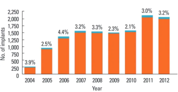
The number of dental implants placed at the Academy for Oral Implantology increased significantly from 2004 to 2012 (over 2,000 implants per year), and the failure rate remained stable at 3.0%±0.7% (Fig. 1). The 402 implant failures (out of 13,147 im- plants placed) were evenly distributed among all of the classes of indication (Table 2). However, single-tooth gaps in the esthetic re- gion of the upper jaw (95.8% survival rate) and completely eden- tulous maxillae (96.2% survival rate) were identified as the most complex situations of implant rehabilitation in our study popula- Table 1. Demographic characteristics of the 4,316 patients treated at the Academy for Oral Implantology in Vienna from 2004 to 2012.
Characteristic Male patients (%) Female patients (%) Age (year)
≤20 2.2 2.0
21–30 4.9 4.2
31–40 9.9 10.3
41–50 23.0 21.2
51–60 22.2 23.7
61–70 25.6 25.2
71–80 9.8 10.2
81–90 2.3 3.1
>90 0.1 0.1
Smokers 19.1 21.2
Osteoporosis 1.2 2.3
Diabetes mellitus 2.4 1.9
Periodontal disease 15.2 16.6
tion (Fig. 2). The estimated implant survival after 8 years was 94.8% in the mandible compared to 92.6% in the maxilla (Table 3) without significant difference (P=0.545). Implants replacing ante- rior teeth (incisors or canines) had a 93.5% survival rate that was similar to the survival rate of implants in the posterior region (93.9%, P=0.450). Narrow-diameter implants (less than 3.75 mm in width) demonstarted a similar survival rate of 95.4% compared to wide implants with a diameter of 5 mm or more (95.8%, P=0.704).
Short dental implants less than 10 mm in length demonstrated a fa- vorable 8-year survival rate of 96.3% and this rate was not signifi- cantly different to the survival of the longer ones (P=0.930).
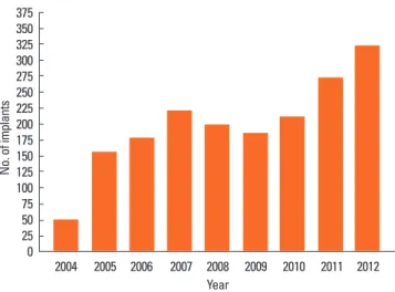
The number of grafting procedures performed to enhance the bone volume at the implant site had also risen since 2004 with 346 bone grafts in 2012 (Fig. 3). Of the 1,917 bone augmentation procedures, 46% occurred in the maxillary sinus (sinus floor eleva- tion surgery), 9% were fresh extraction sockets (alveolar ridge preservation), 32% were done to increase the alveolar ridge width (horizontal augmentation), and 12% were done to increase alveo- lar ridge height (vertical augmentation). Implants placed in the augmented maxillary sinus following sinus floor elevation showed a 4-year and 8-year survival rate of 96.6% and 91.4%, respectively.
Vertical augmentation using autologous onlay bone grafts in cases of reduced alveolar bone height yielded survival rates of 97.0% at 4 years and of 91.9% at 8 years. Horizontal augmentation in cases of knife-edged residual ridges (Atwood [13] class IV) were less problematic than vertical grafts were, considering that the implant survival rates after 4 and 8 years remained consistent at 95.5%
and 95.0%, respectively, and no significant difference between im- plants inserted into augmented bone and native jawbone was found (P=0.617).
The 8-year survival rate of implants placed in smokers was sig- nificantly reduced to 76.5% (P<0.001). This translates into a more than 3-fold increased risk of implant failure among smokers com- pared to nonsmokers. Moreover, the number of cigarettes smoked per day influenced the failure rate, but implant survival was re- duced by only 1% in patients smoking up to five cigarettes a day.
Smoking 6–15 cigarettes doubled the risk of implant failure. Peri- odontal disease was another important factor influencing implant
survival. The 8-year survival rate in patients suffering from peri- odontitis was reduced to 88.6%, which is a 2-fold increased risk of implant failure compared to implants in patients without peri- odontitis (P =0.001). Approximately one third of all treated pa- tients were smokers and/or suffered from periodontitis; therefore, it not surprising that almost half of all implant failures (43%) oc- curred in these at-risk patients (Fig. 4).
In patients suffering from osteoporosis, the 8-year survival rate (94.4%) was not significantly different from the healthy patients (P=0.661). However, the failure rate was 2% higher in the upper jaw of patients with osteoporosis compared to the lower jaw; thus, reduced bone density may be more influential in the mandible than in the spongy bone of the maxilla. By contrast, reduced local bone density at the site of implant placement (defined as class IV according to Lekholm and Zarb [14]: thin layer of cortical bone surrounding a core of low density trabecular bone of poor strength) was associated with a 2-fold increased risk of implant failure (8- year survival, 84.8%) compared to implants in areas of high bone density, but this finding was not statistically significant (P=0.449).
Diabetes is not considered a risk factor for implant survival, if blood sugar levels are effectively controlled. Patients suffering from diabetes demonstrated high long-term survival rates of 95.1% that did not significantly differ from the healthy popula- tion (P =0.928). Despite widespread concerns regarding implant survival in senior citizens, there is no evidence that advanced age poses a risk for implant survival. Patients over 70 years of age had an 8-year survival rate of 95.3% that was similar to those younger than 30 years (96.5%, P =0.575). Thus, the majority of patients qualified for implant therapy and was able to benefit from an im- proved quality of life, which is associated with fixed implant-re- tained prostheses.
DISCUSSION
Overall implant survival at the Academy for Oral Implantology of 97% compares well to long-term results in international scien- Table 2. The number and success rates of dental implants achieved in various classes of indication.
Classes of indication Number of
implants Implant success rate (%)
Free-end gaps in the maxilla 1,816 97.9
Intermediate gaps in the mandible 1,174 97.8
Single-tooth gaps in posterior regions 1,418 97.1
Intermediate gaps in the maxilla 1,774 97.0
Completely edentulous mandible 1,734 97.0
Free-end gaps in the mandible 1,733 96.8
Completely edentulous maxilla 2,845 96.2
Single-tooth gaps in the esthetic region 653 95.8
Total 13,147 97.0
Figure 1. The number of dental implants placed at the Academy for Oral Im- plantology increased significantly in the years 2004 to 2012 (over 2000 im- plants per year) while the failure rate remained stable at around 3%.
2,250 2,000 1,750 1,500 1,250 1,000 750500 2500
No. of implants 3.9%
2.5%
4.4% 3.2%
3.0% 3.2%
3.3% 2.3% 2.1%
2004 2005 2006 2007 2008 2009 2010 2011 2012 Year
tific literature. Charyeva et al. [15] reported a 6-year survival rate of 96%, and Simonis et al. [16] found a cumulative survival rate of 83% after 10–16 years. Moreover, a systematic review by Ber- glundh et al. [17] summarizing the results of 51 studies (8,588 im- plants in 2,675 patients) calculated an overall failure rate of 2.5%
prior to loading and 2%–5% after 5 years. Our analysis of 13,147 implants in 4,316 patients yielded early failures (prior to or within the first year of loading) in 2.1% and late failures in 0.9%.
The risk factor analysis in our study population yielded findings
similar to the results of systematic reviews and meta-analyses. Re- garding implant length, Monje et al. [18] reported that short im- plants had a similar estimated long-term survival; however, peak failure rates of short dental implants occurred earlier (4–6 years) than standard dental implants did (6–8 years). Regarding implant diameter, Sohrabi et al. [19] summarized the survival rates for nar- row implants (3.5 mm or less in diameter) and found they are simi- lar to those reported for standard-width implants. In addition, Strietzel et al. [20] agreed that smoking is a significant risk factor
A B
C D
E F
Figure 2. Implant rehabilitation of the completely edentulous maxilla according to the All-on-4 (Nobel Biocare, Gothenburg, Sweden) flapless protocol. (A) Pre- operative planning procedure using cone-beam computed tomographic scans, (B) postoperative X-ray with impression copings and angulated multiunit abut- ments on tilted distal implants, (C) immediate temporary fixed restoration delivered at the day of surgery, (D) pictures and x-rays at final prosthesis delivery, and (E, F) 5 years after implant placement.
of implant failure with an odds ratio of 2.25; we found a 3-fold increased risk our patient sample. An odds ratio of 3.02 and an av- erage of 0.61 mm more marginal bone loss was found in patients Table 3. Eight-year implant survival rates for potential risk factors.
Variable 95% Confidence interval
Jaw region
Mandible 93.6–96.1
Maxilla 90.3–94.9
Implant position
Incisors/canines 91.0–96.0
(Pre) molars 92.3–95.3
Implant diameter
Narrow (<3.75 mm) 94.1–96.8
Wide (≥5 mm) 93.4–98.1
Implant length
Short (<10 mm) 93.6–98.0
Regular (≥10 mm) 92.3–94.7
Sinus floor augmentation
4 Years 95.5–97.8
8 Years 86.1–96.8
Vertical onlay bone grafts
4 Years 93.7–100
8 Years 81.7–100
Horizontal onlay bone grafts
4 Years 93.2–97.8
8 Years 92.5–97.5
Smoking
Positive 64.4–88.7
Negative 92.5–94.8
Periodontal disease
Positive 82.9–94.3
Negative 92.1–94.5
Osteoporosis
Positive 89.9–98.9
Negative 92.4–94.7
Bone quality
Class I–III 96.6–97.3
Class IV 71.6–97.9
Diabetes mellitus
Positive 91.6–98.5
Negative 92.4–97.0
Age
<30 Years 94.0–99.3
>70 Years 94.1–96.4
The 95% confidence intervals were estimated using the Kaplan-Meier method.
Year 375350
325 300275 250225 200 175150 125100 75 5025 0
No. of implants
2004 2005 2006 2007 2008 2009 2010 2011 2012
Figure 3. The number of grafting procedures to enhance bone volume for implant insertion (total of 1,917 sinus floor elevation operations, horizontal or vertical block augmentations, and socket grafts).
Figure 4. Kaplan-Meier curves (and 95% confidence intervals) for implant survival through 96 months (8 years) in healthy patients (A), smokers (B), and patients suffering periodontal disease (C).
A 100
80 60 40 20
00 10 20 30 40 50 60 70 80 90
Months
Survival rate (%)
100 80 60 40 20
00 10 20 30 40 50 60 70 80 90
B
Survival rate (%)
Months
C 100
80 60 40 20
00 10 20 30 40 50 60 70 80 90
Survival rate (%)
Months
with a history of periodontitis compared to those without a history of periodontitis [21], and we found a 2-fold increased failure risk in our population. Moreover, Bornstein et al. [22] found little evi- dence to support an association of osteoporosis with implant fail- ure. Regarding diabetes, Salvi et al. [23] found that only poorly controlled diabetes may be a risk factor for increased severity (but not extent) in periodontitis; however, lacking evidence to indicate that implant therapy in subjects with diabetes yields long-term outcomes comparable with those of nondiabetic subjects.
Implant failure in augmented bone was not found to be signifi- cantly higher than that in native jawbone. Implants placed in graft- ed sinuses demonstrated a high survival rate of 97% 4 years after sinus floor elevation surgery (sinus-lift). Results in the literature range from 60% to 100% with a mean survival rate of 95% [24- 27]. Implant survival following vertical and horizontal bone aug- mentation was 97% and compares favorably to the results in the literature of 85% and 99% (range, 76% to 100%) after vertical and horizontal onlay block grafting, respectively, and 98% (range, 77% to 100%) after guided bone regeneration procedures [28-35].
Higher long-term survival rates in horizontal bone grafts than those in vertical bone augmentation are in agreement with re- cently published results [36].
Summing up the long-term results of 13,147 dental implants placed at the Academy for Oral Implantology in Vienna the follow- ing conclusions may be drawn: First, the 402 total implant failures were evenly distributed among all of the classes of indication and resulted in an overall implant survival rate of 97%. Second, the edentulous maxilla and the esthetic zone of the upper jaw can be considered more complex and challenging treatment situations.
Third, implants of short length or reduced diameter are routinely used and have comparably high survival rates of more than 95%.
Fourth, bone-grafting procedures may also provide satisfactory, long-term survival rates between 90% and 95% in augmented bone. Fifth, smoking and a history of periodontal disease increases the risk of implant failure by 3-fold and 2-fold, respectively. Last, patients suffering from osteoporosis or diabetes have a favorable implant survival of 95% after 8 years of loading. Advanced age, as well, does not seem to pose a contraindication for the placement of dental implants.
CONFLICT OF INTEREST
No potential conflict of interest relevant to this article was re- ported.
ORCID
Dieter Busenlechner http://orcid.org/0000-0002-6731-6160 Rudolf Fürhauser http://orcid.org/0000-0001-9373-2101 Robert Haas http://orcid.org/0000-0003-0989-5069 Georg Mailath http://orcid.org/0000-0001-8869-4059 Georg Watzek http://orcid.org/0000-0003-4998-2829
Bernhard Pommer http://orcid.org/0000-0002-2617-0353
REFERENCES
1. Pommer B, Zechner W, Watzek G, Palmer R. To graft or not to graft? Evidence-based guide to decision making in oral bone graft surgery. In: Zorzi A, Miranda J, editors. Bone Grafting. Rije- ka: InTech; 2012. p.159-82.
2. Levin L. Dealing with dental implant failures. J Appl Oral Sci 2008;
16:171-5.
3. Palma-Carrio C, Maestre-Ferrín L, Penarrocha-Oltra D, Penarro- cha-Diago MA, Penarrocha-Diago M. Risk factors associated with early failure of dental implants: a literature review. Med Oral Pa- tol Oral Cir Bucal 2011;16:e514-7.
4. Lee JH, Frias V, Lee KW, Wright RF. Effect of implant size and shape on implant success rates: a literature review. J Prosthet Dent 2005;94:377-81.
5. Chiapasco M, Zaniboni M, Boisco M. Augmentation procedures for the rehabilitation of deficient edentulous ridges with oral implants. Clin Oral Implants Res 2006;17 Suppl 2:136-59.
6. Pommer B, Hof M, Fadler A, Gahleitner A, Watzek G, Watzak G.
Primary implant stability in the atrophic sinus floor of human cadaver maxillae: impact of residual ridge height, bone density, and implant diameter. Clin Oral Implants Res 2014;25:e109-13.
7. Diz P, Scully C, Sanz M. Dental implants in the medically com- promised patient. J Dent 2013;41:195-206.
8. Clementini M, Rossetti PH, Penarrocha D, Micarelli C, Bonachela WC, Canullo L. Systemic risk factors for peri-implant bone loss: a systematic review and meta-analysis. Int J Oral Maxillofac Surg 2014;43:323-34.
9. Pommer B, Frantal S, Willer J, Posch M, Watzek G, Tepper G. Im- pact of dental implant length on early failure rates: a meta-anal- ysis of observational studies. J Clin Periodontol 2011;38:856-63.
10. Andersen E, Saxegaard E, Knutsen BM, Haanaes HR. A prospec- tive clinical study evaluating the safety and effectiveness of narrow-diameter threaded implants in the anterior region of the maxilla. Int J Oral Maxillofac Implants 2001;16:217-24.
11. Polizzi G, Rangert B, Lekholm U, Gualini F, Lindstrom H. Brane- mark System Wide Platform implants for single molar replace- ment: clinical evaluation of prospective and retrospective mate- rials. Clin Implant Dent Relat Res 2000;2:61-9.
12. Arisan V, Bolukbasi N, Ersanli S, Ozdemir T. Evaluation of 316 narrow diameter implants followed for 5-10 years: a clinical and radiographic retrospective study. Clin Oral Implants Res 2010;21:
296-307.
13. Atwood DA. Reduction of residual ridges: a major oral disease entity. J Prosthet Dent 1971;26:266-79.
14. Lekholm U, Zarb GA. Patient selection and preparation. In: Br- anemark PI, Zarb GA, Albrektsson T, editors. Tissue-integrated prostheses: osseointegration in clinical dentistry. Chicago: Quin- tessence; 1985. p.199-220.
15. Charyeva O, Altynbekov K, Zhartybaev R, Sabdanaliev A. Long-
term dental implant success and survival: a clinical study after an observation period up to 6 years. Swed Dent J 2012;36:1-6.
16. Simonis P, Dufour T, Tenenbaum H. Long-term implant survival and success: a 10-16-year follow-up of non-submerged dental implants. Clin Oral Implants Res 2010;21:772-7.
17. Berglundh T, Persson L, Klinge B. A systematic review of the inci- dence of biological and technical complications in implant den- tistry reported in prospective longitudinal studies of at least 5 years. J Clin Periodontol 2002;29 Suppl 3:197-212.
18. Monje A, Chan HL, Fu JH, Suarez F, Galindo-Moreno P, Wang HL.
Are short dental implants (<10 mm) effective? A meta-analysis on prospective clinical trials. J Periodontol 2013;84:895-904.
19. Sohrabi K, Mushantat A, Esfandiari S, Feine J. How successful are small-diameter implants? A literature review. Clin Oral Implants Res 2012;23:515-25.
20. Strietzel FP, Reichart PA, Kale A, Kulkarni M, Wegner B, Kuchler I.
Smoking interferes with the prognosis of dental implant treat- ment: a systematic review and meta-analysis. J Clin Periodontol 2007;34:523-44.
21. Safii SH, Palmer RM, Wilson RF. Risk of implant failure and mar- ginal bone loss in subjects with a history of periodontitis: a sys- tematic review and meta-analysis. Clin Implant Dent Relat Res 2010;12:165-74.
22. Bornstein MM, Cionca N, Mombelli A. Systemic conditions and treatments as risks for implant therapy. Int J Oral Maxillofac Im- plants 2009;24 Suppl:12-27.
23. Salvi GE, Carollo-Bittel B, Lang NP. Effects of diabetes mellitus on periodontal and peri-implant conditions: update on associa- tions and risks. J Clin Periodontol 2008;35(8 Suppl):398-409.
24. Chao YL, Chen HH, Mei CC, Tu YK, Lu HK. Meta-regression analy- sis of the initial bone height for predicting implant survival rates of two sinus elevation procedures. J Clin Periodontol 2010;37:
456-65.
25. Esposito M, Grusovin MG, Rees J, Karasoulos D, Felice P, Alissa R, et al. Interventions for replacing missing teeth: augmentation procedures of the maxillary sinus. Cochrane Database Syst Rev 2010;(3):CD008397.
26. Graziani F, Donos N, Needleman I, Gabriele M, Tonetti M. Com- parison of implant survival following sinus floor augmentation
procedures with implants placed in pristine posterior maxillary bone: a systematic review. Clin Oral Implants Res 2004;15:677- 82.
27. Pjetursson BE, Tan WC, Zwahlen M, Lang NP. A systematic review of the success of sinus floor elevation and survival of implants inserted in combination with sinus floor elevation. J Clin Peri- odontol 2008;35(8 Suppl):216-40.
28. Aghaloo TL, Moy PK. Which hard tissue augmentation techniques are the most successful in furnishing bony support for implant placement? Int J Oral Maxillofac Implants 2007;22 Suppl:49-70.
29. Bernstein S, Cooke J, Fotek P, Wang HL. Vertical bone augmenta- tion: where are we now? Implant Dent 2006;15:219-28.
30. Chiapasco M, Casentini P, Zaniboni M. Bone augmentation pro- cedures in implant dentistry. Int J Oral Maxillofac Implants 2009;
24 Suppl:237-59.
31. Clementini M, Morlupi A, Canullo L, Agrestini C, Barlattani A.
Success rate of dental implants inserted in horizontal and verti- cal guided bone regenerated areas: a systematic review. Int J Oral Maxillofac Surg 2012;41:847-52.
32. Donos N, Mardas N, Chadha V. Clinical outcomes of implants fol- lowing lateral bone augmentation: systematic assessment of available options (barrier membranes, bone grafts, split osteoto- my). J Clin Periodontol 2008;35(8 Suppl):173-202.
33. Jensen SS, Terheyden H. Bone augmentation procedures in local- ized defects in the alveolar ridge: clinical results with different bone grafts and bone-substitute materials. Int J Oral Maxillofac Implants 2009;24 Suppl:218-36.
34. Rocchietta I, Fontana F, Simion M. Clinical outcomes of vertical bone augmentation to enable dental implant placement: a sys- tematic review. J Clin Periodontol 2008;35(8 Suppl):203-15.
35. Waasdorp J, Reynolds MA. Allogeneic bone onlay grafts for alve- olar ridge augmentation: a systematic review. Int J Oral Maxillo- fac Implants 2010;25:525-31.
36. Sbordone L, Toti P, Menchini-Fabris G, Sbordone C, Guidetti F.
Implant survival in maxillary and mandibular osseous onlay grafts and native bone: a 3-year clinical and computerized to- mographic follow-up. Int J Oral Maxillofac Implants 2009;24:
695-703.