대 한 방 사 선 의 학 회 지 1992; 28 (3) ‘ 327~331
Journal of Korean Radiological Society, May, 1992
안와 가종양의 전산화단층촬영 소견
부산대학교 의과대학 방사선과학교실
최민연 • 남상화 • 김건일* • 설창효 • 김병수 - Abstract-
CT
Findings of Orbital PseudotumorMin Yun Choi, M.D., 8ang Hwa Nam, M.D., Kun
11
Kim, M.D .• , Chang Hyo 801, M.D., Byung 800 Kim,
M.D.Department of Radiology, College of Medicine , Pusan National University
To evaluate characteristic CT findings of orbital pseudotumor and to define differentialpoints from other pathology.
the authors retrospectively reviewed CT of 19 patients who were prooen to have orb!tal pseudotumor by clinical course and, in some cases. biopsy. A variety of CT findings including extraocular muscle thickening (11 c잃es). stre와‘Y
infiltration of retroorbital fat (11 cases), mass formation (10 cases). optic nerve thickening (6 cases). conjunctival thickening (5 cases). scleral thickening (4 cases). enlarged lacrimal gland (4 cases) and destruction of orb!tal bone (2 cases) were observed.
Thickening of the anterior portion and irregular margin were characteristic findings of extraocular muscle and optic nerve lesions. Mass formation predominantly occurs in the anterior portion of the orbit. In most cases more than two orbital structures are involved by lesion.
Index Words: Orbit. CT. 22.1211
Orbit, inflammation, 22.299
서 론
안와 가종양 (orbital pseudotumor)은 특발성 안와 염증 성 증후군(idiopathic orbital inflammatory syndrome IOIS)의 통상명으로 원인이 확실히 밝혀져 있지 않은 반응 성 병변으로써 연역학적으로 중개되는 질환으로 간주되고 있다. 안와 가종양은 병 리학적으로는 림 프양종양(lymphoid tumor)과 감별이 어려우며 영상진단법으로는 림프양종양,
갑상선 안증과의 감별이 중요한 문제이다. 가종양의 전산화 단층촬영 (이하 CT로 약함)소견은 여러 연구자들에 의해 보 고되어 있으나 국내에서는 이에 대한 보고가 없었다. 저자 는 최근 7년간 가종양으로 진단된 환자의 CT소견을 분석하 여 이 질환의 특징과 타질환과의 감별점을 찾고자 이 연구 를하였다.
*마산의료원 방사선과
*
Department of Radiology. Masan Medical Center대상 및 방법
1984년 7월부터 1991년 7월까지 7년간 안와 CT를 시행하 고1 안와 가종양으로 진단된 19례를 대상으로 하였다. 이중 13례는 안와 가종양을 추정하여 스테로이드를 투여한 후 임 상증상의 호전으로 진단하였으며 나머지 6례는 임상증상의 호전이 없거나 다른종양과의 감별이 어려워서 CT유도하 칩 생검에 의해 병리학적으로 확진되었다. 19례에서 전신질환 의 증거를 찾아볼 수 없었으며 CT상 안와가종양이 의심되 나 스테로이드 (steroid)에 반응이 없고 조직검사를 시행하지 않았거나 조직검사를 실패한 환자는 대상에서 제외되었다
(1-2). 이들의 나이는 147~ 월부터 72세로 다양하였으며 평 균연령은 41세였고 남자가 12명, 여자가 7명 이었다. CT는 5mm의 절편두께로 조영증강후의 횡단상과 직접관상연상
이 논문은 1991년 12월 16일 접수하여 1992 년 2월 28일에 채택되었음
대한방사선의학회지 1992; 28(3) : 327~331
(coronal image)을
얻었다.
CT상 안외근, 시신경, 포도
나머지 7례는 국소형으로 이중
6례는 안와의 前部에, 1례는
막, 결막, 누액션, 안구후부지방의 변화를 각각 관찰하였는 後部에 위치하였다. 종괴의 크기는 10mm에서 67mm의 범 데 안와내 종양이 형성된 경우는 국소형 (localized form)과 위 였으며 평균직경은 28.6mm였다.미만형 (diffuse form)으로 나누고 국소형인 경우는 종괴에 외안근 : 16례중 11례에서 한개 이상의 외안근 비후가 있 포함된 구조물이 병변에 침범된 것으로 간주하였으며 미만 으며 5례에서는 2개 이상의 비후가 있어서 총 19개의 외직 형인 경우는 각각의 구조물의 변화를 관찰하지 않고 따로 근이 비후되어 있었다. 그러나 양측 안와 외안근을 동시에
분류하였다. 침범한 경우는 없었다. 외안근중 내직근의 비후는 6례에서,
안외근의 병변은 4개의 직근을 관찰하였으며 두께는 내외 하직근은 5례에서, 외직근과 상직근은 각각 4례에서 관찰되 직근은 횡단상에서 상하직근은 관상면상에서 가장 두꺼운 었다. 상직근과 하직근이 침벙된 경우는 내직근과 외직근에 부위를 측정하였으며 시신경의 두께는 횡단상에서 가장 두 비해 평균직경이 두꺼운 양상을 보였다(Table 2). 팽대부와 꺼운 부위를 측정 하였다.
결 과
안와 가종양으로 확진된 19례의 안와 CT를 분석하여 나 타난 병변의 종류와 빈도를 표로 정리하면 다음과 같다 (Table 1).
종괴형성 : 19례중 10례에서 안와내 종괴를 관찰할 수 있 었는데 이중 3례는 안와전체를 차지하는 미만형 종괴였으떠
Table 1. Types and lncidence ofOrital Lesions in 19 Pa- tients
Type No. of case(%}
Mass Diffuse Localized Muscle thickening Streaky infiltration of fat Optic nerve thickening Conjunctival thickening Scleral thickening Enlarged lacrimal gland Bone destruction
10(52.6}
3(15.8) 7(36.8) 1l(57.9}
11(57.9) 6(31.6) 5(26.3) 4(21.1}
4(21.1) 2(10.5}
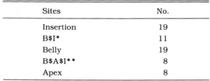
근부착부위는 비후된 19개의 외직근 모두에서 비후를 보였 으나 첨부는 전체 중 약 반(8/19)에서만 비 후를 보였다 (Table 3). 비후된 외직근의 외연은 19개의 근중 17개에서 불규칙한 양상을 관찰할 수 있었다 (Fig. 1).
시신경 : 16례중 6례에서 시신경초의 비후를 볼 수 있었으
Table 2. Prevalence of 4 Rectus Muscle Involvement and Their Thickness
Muscles No. Mean Thickenss(mm)
Medial rectus 6 6.0
Lateral rectus 4 6.3
Inferior rectus 5 7.8
Superior rectus 4 7.8
Table 3. Sites of Muscle Thickening
Sites No
Insertion B$I*
Belly
9 l 9 8 8 ,,‘
1li
--‘
B$A$I* * Apex
B$I* : belly and tendon insertion portions
B$A$I* * : belly. apex and tendon insertion portions
a b
Fig. 1. a. Post-contrast axial CT im- age in 42-year-old male patient.
Left lateral rectus shows thickening and marginal irregularity in entire portions. Streaky infiltrations in ad- jacent retrobular fat are noted. b. Coronal image of patient shows enlargment of left superior. inferior.
and lateral rectus muscles.
- 328-
최민연 외 . 안와가종앙의 전산화단층촬영
Fig.2. Post-contrast axial CT image in 10-year-old female patient.
Left optic nerve is thickened in anterior portion and the scleral sur- face is diffusely enhanced and thickened
Fig. 3. Post-contrast‘axial CT image in 46-year-old male patient.
Diffuse thickening and enhancement of sclera and conjunctiva are noted in right orbit. Irregular streaky infiltra- tions in retrobulbar fat are noted.
2 3
a b
• “‘-
-
‘
‘‘ . . .‘ .. ,- - ‘ ’
‘ / ’ ‘ a‘、““
‘
-‘
,. ." .'
.,
. , •• ‘.1::- • “ - “‘ ‘ · 、 ....,-.. ~~‘
“
,.、
“ ~’ ‘ • • ‘
~ .""",..’ - .-ι. 、껴
-
ι‘ ..
“ - ‘
‘ . .,.
- -
'- J. ;
‘ .. 、
’‘
,
‘
.‘-
--‘ ‘ • • ‘ ‘
- .
‘"" .
c
Fig. 4. 49-year-old male patient presented with right exophthalmos for 8 years. CT guided biopsy was done to rule out malignancy, and revealed pseudotumor of right orbit
a. Post contrast axial CT image shows diffuse orbital mass, destructing the right lamina papyracea of ethmoid bone and extending into ethmoid air cells.
b. Coronal image of same patient shows diffuse orbital mass. destructing right ethmoid bone and l1oor. and exten- ding into ethmoid‘ nasaI cavity and maxillary antrum.
c. Microphotograph of the biopsy shows diffuse infiltration of lymphoplasma cell‘ histiocytes and fibloblasts with background of hyalinized fibrous connective tissue.(H&E. x 100)
며 이중 화안이 4례 우안이 2례였으며 모두 일측성이었다.
비후된 시신경의 두께는 8-12mm의 범위로 평균 9.7mm였 다. 6례중 3례는 시신경전체의 비후가 있었으며 1례는 시신 경의 前部와 중간부가, 2 례는 前部만 비후되어 주로 시신경 의 前部가 비후되는 양상을 보였다(Fig. 2),
누액선 : 4례에서 누액선이 커져 있었으며 이중 l례는 양 안의 누액선이 커져 있었다.
공막 및 결막 :4례에서 공막의 비후를 볼 수 있었는데 비 후의 정도는 5mm-lOmm 범위였으며 평균 7.6mm였다. 5 례에서는 결막의 비후도 볼 수 있었다(Fig. 3).
지방 : 안구후부의 지방의 변화는 11례에서 나타났는데 안 구후부지방내에 작은 선조가 증가되어 있었으며 다소 지저
분하게 보이며 음영이 증가된 양상을 보였다. 안와 가종양은 아직까지 원인이 정확히 규명되지 않았으 안와글 :2례의 환자에서 안와골 파괴를 볼 수 있었다. 이 나 항원에 대한 자가 면역반응으로 간주되는 질환으로써(3) 들은 CT 소견상 안와전체를 채우는 종괴를 형성하였으며 병리학적으로 비정형적인 다형원형세포, 임파구, 원형질세 안와골의 파괴가 있어 CT 유도하 침생겁을 하여 확진되었 포, 대식세포, 호산구등의 침윤을 주로 나타내며 악성임파 다. 1례는 사골의 안와판(I amina papyracea),사골통 및 상 종의 임파양증식과 감별이 힘든 경우가 많다(4), 또한 임상 악골의 파괴를 관찰할 수 있었으며(Fig. 4) 다른례에서는 접 적으로도 만성 혹은 아급성인경우에는 염증소견이 거의없기 형골의 대익 (greater wing)부분에 골파괴(destruction) 및 때문에 임상적 진단이 어려운 경우가 대부분이다. 따라서 미란 (erosion)을 관찰할 수 있었다. 가종양의 진단에 있어서 방사선학적방법이 중요한 역할을 미만형종괴를 보인 3례를 제외한 16례에서 병변이 침윤된 구조물의 숫자를 관찰한 결과 단 2례만이 1가지 구조물만의 병변이었으며 4례는 2가지 구조물이, 5례는 3가지 구조물 이, 3례는 4가지 구조물이, 2례는 5가지 이상의 구조물을 침범하였다. 따라서 안와전체를 침범한 미만형종괴인 3례를 포함하면 전체 19례중 17례(89.5%)에서 2가지 이상의 병변 을, 13례(68.4%)에서 3가지 이상의 구조물의 병변을 보였 다.
고
*f
2대한방사선의학회지 1992; 28(3) : 327"'-'331
하는 경우가 않으며 CT와 최근에는 자기공명영상술이 방사 선학적진단에 있어서 중심된 진단방법이다. 가종양은 안와 내 여러가지 구조물을 동시에 침범하며 한구조물에서 다른 구조물로 병변이 쉽게 파급되는 특정을 가지고 있는 것으로 알려져있으며, 이런 다양한 명변을 방사선학적으로 구분하 기 위하여 근염형, 누선염형, 공막주위염형, 신경초염형과 종괴형성형 등으로 분류하고 있다(2, 5-8). 그러나 개별 구 조물의 병변이 CT소견상 특정적인 소견을 보이는 것이 아 니기 때문에 임파종, 갑상선안증을 포함한 타질환과의 감별 이 어려운 경우가 많다. 이런 경우 안와 가종양은 스테로이 드에 빠른 효과를 보이기 때문에 이 약제의 사용이 진단적 시험으로 사용되기도 하며 생검에 의한 조직검사는 스테로 이드에 효과가 없는 경우에만 시도되어야만 한다고한다(6,
9-10).
가종양이 외안근을 침범하는 경우 갑상선안증과의 감별이 중요한 문제이다. 갑상선안증인 경우 대개 양측성이며 가종 양은 일측성인 것으로 알려져 있으나 갑상성안증에서도 일 측성 병변이 2-50%에서 나타날 수 있으며 가종양에서도 양 측성 병변이 10-50%에서 나타난다고 보고되어 있기 때문에 병변의 일측성 양측성여부는 감별진단에 있어서 의의가 없 는 것으로 사료된다(1l-14). 병변에 따른 각각의 외안근에 대한 침벙빈도의 문제에 있어서도 Alper등(1 5) 은 가종양의 경우 하직근과 외 직근의 침 범 이 가장 빈번하다고 하였으나 저자의 예에서는 4개의 외직근의 침범빈도는 대동소이 하였 다. Trokel 등(1 6)은 비후된 외안근 외연의 평활성 여부가 갑상선안중과의 감별에 중요하다고 하였다. 저자의 예에서 도 비후된 19개의 외직근중 17개(89.5%)에서 외연이 불규 칙한 양상을 볼 수 있었다. 외안근의 비후가 생기는 부위에 따라 감별점을 찾고자 하는 시도가 있었는데 갑상선안증에 서는 근부착 부위에서는 비후가 되지 않는다고 알려져 있으 며 가종양에서는 이부위의 비후가 올 수 있는 것으로 알려 져 있다(1). (16-18). 저자는 가종양에 의한 외안근의 근부 착부, 팽대부와 첨부의 비후양상을 관찰한 결과 근부착부와 팽대부는 전례에서 비후되어 있었으나 첨부는 대상의 약 반 에서만 비후를 볼 수 있었다. 따라서 가종양에 의한 외안근 염의 특정은 근부착부와 팽대부 즉 前部의 비후를 항상 보 이며 외연이 불규칙한 점이라고 할 수 있다.
가종양에 의한 안와내 종괴형성이 있는 경우 안와내 종양 과 감별이 힘든 경우가 많다(1. 19), 안와내 종괴형성의 빈 도는 David 등(2) 은 33% 에서. Nugent 등(5)은 16례중 2례 에서 종괴를 보였다고 보고를 하였으나 저자의 예에서는 19 례중 10례(52.6%)로 높은 빈도에서 관찰 할 수 있었다. 이 는 국내에서 발생한 가종양의 특징인지 혹은 병증이 경과된 기간이 다른데 따른 표본의 차이에 의한 것인지는 분영치 않으며 이에 대한 연구가 더 있어야 할 것으로 사료된다. 그
러나 저자의 예에서는 안와 전체를 차지한 종괴인 미만형을 제외한 국소형 7례중 6례가 안와의 前部에 위치한 것으로 나타나 이것도 안와가종양에 의한 종괴 형성에 있어서 발생 위치의 특정으로 간주할 수 있을것으로 사료된다. 시신경초 염의 경우에 있어 신경이 비후되는 부위에 대한 관찰은 지 금까지 보고 된 적이 없으나 저자의 예에서는 시신경의 前 部는 병변이 있는 경우 모두 비후되어 있었다.
안와 가종양은 병변이 여러가지 구조물을 통시에 침범하 는 특정을 가지고 있는 것으로 알려져 있다(4). (20). 처자는 미만형으로 발생한 3례를 제외한 16례에서 안와내 각각의 구조물이 침범된 숫자를 관찰한 결과 단 2례만이 한가지 구 조물의 병변으로서 전체 19례중 17례(89.5%)에서 2가지 이 상의 병변을 볼 수 있었다. 따라서 가종양이 의심되는 경우 뚜렷이 보이는 병변외에 다른 구조물에 병변이 있는지 여부 를 세심히 관찰하는 것이 중요할 것으로 사료된다.
결론적으로 안와 가종양의 CT소견은 외안근의 비후, 안 구후부지방의 지저분한 변화, 종괴형성, 시신경의 비후, 공 막 및 결막의 비후와 골파괴 등이었다. 외안근과 시신경의 병변의 경우 前部가 주로 비후되고 외연이 불규칙한 점, 종 괴가 있는 경우 주로 안와의 전부에 발생하는 점과 명변이 2가지이상의 구조물을 동시에 침범하는 점이 안와 가종양의 CT소견상의 중요한 특정이었다.
참고문헌
1. Flanders AE. Mafee MF. Rao VM, Choi KH. CT characteristics of orbital pseudotumors and other orbital inflammatory processes. Journal of Com- mputer Assisted Tomograohy 1989; 13:40-47 2. David LH. Robert MQ. Gray WA. Computed
tomography and ultrasound in the evaluation of or- bital infection and pseudotumor. Radiology 1982;
142:395-401
3. Wilner HI. Cohn EM. KlingG, Jampel R5. Computer assisted tomography in experimentally induced or- bital pseudotumor. J Comput Assist Tomogr 1978;
2:431-455
4. Blodi FC. Gass JDM. 1nflammatory orbital pseudotumor of the orbit. Br J Ophthalmol 1968;
52:79-93
5. Nugent RA. Rootman J. Robertson WD. Lapointe J5. Harrison PB. Acute orbital pseudotumors:
Classification and CT features. AJR 1981;137:
957-962
6. Coleman DJ. Jack RL. Jones 15. Frazen LA:
Pseudotumors of the orbit. Arch Ophthalmol 1972;
- 330-
최민연 외 : 안와가종앙의 전산화단층촬영
88:472-480 grave diseae. In: Thompson HS. Williams. eds.
7. Som PM. Inf1ammatory disease. In: Bergeron RT. Topics in Neuro-Ophthalmology. Baltimore:
eds. Head and Neck Imaging. 2nd edition. St. Louis: Williams and Willkins. 1979:347-368
Mosby-Yearbook Inc. 1991:781-792 16. Trokel SL. Hilal SK. Recognition and differential 8. Curtin HD. Pseudotumor. The Radiologic Clinics of diagnosis of enlarged extraocular muscles in com-
North America. Philadelphia: W.B.Saunders Com- puted tomography. AM J Ophthalmol
pany. 1987;583-599 1979;87:503-512
9. Grove AS. Evaluation of exophthalmos. N Engl J 17. Trokel SL. Hilal SK. Submillimeter resolution CT Med 1975;292:1005-1013 scanning of orbital disease. Ophthalmology 10. Jellinek EH. The orbital pseudotumor syndrome (Rochester) 1980;87:412-417
and its differentiation from endocrine ex- 18. Trokel SL. Jakobiec FA. Correlation of CT scann- ophthalmos. Brain 1969;92:35-58 ing and pathologic features of ophthalmic 11. Bullock W. Reeves RJ. Unilateral exophthalmos. Graves’disease. Ophthalmology (Rochester) 1981;
Am J Roentgenol 1959;82:290-299 88:553-564
12. Werner S. Euthyroid patients with early eye signs 19. Forbes GS. Earnest F. Waller RR. Computer of Graves disease. Am J Med 1955; 18:608-612 Tomograpth of orbital tumors. Inc1uding late- 13. Bowden AN. Rose FC. Dysthyroid exophthalmos. generation scanning techniques. Rodiology 1982;
Proc R Soc Med 1969;62:13-15 142:387-394
14. Jackson H. Pseudotumor of the orbit. Br J 20. Enzman D. Donaldson SS. Marshal WH. Kriss JP. Ophthalmol 1958;42:212-224
15. Alper MG. Computerized tomography (CT)in diagnosis of inf1ammatory orbital pseudotumor and
Computed tomography in orbital pseudotumor(idiopathic orbital inf1ammation) Radiology 1976; 120:597-601
29th Congress European Society of Pediatric Radiology (ESPR)
venue: Hotel Hilton Budapest
,Hungary.
contact: D
r.Bela Lombay (pres.)
,Borsod County Hosp
.,Ped. Rad.
,P
.O.Box 188
,3501 Miskolc
,Hungary.
(Tel: 36-46-2121
1;Fax
:36-46-23694) 1992/04/27
-018th In t. Symposium Radionuclides in Nephro-Urology
venue
:Chester
,United Kingdom
contact: Mr. P.H. 0
’Reilly
,Dept. of Urology
,Stepping Hill Hospital
,SK2 7JE Cheshire
,Unite d Kingdom.
(Tel: 061-419 5484; Fax
: 06-419 5699)1992/05/06-08
92nd Meeting American Roentgen Ray Society
venue: Marriot World Center Orlando
,Florida
,USA.
contact: American Roentgen Ray
,Socie ty
,1891 Preston White Drive
,VA 22091 Reston
,USA.
(Tel
:703-6488992; Fax:
1992/05/10-15
Radiology & Oncology 92
venue: Int. Convention Centre
,Birmingham
,United Kingdom
.contact: British Institute of. Radiology
,36 Portland Place
,W1N 4AT London
,United Kingdom (Tel: 071-5804085; Fax: 071-255
3209)1992/05
/18-20
39th Annual Meeting Society for Nuclear Medicine
venue:
L.A. Convention Center Los Angeles.
California,USA contact: Soc
.of Nu
c1ear Medicine.136 Madison Ave .. 8th
fl ..NY 10016
NewYork.
USA.(T
el: ; Fax:1992/06/09-12
Car
’92
,Computer Assisted Radiology
venue: Baltimore
,Maryland
,USA.
contact:
Prof. Heinz u. Lemke. Univ. Klinikum. Raum 1005.
Augustenburger Platz
1.D-1000 Berlin 65
,Germany
.(Tel
: 49-30 45052044; Fax: 49-30 45052043)1992/06/14-17
3rd Annual Meeting Eur. Soc. of Gastrointestinal Radiologists (ESGR)
venue: Hotel Beach Regency Nice
,France.
contact: SOCFI
-ESGR.
14 rue Mandar
,75002 Paris
,Fra nce
.(Tel: ; Fax: 1-40260444)
1992/06/22-24
- 332