대 한방사선 의 학회 지 1991 ; 27 (2) : 267"-270 Journal of Korean Radiological Society, March, 1991
간외담관의 원발성 간세포암
고려대학교 의과대학 방사선과학교실
정석태·함수연·박철민·김정혁·차인호·정규병·서원혁·이창홍*
- Abstract-
Primary Hepatocellular Carcinoma in Extrahepatic Bile Duct
Seok Tae Jeong, M.D., 800 Youn Ham, M.D., Cheol Min Park, M.D., Jung Hyuk Kim, M.D., In Ho Cha, M.D., Kyoo Byung Chung, M.D., Woon Hyuck 8uh, M.D., Chang Hong Lee, M.D. *
Department of Radiology, College of Medicine, Korea Vniversity
Obstructive jaundice due to hepatocellular carcinoma in an extrahepatic bile duct. without a mass lesion in the liver parenchyma. is extremely rare.
We experienced two cases of primary hepatocellular carcinoma arising from an extrahepatic bile duct: one in a 53-year-old man whose a-fetoprotein value was 800 ng/ml, and another in a 39-year-old woman. in whom the mass lesion was found to be attached to an extrahepatic bile duct.
These tumors had a well-marginated sausage-like shape on CT and US. and the contrast media passed freely along their margins on both PTC and ERCP.
Recurrences ofthese tumors were observed in the extrahepatic bile duct 6 and 2 months after surgery. respec디vely.
Index Words: Bile duct radiography. contrast media 76.1227 Bile duct. CT 76.1211
Bile duct. US 76.12981 Bile duct neoplasm 76.329
간세포암에 의해 이차적으로 간외담관이 막혀 폐쇄성 황달이 초래되는 예는 1947 년 Mallory(l)에 의해 처음 보고된 이래 적지 않은 예가 보고되었다(2).
그러나 간실질 내에는 종괴병소가 없으연서 간외담관 내에 원발성으로 간세포암이 생기는 경우는 매우 회귀해 서 그 보고가 극히 드물다 (3-6).
저자들은 최근 부속병원에서 방사선학적 및 수술 소견 상 간실질 내에는 종괴가 없으면서, 폐쇄성 황달을 동반 한 간외담관의 원발성 간세포암 2 례를 경험하였기에 문 헌고찰과 함께 보고하는 바이다.
증례 보고 증례 1.
53 세 남자가 황달과 상복부 동통을 주소로 내원하
*고려대학교 의과대학 내과학교실
였다. 주요 검사실 소견으로 혈청내 총 빌리루빈치는 22.4 mg/dl 였고 직 접 빌리 루빈은 17_3mg/dl 였다.
SGOT/SGPT 는 45/40, 알칼라인 인산화 효소치는 216 μ/dl 이었으며 , a -fetoprotein:은 800 ng/ml 였다.
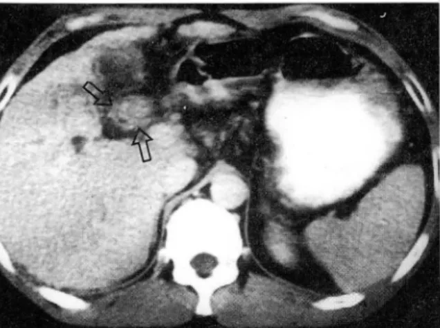
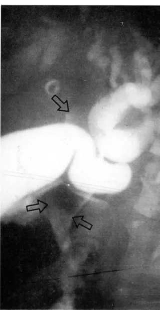
방사선학적 소견은 전산화 단충촬영술상 (Fig. 1) 간내 담관의 확장과 총수간관 내에 조영제 증강이 되지 않는 연조직 종괴가 발견되었고, 종괴주위와 간실질 내에는 침윤성, 또는 전이성 병소가 없었다. 경피경간담관조영 술상(Fig. 2) 총수간관 내에 3X4 cm 정도의 소세지 모 양의 충만결손이 있었으며, 종괴와 담관벽 사이로 조영 제가 흘러 내렸고 총수담관은 정상이었다.
수술 소견상 심한 간경화증이 있었으나 촉지되는 종괴 는 없었다. 총수간관내에는 담관벽과 유리된 3X4 cm 의 연조직 종괴가 있었으며, 이 종괴는 담즙으로 채색되
*Department of Internal Medicine. College of Medicine, Korea University
이 논문은 1990 년 6 월 12 일 접수하여 1991 년 l 월 21 일에 채택되었음
- 267-
대한방사선의학회지 1991 ; 27 (2) : 267"-'270
Fig. 1. CT scan shows well marginated soft tissue mass within the common hepatic duct (arrows).
Fig. 2. PTC shows intrahepatic duct dilatation and large filling defect in common hepatic duct (arrows). Note the downward passage of contrast dye along the margin of mass.
Fig. 3. Pathologic specimen demonstrates <<.fetoprotein secreating tumor cells which is typical finding of hepatocellular carcinoma.
어 있었고 쉽게 부수어졌다.
조직병리학적 소견상 (Fig. 3) <<-fetoprotein을 분비하 는 전형적인 간세포암으로 확진되었다.
수술후 6 개월에 실시한 T- 관 담관조영술에서 간공장 문합술 부위에 거대한 충만결손이 보여 재발이 확인되 었다.
증례 2.
39 세 여자가 황달과 우상복부의 동통을 주소로 내원하 였다. 주요 검사실 소견으로는 총 빌리루빈치는 12.
2 mg/dl, 직접 빌리루빈치 는 9.7 mg/dl 였고, SGOT /SGPT는 27/41, 알카라인 인산화 효소치는
88 μ/dl 였다.
방사선학적 소견은 초음파 검사상 (Fig. 4) 간외담관 내에 비교적 경계가 명확한 고반향성의 소세지 모양의 종물이 관찰되었다. 전산화 단충촬영술상(Fig. 5) 간내
Fig. 4. US shows sausage like hyperechoic mass in ex- trahepatic duct (arrows).
Fig. 5. CT scan shows well defined soft tissue mass within the distal common bile duct (arrows).
- 268-
정석태 외 : 간외담관의 원빌성 간세포암
담관이 확장되어 있었으며, 간외담관내에 경계가 명확한 총수간관 부위에 충만결손이 보여 재발이 확인되었다.
연조직 종괴가 있었고, 간실질 내에서는 종괴를 찾을 수 없었다. 내시경적 역행성 담관조영술상 (Fig. 6) 간외담 관의 전장에 이르는 충만결손이 보였다. 그라고 이 종괴 의 변연을 따라 조영제가 위로 올라가서 담낭과 확장된 간내담관을 충만시켰다.
수술 소견상 총수담관은 지 름이 2cm 정 도로 확장되 어 있었고, 간외담관내에 담관벽에 부착된 소세지 모양의 노란 연조직 종괴와 괴사물이 확인되었다. 그러나 간실 질 내에서는 종괴가 발견되지 않았다.
조직병리학적 검사상 특징적인 간세포암으로 확진되 었다.
수술후 2개월에 실시한 T- 관 담관조영술 (Fig.7) 에서
Fig.6. ERCP shows large filling defect in EHD (arrows).
Filling of dilated intrahepatic ducts and gall bladder are noted.
고 찰
간세포암이 담도 내에서 성장하는 예는 극히 드물어서 1976 년 Lin(7)은 간세 포암 322 례 중 단지 4 례 에 서 담도 내 종양성장이 있었다고 보고하였고, 이를 임상적으로
“icteric hepatoma"라고 분류하였다. 또 1982 년 Kojiro(2) 둥은 그 빈도가 9% 라고 보고하였다. 이 경우 간세포암이 간외담관을 폐쇄시켜 황달을 초래하게 되는 기전은 아직 확실히 밝혀지지는 않았지만 첫째, 전이된 종양이 담관 내에서 성장하여 간외담관을 폐쇄시킬 가능
성 (3 , 8) 둘째, 괴사된 종양 조직의 색전에 의해 담관이
막힐 가능성 (7) 셋째, 근위부의 미세 병소에 의한 출혈이 나 담즙 흐름퉁의 액상 수송 중 좌우 간내담관 분지 부 위에 정착한 후 다수화 될 가능성 (4 , 10 , 11)둥이 제시되 고 있다.
그러나 간실질 내에 종괴가 없이 간외담관에 원발성으 로 간세포암이 생기는 예는 더욱 회귀하여서 그 보고가
Fig. 7. Two months after operation. T-tube cholangiogram reveals the tumor recurrence in cornmon hepatic duct (arrows).
- 269-
대한방사선의학회지 1991 ; 27 (2) : 267~270
극히 드물며 (3-6) 그 발생 기 전도 아직 밝혀 진 바가 없다.
이제까지 보고된 담관 내 간세포암의 공통적 소견은 외견상 간실질 내에 종괴가 없으며, 황달이 예상되는 막 힘정도에 비해 심하지 않거나 약간 변동하는 점, 담관을 막고 있는 종괴의 변연을 따라 조영제가 상하로 비교적 잘 통과되는 점, 종괴는 경계가 분명한 충만결손으로 나 타나며 담관벽과 유리된 양상이 보이는 점, 그리고 a -fetoprotein 의 상숭이 없다는 점 퉁이 다 (6).
그러나 본 증례 중 한 례에서는 a -fetoprotein 이 800 ng/ml로 높은 수치를 보였으며, 수술 소견상 한 례 에서는 종괴가 담관벽과 유리되어 있었으나, 다른 례에 서는 담관벽에 부착되어 있는 양상을 보였다.
담관 내에 간세포암은 진단이 어려워서 수술 전, 혹은 수술 후에 담관 내종괴가 간세포암임을 예측하지 못하는 경우가 대부분이며, 본 증례와 같은 경우에는 Klatskin tumor로 오진할 가능성이 클 것으로 생각된다. 그러나 간외담관 내 간세포암은 상술한 바와 같은 특징적인 소 견이 있어 Klatskin tumor와의 감별진단에 도움이 되리 라고 본다.
결론적으로 저자들은 간실질 내에는 종괴가 없으며,
간외담관 내에 소세지 모양의 충만결손의.양상으로 보이 고, 종괴의 변연올 따라 조영제가 상하로 비교적 잘 통 괴되고, 조기에 담관 내 재발이 확인된 간외담관의 원발 성 간세포암 2 례를 경험하였기에 보고하는 바이다.
잠고문헌
1. Mallory TB. Case records of the Massachusetts
\~
General Hospital. Case 33441. N Engl J Med 1947;
237:673-676
2. Kojiro M. Kawabata K. kawano Y et al.
Hepatocellular carcinoma presenting as intrabile duct tumor growth: A c1inico-pathologic study of 24 cases. Cancer 1982; 49:2144-2147
3 남궁성, 서정돈, 이창홍 동. 원발성 간암에 합병한 폐 쇄성 황달 l 례. 대한의학협회지 1970;13 : 589-573 4. Brand WN. Biandt W. Sprayregan S. Extrahepatic
biliary tract obstruction secondary to a hepatoma- containing blood c10t in the common bile duct. Am J Dig Dis 1976; 21:905-909
5. Wind G. Futterman S. Obstructive jaundice as secondary to hepatoma. Case report and literature review. Am J Gastroenterol 1977; 67:80-83 6. 송치성, 박인애, 최상운. 총수간관에 발생한 간세포암
1 례 보고. 대한방사선의학회지 1989;25 : 573-576 7. Lim TY. Tumors of the liver. In: Bockus HL. ed.
Gastroenterolo밍r. Philadelphia: WB saunders. 1976;
522-533.
8. Dickimson SJ. Santulli TV. Obstruction of common bile duct by hepatoma. Surgery 1962; 52:800-802 9. Gerson CD. Schnlla RA. Hepatoma presenting as ex-
trahepatic biliaπ obstruction. Amer J Dig Dis 1969;
14:42-44
10. Fisher ER. Creed DL. Clot formation in the common duct. Arch. Surg. 1956;73:261-265
11. Rudstrom P. Hemobilia in mallignant tumors ofthe Liver. Acta Chir Scand 1951; 101:243-246.
-
‘