Korean J Gastroenterol Vol. 62 No. 2, 122-125 http://dx.doi.org/10.4166/kjg.2013.62.2.122 pISSN 1598-9992 eISSN 2233-6869
CASE REPORT
Korean J Gastroenterol, Vol. 62 No. 2, August 2013 www.kjg.or.kr
점막하 종양의 양상을 보인 조기 점액성 위암 1예
유찬희, 박선자, 박무인, 문 원, 김형훈, 이준식, 송준영, 장희경
1고신대학교 의과대학 내과학교실, 병리학교실1
Submucosal Tumor-like Early-stage Mucinous Gastric Carcinoma: A Case Study
Chan Hui Yoo, Seun Ja Park, Moo In Park, Won Moon, Hyung Hun Kim, Jun Sik Lee, Jun Young Song and Hee Kyung Jang1 Departments of Internal Medicine and Pathology1, Kosin University College of Medicine, Busan, Korea
Mucinous gastric carcinoma (MGC) is an unusual histologic subtype, and early detection of MGC is very rare. Early-stage MGC appears as an elevated lesion resembling a submucosal tumor (SMT) due to abundant mucin pools in the submucosa or mucosa. We report a rare case of SMT-like early-stage MGC. Tumor type was predicted preoperatively based on characteristic endoscopic findings, in which an SMT-like mass was observed at the gastric fundus. The tumor was covered by nearly normal mucosa, but with an opening allowing for the passage of copious mucus discharge. A total gastrectomy with Roux-en-Y esoph- agojejunostomy was subsequently performed. Histopathology of the tumor revealed early-stage (lamina propria) mucinous adenocarcinoma. (Korean J Gastroenterol 2013;62:122-125)
Key Words: Stomach; Neoplasms; Mucins
Received January 14, 2013. Accepted February 1, 2013.
CC This is an open access article distributed under the terms of the Creative Commons Attribution Non-Commercial License (http://creativecommons.org/licenses/
by-nc/3.0) which permits unrestricted non-commercial use, distribution, and reproduction in any medium, provided the original work is properly cited.
교신저자: 박선자, 602-702, 부산시 서구 감천로 262, 고신대학교 의과대학 내과학교실
Correspondence to: Seun Ja Park, Department of Internal Medicine, Kosin University College of Medicine, 262 Gamcheon-ro, Seo-gu, Busan 602-702, Korea. Tel:
+82-51-990-5205, Fax: +82-51-990-3005, E-mail: [email protected] Financial support: None. Conflict of interest: None.
INTRODUCTION
Mucinous gastric carcinoma (MGC) is a very rarely found histologic subtype.1,2 The reported incidence of MGC among all gastric carcinomas is 3% to 5%.3,4 These tumors are most- ly in an advanced stage and rarely in an early stage. The re- ported incidence of early MGC among early gastric carcino- mas is only 0.3% to 1.0%.5 We report a rare case of sub- mucosal tumor (SMT)-like early-stage MGC, which was pre- dicted preoperatively based on characteristic endoscopic findings.
CASE REPORT
A 40-year-old asymptomatic woman presented for further
evaluation of a gastric lesion discovered on endoscopy dur- ing a routine health examination. The patient had no sig- nificant past medical history. Results of the physical exami- nation and laboratory testing performed on admission were within normal limits. Endoscopic examination revealed an SMT-like mass at the gastric fundus. The tumor was covered by nearly normal mucosa, but with an opening allowing for the passage of copious mucus discharge (Fig. 1).
Endoscopic ultrasound showed a rounded, protuberant, heterogenous and hyperechoic mass with multiple anechoic cystic spaces. The mass was restricted to the mucosa, with no evidence of invasion into the submucosal layer (Fig. 2). A contrast-enhanced abdominal CT scan revealed a 25×23 mm polypoid intraluminal mass with heterogenous enhance- ment and partial mucosal irregularity at the gastric fundus
Yoo CH, et al. Submucosal Tumor-like Early-stage Mucinous Gastric Carcinoma 123
Vol. 62 No. 2, August 2013 Fig. 1. Esophagogastroduodenoscopy showed a submucosal tumor-
like mass at the gastric fundus. The mass was covered by nearly normal mucosa, with an opening allowing for the passage of copious mucus discharge.
Fig. 2. Radial endoscopic ultrasound findings. A 26×23 mm round, elevated, heterogenous and hyperechoic mass with multiple anechoic cystic spaces was observed. The lesion did not invade into the submucosal layer.
Fig. 3. Contrast-enhanced abdominal CT scan revealed a 25×23 mm polypoid intraluminal mass with heterogenous enhancement and partial mucosal irregularity at the gastric fundus.
(Fig. 3). CT and endoscopic ultrasound showed no findings suggestive of lymph node spread or other metastases.
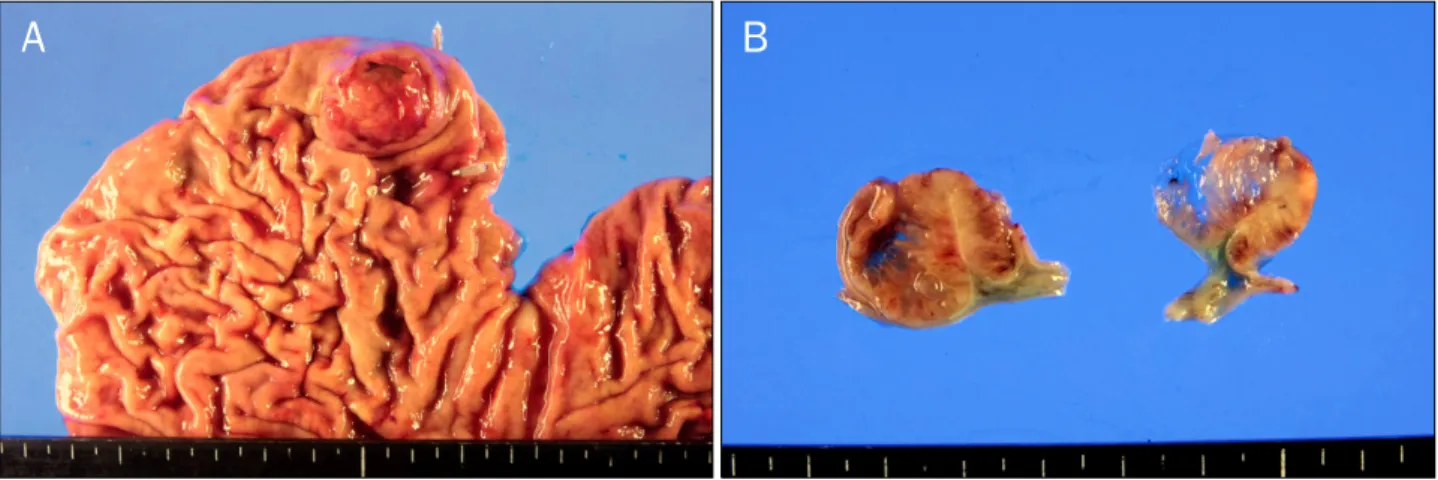
Biopsy specimens obtained from the stripped mucosa and the tumor surface showed a signet ring carcinoma with atyp- ical foveolar cells. A total gastrectomy with Roux-en-Y esoph- agojejunostomy was performed. The well-demarcated mass was within the fundus (Fig. 4A). On cross-sectional views, mu- cinous material was abundant (Fig. 4B). Histopathology of the tumor revealed a well-differentiated MGC with mucin
pools lining the tubular adenocarcinomatous epithelium as well as a mixed signet ring cell carcinoma. Tumor invasion was limited to the lamina propria (Fig. 5).
DISCUSSION
Adenocarcinoma of various organs sometimes produces a mucinous component. Mucinous adenocarcinoma occurs more frequently in the colorectum than in the stomach. The incidence of mucinous colorectal adenocarcinoma is 5.7% to 11.7% of colorectal cancers.1 According to the Japanese Research Society for Gastric Cancer, mucinous carcinoma is defined as a morphologic subtype of adenocarcinoma in which more than 50% of the tumor is composed of mucin.2 Another variant, signet ring cell carcinoma, also produces abundant mucin that is intracytoplasmic rather than extracytoplasmic.2 MGC is a very rarely found histologic subtype. The reported incidence of MGC among all gastric carcinomas is 3% to 5%.3,4 As it is typically diagnosed in ad- vanced stages, MGC that is restricted to the mucosa is very rare. The reported incidence of early MGC among early gas- tric carcinomas is only 0.3% to 1.0%.5
There are several hypotheses regarding the origin of MGCs. Mucinous carcinoma is thought by some to arise ini- tially as a typical adenocarcinoma, and later becomes muci- nous with tumor progression. This is consistent with the find- ing that intraluminal excretion of mucin decreases and intra- mural accumulation of mucin increases in accordance with tumor invasion through the gastric wall.3,5,6
124 유찬희 등. 점막하 종양의 양상을 보인 조기 점액성 위암
The Korean Journal of Gastroenterology
Fig. 4. Gross findings. (A) A well-demarcated fungating mass was present in the gastric fundus, measuring 25×23×20 mm in size. (B) In cross-sections of the tissue, the tumor was found to be confined to the mucosa and contained a large amount mucinous material.
Fig. 5. Microscopic findings (H&E). (A) Well-differentiated mucinous gastric carcinoma with mucin pools lining the tubular adenocarcinomatous epithelium (×40). (B) Mucinous gastric carcinoma confined to the mucosa was characterized by extracellular mucin in the lamina propria (×40).
Epithelial tumors in the stomach sometimes exhibit a mac- roscopic appearance similar to submucosal tumors. But an adenocarcinoma showing features of SMT is unusual.7
Although the incidence of early MGC is rare, that of early MGC resembling SMT on macroscopic appearance is not uncommon. Gross features include abundant extracellular mucin and expansive growth in the mucosal or submucosal layers.5
Adachi et al.3 conducted a clinicopathologic study of MGC.
They found that the biologic behavior of MGC was similar to that of nonmucinous gastric carcinoma (NGC) and that the prognosis was basically determined by the histologic sub- type, not by the mucin content. The tumor size, location, depth of invasion, lymph node metastasis, and stage at diag- nosis were all similar between the MGC and NGC. The out-
comes of patients with MGC were not less favorable com- pared with those of common gastric carcinoma. However, the lesions were mostly advanced carcinomas and rarely early carcinoma in MGC.3
When early stage mucinous and non-mucinous tumors were previously compared, tumor size, presence of lymph node metastasis, and patient outcomes were not sig- nificantly different. While the mucinous histologic subtype it- self was not an independent prognostic factor in patients with gastric carcinoma on multivariate analysis, the post- operative prognosis in early-MGC patients was excellent.
Poor outcomes in cases of MGC were typically due to later de- tection at an advanced stage rather than the mucinous histo- logic subtype.4-6
In this case, we diagnosed MGC at a very early stage. The
Yoo CH, et al. Submucosal Tumor-like Early-stage Mucinous Gastric Carcinoma 125
Vol. 62 No. 2, August 2013
tumor was characterized by macroscopic elevation resem- bling SMT with an opening allowing for the passage of copi- ous mucus discharge. This patient underwent total gas- trectomy with Roux-en-Y esophagojejunostomy, and now has a favorable prognosis based on previous analyses of out- comes in patients with this tumor subtype and stage at the time of detection.
REFERENCES
1. Kawamura H, Kondo Y, Osawa S, et al. A clinicopathologic study of mucinous adenocarcinoma of the stomach. Gastric Cancer 2001;4:83-86.
2. Fang WL, Wu CW, Lo SS, et al. Mucin-producing gastric cancer:
clinicopathological difference between signet ring cell carcino-
ma and mucinous carcinoma. Hepatogastroenterology 2009;
56:1227-1231.
3. Adachi Y, Mori M, Kido A, Shimono R, Maehara Y, Sugimachi K.
A clinicopathologic study of mucinous gastric carcinoma.
Cancer 1992;69:866-871.
4. Wu CY, Yeh HZ, Shih RT, Chen GH. A clinicopathologic study of mucinous gastric carcinoma including multivariate analysis.
Cancer 1998;83:1312-1318.
5. Yasuda K, Shiraishi N, Inomata M, Shiroshita H, Ishikawa K, Kitano S. Clinicopathologic characteristics of early-stage muci- nous gastric carcinoma. J Clin Gastroenterol 2004;38:507-511.
6. Adachi Y, Yasuda K, Inomata M, Shiraishi N, Kitano S, Sugimachi K. Clinicopathologic study of early-stage mucinous gastric carcinoma. Cancer 2001;91:698-703.
7. Kim JH, Jeon YC, Lee GW, et al. A case of mucinous gastric ad- enocarcinoma mimicking submucosal tumor. Korean J Gastro- enterol 2011;57:120-124.