I. 서론
혐기성 흑색세균의 하나인Prevotella intermedia 는 성인형 치주염이나1,2)ANUG3), 임신성 치은염4) 등과 같은 여러 가지 종류의 치주질환과 치수 및 치 근단 질환에서의 강력한 원인균 중 하나인 것으로 알려져 왔다. 그런데 흥미롭게도 이 세균은 건강한 치주를 가진 성인은 물론5)아직 치아가 나지 않은 어 린이의 구강 내에서도 발견되고 있으며6,7), 이러한 결과들은 이 세균종이 적어도 두 가지 이상의 이질 성을 가지고 있음을 시사하는 것이라고 생각할 수 있다.
Prevotella intermedia의 intraspecies heterogeneity 는 Johnson과 Holdeman의 연구에서부터 증명되어 지기 시작했는데, 이들은Prevotella intermedia가 두 가지의 DNA homology group을 가지고 있음을 처음 으로 밝혀냈고8), 이어서 Gmür와 Guggenheim 등은 monoclonal antibody를 이용하여Prevotella inter- media를 3개의 serogroup(I, II, III)으로 세분할 수 있 다고 하였으며9), Shah와 Gharbia는 multilocus enzyme electrophoresis를 이용하여Prevotella inter- media의 두 가지 homology group의 존재를 확인한 후 이들을 각각 Prevotella intermedia와 Prevotella
nigrescens로 세분할 것을 제안하였다10).
현재까지의 연구 결과를 종합해볼 때,Prevotella intermedia와Prevotella nigrescens는 DNA homolo- gy(homology group 4197은Prevotella intermedia를, 그 리 고 homology group 8944는 Prevotella nigrescens를 나타낸다.)8), serology(serogroup I은 Prevotella intermedia를, serogroup II와 III는 Prevotella nigrescens를 나타낸다.)9), multilocus enzyme electrophoresis10), sodium dodecyl sulfate- polyacrylamide gel electrophoresis(SDS-PAGE)에 의 한 protein profile11), 그리고 oligonucleotide probe12) 나 genomic DNA를 이용한 hybridization13,14) 등과 같은 여러 가지 방법을 통하여 분리될 수 있었으며, 최근에 도입된 Polymerase Chain Reaction(PCR) 방 법은 두 세균을 보다 분명하고도 신속하게 분리할 수 있게 해주었다.15,16)Prevotella intermedia를 나타 내는 type strain은 ATCC 25611이며 Prevotella nigrescens를 나타내는 type strain은 ATCC 33563이 다.
Prevotella intermedia와Prevotella nigrescens가 세 분된 이후 두 세균의 특징에 대한 다양한 연구 및 이 들 세균과 각종 치주질환과의 관계를 규명하기 위한 연구들이 활발하게 진행되어져 왔다. Gharbia 등은
한국인 치주 감염 환자에서의 Prevotella intermedia 와 Prevotella nigrescens 의 발현빈도
백승엽·구 영·류인철·함병도·한수부·최상묵·정종평 서울대학교 치과대학 치주과학교실
대한치주과학회지 : Vol. 30, No. 2, 2000
* 이 연구는 1997년도 서울대학교병원 대형공동연구비(03-1997-090-3) 지원에 의한 결과임
정상치주조직, 치주병소 및 근관병소로부터 분리 동 정 된 Prevotella intermedia를 다 시 세 분 하 여 Prevotella intermedia와Prevotella nigrescens의 비율 을 조 사 하 였 는 데 , 정 상 치 주 조 직 에 서 채 취 된 Prevotella intermedia의 대 부 분 은 Prevotella nigrescens였고 치주병소에서 채취된 Prevotella intermedia의 대부분은Prevotella intermedia였으며 (70.7%), 근관병소에서는 26.7%만이 Prevotella intermedia였다고 보고한바 있다.11)
이와는 약간 다른 결과를 나타내는 연구도 있었는 데, Teanpaisan 등은 치주염 환자의 염증부위와 건강 한 부위, 그리고 치주적으로 건강한 환자의 치은열구 에서의 흑색세균의 발현빈도를 조사한 결과, 건강한 환자에서의 결과는 앞에서 언급한 Gharbia 등의 연 구 결과와 유사하게Prevotella intermedia의 발현빈 도가 18%, Prevotella nigrenscens의 발현빈도가 31%
로 나타났으나, 치주염환자의 염증부위에서는 Prevotella nigrenscens의 발현빈도가 Prevotella intermedia보다 두 배 이상 더 높은 것으로 나타났으 며, 건강한 부위에서는Prevotella intermedia의 발현 빈도가 더 높은 것으로 나타났다.17)
본 연구에서는Prevotella intermedia와Prevotella
nigrescens의 감염치주조직 및 정상치주조직에서의
발현 빈도를 16S ribosomal DNA sequence에 기초한 Polymerase Chain Reaction(PCR) 방법을 이용하여 측정, 비교하고자 한다.
II. 실험 재료 및 방법
1. Sample 채취서울대병원에 내원한 환자들 중 최근 6개월 이내 에 항생제를 복용한 경험이 없으며 치주치료를 받지 않은 100명의 성인형치주염 환자 및 50명의 건강한 치주를 가진 환자를 대상으로 하였다. 이때 감염치 주낭은 5mm 이상의 pocket depth를 가지면서 탐침 시 출혈이 나타나는 부위로 한정하였다. sample을 채취하고자 하는 치아의 주위를 방습하고 치은 연상 치태 및 음식물 잔사를 완전히 제거한 후, 무균 처리 된 #35 paper point를 치은연하에 저항감이 느껴질 때까지 삽입한 후 30초간 기다렸다가 꺼낸 다음, 미 리 준비된 2ml의 VMGA III 용액에 담아서 10% H2,
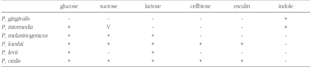
Table 1. Biochemical characteristics of BPB
glucose sucrose lactose cellbiose esculin indole
P. gingivalis - - - - - +
P. intermedia + V - - - +
P. melaninogenicus + + + - - -
P. loeshii + + + + + -
P. levii + - + - - -
P. oralis + + + + + -
Figure 1. PCR procedure 94°C, 5min 94°C, 1min
55°C, 1min 33 cycles
72°C, 1min 30s 72°C, 10min
4°C,
∞
10% CO2, 80% N2로 구성된 혐기성 배양기로 옮겼 다.
2. 균의 배양과 동정
배양기 내에서 단계적으로 10배씩 희석하여 1000 배의 희석액을 만든 후 이중 100㎕를 hemin과 menadione이 포함된 가토혈액한천배지에 접종하였 다. 37°C에서 10일간 배양하여 키워진 colony 중 black-pigmented bacteria를 선택하여 BHI 배양액에 서 키운 후 Gram 염색 및 biochemical test(Table 1) 를 통하여Prevotella intermedia로 판정된 균들을 액 체질소탱크에 보관하였다.
3. 중합효소연쇄반응
분리 배양된 균주 중 생화학적 검사에서Prevotella
intermedia로 동정된 세균에 한하여 중합효소연쇄반
응법을 이용하여Prevotella intermedia와 Prevotella nigrescens를 구 분 하 였 다 . 생 화 학 검 사 에 서 Prevotella intermedia로 동정된 균주를 BHI 배양액 에서 배양한 후 균을 원심분리하여 pellet을 모았다.
pellet에 5배의 guanidine 용액을 넣어 잘 섞은 후 동 량의 phenol과 chloroform을 넣어 DNA를 추출하였 으며, EtOH로 DNA를 침전시켜 순수한 DNA만을 분 리해내었다. 분리된 DNA로부터 70% EtOH를 이용 하여 염을 제거한 후 TE(pH 8.0)에 DNA를 녹여 4oC 에서 보관하였다.
DNA의 농도를 UV-spectrophotometer를 사용하여 측정한 후 중합효소연쇄반응을 실시하였다. 기존에 고안되어 있는 primer design18)을 이용하여 제작된 primer와 앞에서 추출했던 세균의 DNA를 AccuTM
Power Premix kit에 혼합한 후 PCR machine에서 그 림 1의 과정을 시행하였다. PCR을 통해 증폭된 DNA 를 ethidium bromide가 첨가된 1.0% agarose gel에 서 80V로 40분간 전기영동 시키고 UV transillumina- tor를 이용하여 각각의 band 위치를 확인하였다.
size marker로는 100bp DNA ladder(Life technolo- gies Co. U.S.A.)를 사용하였으며 polaroid camera (Seoulin scientific Co. LTD., Korea)로 촬영하였다.
III. 결과
감염 치주낭으로부터 채취한 총 100개의 sample 중 39개의 sample에서 BPB가 관찰되었으며 이 중 24개 가 Prevotella intermedia로 동 정 되 었 다 . Prevotella intermedia로 동정된 균주들을 중합효소 연쇄반응을 시킨 후 agarose gel 상에 나타난 결과를 관찰하였더니, 24종의 Prevotella intermedia 중 2종 의 균주가 Prevotella nigrescens의 primer와 반응을 하여 830bp에서 밴드를 나타내었고, 21종의 균주가 Prevotella intermedia 의 primer와 반응을 하여 240bp에서 밴드를 나타냈다. 그러나 나머지 1종의 균주는 두 종류의 primer 모두에서 밴드를 나타내지 않았다.
정상 치주조직으로부터 채취한 총 50개의 sample 중 9개의 sample에서 BPB가 관찰되었고 이 중 2개 가Prevotella intermedia로 동정되었으며, 이들을 중 합효소연쇄반응 시킨 결과 하나는 Prevotella inter- media이며 나머지 하나는Prevotella nigrescens인 것 으로 나타났다.
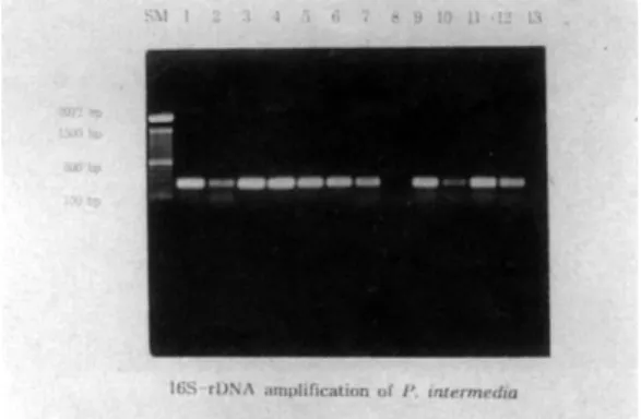
Figure 2와 3은 PCR 및 전기영동을 시행하고 나서 Prevotella intermedia와Prevotella nigrenscens로 분 리된 결과를 보여주는 사진이며, Table 2는 이번 실
Table 2. The frequency of P.intermedia and P.nigrescens in infected periodontal tissue and normal periodon- tal tissue
Total samples BPB detecting P.intermedia P.nigrescens samples detecting samples detecting samples
Infeted periodontal tissue 100 39 21 2
Normal periodontal tissue 50 9 1 1
험 결과를 요약하여 나타낸 것이다.
IV. 총괄 및 고찰
BPB는Actinobacillus actinomycetemcomitans와 더불어 여러 가지 종류의 치주질환의 주요 원인균인 것으로 알려져 왔으며, 이들에 대한 다양한 연구는 질병의 원인규명과 치료법 개발에 있어서 필수적인 과정이라 할 수 있다.
Bacteroides는 화학적 및 생화학적 방법과 가장 최 근의 유전학적 방법 등에 이르기까지 새로운 분류방 법이 개발됨에 따라 많은 분류학적 변화를 거치게 되었고, 현재까지는 cytophaga-flavobacter-bac-
teroides(CFB) phylum의 subgroup으로 약 30여종이 발견되었으며, 앞으로도 새로운 종들이 계속 발견될 가능성이 있는 것으로 보인다. 이전까지 BPB는 genus Bacteroides 내에서 분류되었으나 Shah와 Collins는 genus Bacteroides를 다음과 같은 3가지 genera로 나눌 것을 제안하였다; 1)saccharolytic, non-pigmenting species로 구성되는 Bacteroides (e.g. B. fragilis), 2)asaccharolytic, black- pigmenting species로 구성되는 Porphyromonas (e.g. P. gingi- valis), 3)saccharolytic, black-pigmenting species로 구성되는 Prevotella (e.g. P. intermedia)19). 모든 Porphyromonas species와 6종류의 Prevotella species는 protoporphyrin 및 protoheme으로 이루어 지는 갈색 혹은 흑색 pigment를 만들어낸다.20)
Prevotella intermedia는 gram-negative, obligately anaerobic rod로서 치주질환이나 다양한 구강감염조 직에서 흔하게 검출되는 강력한 병원균인 것으로 알 려져 왔다. 그러나, Actinobacillus actinomycetem- comitans나 Porphyromonas gingivalis와 달리 Prevotella intermedia는 염증조직뿐만 아니라 건강 한 조직에서도 자주 발견되는바, 기회감염성 병원균 인 것으로 받아들여지고 있고, 다른 병원균들의 존재 와 밀접한 상관관계는 특히 긴밀하여Fusobacterium nucleatum이 발견되지 않는 곳에서는 Prevotella intermedia도 발견되지 않는다는 보고도 있었다.21)
Prevotella nigrescens는 1992년 Shah 와 Gharbia의 연구에 의해Prevotella intermedia로부터 독립 분류 되었다. Prevotella nigrescens는 gram-negative, non- sporing, obligately anaerobic, rod 모양의 세균이다.
Broth culture상에서 0.3 내지 0.7㎛의 폭에 1 내지 2
㎛ 깊이까지 자란다. Blood agar 상에 나타나는 3일 후의 surface colony는 0.5 내지 2mm의 직경에 원형 이고 low convex하며 갈색 내지 흑색을 나타낸다.
Pigmentation은 colony의 주변부에서 주로 나타나고 중심부는 크림색 내지는 암갈색을 띤다.
Glucose가 함유된 broth에서의 주요한 효소산물 은 acetic, isobutyric, isovaletic, 그리고 succinic acid 등이다. Dextran과 glucose, maltose, sucrose의 발효 는 대부분의 strain에서 나타나나 fructose나 glyco- Figure 2. 16S-rDNA amplification of Prevotella
intermedia
Figure 3. 16S-rDNA amplification of Prevotella nigrescens
gen, insulin의 발효는 편차가 크다.10)
Prevotella intermedia의 독성은 Porphyromonas
gingivalis의 독성에 미치지는 못하지만 독성인자인
것으로 여겨지는 몇 가지의 표면구조(surface prop- erty)를 가지고 있는데, lipopolysaccharide, capsule, 그리고 부착을 매개하는 surface appendage 등이 그 것이다. 그러나Prevotella nigrescens에 대한 연구는 아직 많이 이루어지지 않아Prevotella intermedia와 Prevotella nigrescens의 차이에 대하여 분명하게 밝 혀진 것은 아직 없는 형편이다. Prevotella interme- dia의 surface appendage가 몇 가지 다른 type으로 세분될 수 있다는 보고가 있었고22), negative staining 을 시행한 결과Prevotella intermedia에서는 발견되 지 않 는 fibriae-like projection을 Prevotella nigrescens에서는 관찰할 수 있었으며 EM 상에서는 Prevotella nigrescens의 capsular layer가 Prevotella
intermedia보다 덜 돌출되어 보인다는 연구 결과도
발표되었다11). 그러나 이 정도만으로는 Prevotella intermedia와Prevotella nigrescens의 차이를 설명하 기는 어려우며 따라서 이 부분에 있어서 앞으로 지 속적인 연구가 뒷받침되어져야 할 것이다.
Prevotella intermedia와 Prevotella nigrescens의 transmission과 관련된 연구들도 많이 이루어졌는데, 성인과 성인, 혹은 성인과 어린이 사이에서는 이들 세균이 쉽게 전달될 수 있지만, 가족의 범위를 넘어 서는 transmission은 나타나지 않는 것으로 알려지고 있다23,24).
Prevotella intermedia와 Prevotella nigrescens의 site specificity는 그 동안의 많은 연구들에 의해 그 윤곽이 드러나고 있는데, 1994년 Gharbia 등은 정상 치주조직과 치주병소 및 근관병소에서의Prevotella intermedia와Prevotella nigrescens의 분포를 조사한 결과 정상치주조직과 근관병소에서는 Prevotella
nigrescens가 다수를 차지하며 치주병소에서는
Prevotella intermedia가 다수를 차지한다고(70.7%) 보고하였고11), 1997년 배 등은 근관감염부로부터 Prevotella intermedia와Prevotella nigrescens의 발현 빈도를 조사한 결과 대부분이Prevotella nigrescens 였다고 보고함으로써(73.2%) 앞의 실험의 결과를 뒷
받침해주었다25). 반면 1995년 Teanpaisan 등이 치주 조직에서 검출된 BPB중 Prevotella intermedia와 Prevotella nigrescens의 비율을 조사한 결과, 건강한 성인에서는 Prevotella nigrescens의 비율이 월등히 높게 나타났으나 성인형치주염 환자의 염증부위 및 건강한 부위에서는 각각 Prevotella nigrescens와 Prevotella intermedia의 비율이 더 높은 것으로 나타 남으로써17), 치주조직에서Prevotella intermedia와 Prevotella nigrescens의 site specificity는 아직까지 이론의 여지가 남아있는 것으로 여겨진다.11,17,26,27-29)
이번 실험에서 치주염환자로부터 채취한 총 100 개의 sample 중 39개에서 BPB가 검출되었으며 생화 학적 검사 결과 이들 중 24개가Prevotella intermedia 인 것으로 확인되었고, 이 균주들을 16s 라이보좀 유 전자 DNA 중합효소연쇄반응 증폭법을 이용하여 분 리 동정한 결과 24개의 sample 중 21개가Prevotella intermedia이며 2개가 Prevotella nigrescens인 것으 로 나타났으며, 나머지 1개의 sample은 생화학적 검 사 과정에서 나타난 오류였던 것으로 추정된다. 이 러한 결과는 앞에서 언급된 바 있던 Gharbia 등의 연 구 결과와 유사한 것이다11). 하지만 이러한 결과를 통 해 , 치 주 질 환 의 진 행 에 있 어 서 Prevotella nigrescens보다Prevotella intermedia가 더 중요한 원 인균으로 작용하는 것이라는 결론을 도출해내기에 는 약간의 무리가 있는 것으로 보인다. Teanpaisan 등의 연구 결과 성인형 치주염 환자의 염증활성화 부위에서는Prevotella nigrescens가 Prevotella inter- media보다 훨씬 많이 검출되었는데(37.7% : 15.5%), 이때 염증활성화 부위를 홍반이나 농양을 수반하는 경우로 제한하였던 것을 고려한다면, 강력한 병원균 인 Porphyromonas gingivalis와는 달리 Prevotella intermedia나Prevotella nigrescens는 주변 환경의 변 화에 따라 그 발현 빈도와 역할이 달라질 수 있는 기 회감염성병원균으로 작용하고 있기 때문인 것으로 생각되어진다17).
건강한 치주조직으로부터 채취된 총 50개의 sam- ple 중에서 Prevotella intermedia와 Prevotella nigrescens가 겨우 1개씩만 검출된 것은 예상하지 못 했던 결과이다. 단순히 인종간의 차이나 실험과정
상의 오류 때문이라고 보기에는 이전의 결과들과 너 무 많은 차이가 나기 때문이다. 그러나 이번 실험에 서 적용된 엄격한 기준을 고려한다면, 어느 정도는 예상 가능한 결과라고 할 수도 있을 것이다. 즉, 건강 한 치주를 가진 성인을 선정함에 있어서, 가족간에 흔히 나타날 수 있는 transmission에 의한 오류를 없 애고 false positive effect가 나타날 가능성을 낮추기 위하여, 같이 살고 있는 가족들 중에 잇몸질환을 앓 고 있는 사람이 없으면서 전 치아에 걸쳐서 건강한 치주조직을 가지고 있는 사람만을 대상으로 하였는 데, 이러한 조건들은 결국 이 세균들의 발현빈도를 현저하게 낮게 나타나도록 만들 수밖에 없으리라고 생각한다.
한편 전체적으로 BPB의 검출빈도는 다른 많은 연 구들과 비교할 때 상당히 낮은 수준인 것으로 나타 났는데 이는 치주질환 원인균의 인종간 차이에 의한 결과인 것으로 생각된다. Actinobacillus actino- mycetemcomitans균의 백혈구 독성 인자 중 강한 독 성인자를 생산하는 JP-2 like gene의 발생빈도가 인 종에 따라서 많은 차이를 나타낸다는 연구결과30)를 원용하여 생각해본다면, 앞으로 Prevotella interme- dia와Prevotella nigrescens의 인종간 차이에 대해서 도 다양한 연구가 이루어져야 할 것이다.
Prevotella intermedia와 Prevotella nigrescens는 multilocus enzyme electrophoresis나 monoclonal antibody, protein profile 등을 이용하여 분리, 동정 될 수 있으나 이들 중 두 종을 분간할 수 있는 표현형 검사방법은 없으며, 시간이 지나치게 오래 걸리고 그 결과를 판독하기가 어렵다는 단점을 가지고 있다.
그러나 1991년 Gurtler 등이 도입한 16s 라이보좀 유 전자 DNA의 중합효소연쇄반응 증폭을 이용한 세균 동 정 법 은 Prevotella intermedia와 Prevotella
nigrescens를 분리하는 데 있어서 신속하면서도 확
실한 방법으로 인정받고 있다15,16). 본 실험에서도 중 합효소연쇄반응법을 통하여 두 가지 세균 종을 매우 명확하게 구분할 수 있었다.
한국인을 대상으로 치주낭에서의Prevotella inter- media와Prevotella nigrescens의 발현빈도를 조사하 는 것은 이번 실험이 처음이었는데, 인종적 특이성이
나 식생활 및 문화적 차이 등에도 불구하고 서구인 들을 대상으로 이루어졌던 실험결과와 크게 다르지 는 않은 것으로 나타났으나, 통계적 유의성을 비교하 기에는 아직까지 자료가 충분하지 않은 실정이다.
Prevotella intermedia와 Prevotella nigrescens가 site specificity 라는 특성을 가지고 있는 것으로 밝혀 져 있는 가운데, 더 나아가서 여러 가지 종류의 치주 질환간에, 그리고 동일한 치주질환의 심도별 차이에 따르는 두 세균의 발현빈도를 비교하는 것은 두 세 균의 정체성을 파악하고 각종 치주질환에서의 역할 을 규명하는 데에 있어서 중요한 정보를 제공하는 단초가 될 수 있을 것이다.
또한 감염근관 내에서는 Prevotella nigrescens가 Prevotella intermedia보다 월등하게 높은 비율로 검 출된다는 여러 연구들의 결과를 고려해 볼 때, 근관- 치주 복합감염병소에서 두 세균의 발현 빈도를 비교 해 보는 것도 그 병소의 기원을 밝히는데 도움이 될 수 있으리라는 가정 하에 매우 의미 있는 연구가 될 수 있으리라 생각한다.
V. 결론
본 실험에서는 한국인의 감염치주조직과 정상치 주조직에서의 Prevotella intermedia와 Prevotella nigrescens의 발현빈도를 조사하기 위하여, 최근 6개 월 이내에 항생제를 복용한 경험이 없으며 치주치료 를 받은 경험이 없는 성인형치주염 환자 및 건강한 치주를 가진 환자를 대상으로 paper point를 이용하 여 sample을 채취하였다. 여기에서 채취된 세균들을 혐기성 배양기에서 배양하였고 이들 중 biochemical test 결과 Prevotella intermedia로 동정된 세균들을 중합효소연쇄반응 시킴으로써Prevotella intermedia 와 Prevotella nigrescens를 분리, 동정하였다. 이와 같은 과정을 통하여 한국인의 감염치주조직과 정상 치주조직에서의 Prevotella intermedia와 Prevotella
nigrescens의 발현빈도에 관한 다음과 같은 결론을
얻었다.
1. 본 실험의 결과, 100개의 치주감염부로부터 채취 한 100개의 sample 중 39개 sample (39%)에서
BPB가 동정되었다.
2. 본 실험의 결과, BPB가 동정된 39개 sample 중 24개의 sample이Prevotella intermedia로 동정되 었다.
3. 본 실험의 결과, Prevotella intermedia로 동정된 24개의 sample 중 21개는 Prevotella interme- dia(87.5%), 2개는 Prevotella nigrescens인 것 (8.33%)으로 판명되었다.
4. 본 실험의 결과, 50개의 정상치주조직으로부터 채취한 50개의 sample 중 9개 sample에서 BPB 가 동 정 되 었 으 며 , 이 들 중 1개 sample이 Prevotella intermedia, 1개 sample이Prevotella nigrescens인 것으로 판명되었다.
VI. 참고문헌
1. Slots J, Bargd L, Wikstrom M, Dahlen G. The occurrence of Actinobacillus actinomycetem- comitans, Bacteroides gingivalis and Bacteroides intermediusin destructive periodontal disease in adults. J Clin Periodontol 1986;13:570-577 2. Wennstrom JL, Dahlen G, Svensson J, Nyman S.
Actinobacillus actinomycetemcomitans, Bacteroides gingivalis and Bacteroides inter- medius: predictors of attachment loss? Oral Microbiol Immunol 1987;2:158-163
3. Loesche WJ, Syed SA, Laughon BE, Stoll J. The bacteriology of acute necrotizing ulcerative gin- givitis. J Periodontol 1982;53:223-230
4. Kornman KS, Loesche WJ. The subgingival microbial flora during pregnancy. J Periodont Res 1980;15:111-122
5. Kononen E. Pigmented Prevotellaspecies in the periodontally healthy oral cavity. FEMS Immunol Med Microbiol 1993;6:201-206
6. Frisken KW, Higgins T, Palmer JM. The inci- dence of periodontopathic microorganism in young children. Oral Microbiol Immunol 1990;5:43-45
7. Kononen E, Asikainen S, Saarela M, Karjalainen J, Jousimies-Somer H. The oral gram-negative anaerobic microflora in young children: longitu- dinal changes from edentulous to dentate mouth. Oral Microbiol Immunol 1994;9:136-141 8. Johnson JL, Holdeman LV. Bacteroides inter-
medius comb. nov. and descriptions of Bacteroides corporis sp. nov. and Bacteoides leviisp. nov. Int J Syst Bacteriol 1983;33: 15-25 9. Gmur R, Guggenheim B. Antigenic geterogene-
ity of Bacteroides intermediusas recognized by monoclonal antibodies. Infect Immun 1983;42:459-470
10. Shah HN, Gharbia SE. Biochemical and chemi- cal studies on strains designated Prevotella inter- media and proposal of a new pigmented species, Prevotella nigrescenssp. nov. Int J Syst Bacteriol 1992;42:542-546
11. Gharbia SE, Haapasalo M, Shah HN et al.
Characterization of Prevotella intermediaand Prevotella nigrescensisolates form periodontic and endodontic infections. J Periodontol 1994;65:56-61
12. Dix K, Watanabe SM. et al. Species-specific oligodeoxynucleotide probes for the identifica- tion of periodontal bacteria. J Clin Microbiol 1990;28:319-323
13. Fukushima H, Moroi H, Inoue J et al.
Phenotypic characteristics and DNA relatedness in Prevotella intermediaand similar organism.
Oral Microbiol Immunol 1992;7:60-64
14. Moncla BJ, Strockbine L, Braham P, Karlinsey J, Roberts MC. The use of whole-cell DNA probes for the identification of Bacteroides intermedius isolates in a dot blot assay. J Dent Res 1988;67:1267-1270
15. Gurtler V, Wilson VA, Mayall BV. Classification of medically important clostridia using restriction endonuclease site differences of PCR-amplified
16S rDNA. J Gen Microbiol 1991;137:2673-2679 16. Susan E. Milsom, Susan V. Sprague, D.
Dymock. Rapid differentiation of Prevotella intermedia and Prevotella nigrescens by 16S rDNA PCR-RFLP. J Med Microbiol 1996;44: 41-43 17. Teanpaisan R, Douglas CWI, Walsh TF.
Characterization of black-pigmented anaerobes isolated from diseased and healthy periodontal sites. J Periodont Res 1995;30:245-251
18. Bae KS. Baumgartner JC. Watkins BJ, Tian X.
Association of black-pigmented bacteria with endodontic infections. Unpublished.
19. Shah HN, Collins DM. Prevotella, a new genus to include to Bacteroides melaninogenicusand related species formerly classified in the genus Bacteroides. Int J Syst Bacteriol 1990;40:205-208 20. Van Winkelhoff AJ. Black-pigmented
Bacteroides in human oral diseases. Amsterdam:
Free University Press. 1986
21. Raouf Wahab Ali, Nils Skaug, Rune Nilsen.
Microbial associations of 4 putative periodontal pathogens in Sudanese adult periodontitis patients determined by DNA probe analysis. J Periodontol 1994;65:1053-1057
22. Leung KP, Fukushima H, Sagawa H, Walker CB, Clark WB. Surface appendages, hemagglutina- tion, and adherence to human epithelial cells of Bacteroidis intermedius. Oral Microbiol Immunol 1989;4:204-210
23. Matto J, Saarela M, Jousimies-Somer H, Torkko H, Asikainen S. Distribution and genetic analysis of oral Prevotella intermedia and Prevotella nigrescens.Oral Microbiol Immunol 1996;11:96- 102
24. Van Steenbergen TJM, Basch-Tijihof CJ, Petit MDA, Van de Velden. Intra-familial transmission and distribution of Prevotella intermediaand Prevotella nigrescens. J Periodont Res 1997;32:345-350
25. Bae KS, Baumgertner JC. Occurrence of Prevotella nigrescensand Prevotella intermedia in infections of endodontic origin. J Endod.
1997;23:620-623
26. Gmur R, Guggenheim B. Interdental supragingi- val plaque-a natural habitat of Actinobacillus actinomycetemcomitans, Bacteroides forsythus, Campylobacter rectus, and Prevotella nigrescens. J Dent Res 1994;73:1421-1428 27. Dahlen G, Wikstrom M, Renvert S, Gmur R,
Guggenheim B. Biochemical and serological characterization of Bacteroides intermedius strains isolated from the deep periodontal pock- et. J Clin Microbiol 1990;28:2269-2274
28. Moore LVH, Moore WEC, Cato et al.
Bacteriology of human gingivitis. J Dent Res 1987;66:989-995
29. Moore WEC, Moore LH, Ranney RR, Smibert RM, Burmeister JA, Schenkein HA. The microflora of periodontal sites showing active destructive pro- gression. J Clin Periodontol 1991;18:729-739 30. Haubek D, Dirienzo JM, Tinoco EMB,
Westergaard J, Lopez NJ, Chung CP, Poulsen K, Kilian M. Racial tropism of highly toxic clone of Actinobacillus actinomycetemcomitansassociat- ed with juvenile periodontitis. J Clin Microbiol 1997;35:3037-3042
-Abstract-
The Frequency of Detecting Prevotella intermedia and Prevotella nigrescens in Korean Adult Periodontitis Patients
Seung-Yup Peck, Young Ku, In-Cheol Rhyu, Byung-Do Hahm Soo-Boo Han, Sang-Mook Choi, Chong-Pyoung Chung
Department of Periodontology, College of Dentistry, Seoul National University
Prevotella intermediahas been implicated as a potent pathogen in many kinds of periodontal, pulpal and periapical diseases. However, it has been isolated from periodontally healthy adults and from edentulous chil- dren as well.
The intraspecies heterogeneity of Prevotella intermediahas been demonstrated in early studies and finally Shah & Gharbia confirmed the existence of 2 DNA homology groups and proposed dividing Prevotella inter- mediainto 2 species, Prevotella intermediaand Prevotella nigrescens.
This study was designed to examine the frequency of Prevotella intermediaand Prevotella nigrescensin dis- eased periodontal pockets and healthy gingival sulcus of Korean people by PCR based on 16s ribosomal DNA sequence.
One hundred adults who had adult periodontitis but not taken any periodontal treatment or antibiotics dur- ing previous 6 months and 50 adults who had healthy periodontal tissue were selected for this study.
The sulcular fluid was collected into VMGA by sterilized paper point and diluted to 1,000 times in anaerobic chamber. 100㎕ of sample was cultured in 37℃ for 10 days. Among the bacterial colonies, BPB were selected and cultured in BHI broth and then Prevotella intermediawas identified through Gram staining and biochemi- cal test. Identified Prevotella intermediawas cultured again and centrifuged. DNA was extracted from the pellet using several reagents. PCR was performed by previously designed primer.
The results were followed.
1. BPB were isolated from 39 of 100 samples of diseased periodontal pockets(39%).
2.Prevotella intermediawas identified from 24 of 39 BPB samples.
3. Among 24 Prevotella intermedia, 21 were confirmed as Prevotella intermedia(87.5) and 2 were confirmed as Prevotella nigrescens(8.33%).
4. BPB were isolated from 9 of 50 samples of periodontally healthy patients. Among them only two were identified as Prevotella intermedia, that is, one was confirmed as Prevotella intermediaand the other was Prevotella nigrescens.
Key words : Prevotella intermedia, Prevotella nigrescens, Polymerase chain reaction