CASE REPORT
급성 위확장증으로 발현된 신경성 식욕부진증 환자에서 발생한 위 기종 1예
김태윤, 김흥업, 송현주
제주대학교 의학전문대학원 내과학교실
A Case of Gastric Emphysema in Anorexia Nervosa Presenting as Acute Gastric Distension
Taeyun Kim, Heung Up Kim and Hyun Joo Song
Department of Internal Medicine, Jeju National University School of Medicine, Jeju, Korea
Gas within the gastric wall is an alarming finding and a rare condition. Clinically, this condition is divided into two entities;
Gastric emphysema and emphysematous gastritis. These two diseases should be differentiated because they are characterized by different clinical symptoms, possible etiology, treatment and prognosis. While emphysematous gastritis is a severe condition with high mortality, gastric emphysema is asymptomatic and usually has benign course. Rarely, anorexia nervosa and bulimia nervosa have been discribed to be associated with acute gastric distension and duodenal obstruction induced by superior mesentery artery syndrome. So, gastric emphysema could be accompanied by acute gastric distension induced by anorexia nervosa. We report a rare case of gastric emphysema in a patient with anorexia nervosa presenting as superior mesenteric artery syndrome with relevant literatures. In this case, the gastric emphysema was improved without surgical intervention after nasogastric tube for decompression and feeding insertion in the fourth portion of the duodenum. (Korean J Gastroenterol 2012;60:315-319)
Key Words: Emphysema; Emphysematous gastritis; Anorexia nervosa; Superior mesenteric artery syndrome; Gastric dilatation
Received August 24, 2011. Revised September 27, 2011. Accepted September 28, 2011.
CC This is an open access article distributed under the terms of the Creative Commons Attribution Non-Commercial License (http://creativecommons.org/licenses/
by-nc/3.0) which permits unrestricted non-commercial use, distribution, and reproduction in any medium, provided the original work is properly cited.
교신저자: 김흥업, 690-767, 제주시 아란13길 15, 제주대학교병원 내과
Correspondence to: Heung Up Kim, Department of Internal Medicine, Jeju National University Hospital, 15 Aran 13-gil, Jeju 690-767, Korea. Tel: +82-64-717-1363, Fax: +82-64-717-1131, E-mail: [email protected]
Financial support: None. Conflict of interest: None.
서 론
위벽 내에 공기가 존재하는 질환은 매우 드문 질환으로 원 인은 크게 비 감염성 요인인 위기종(gastric emphysema)과 세균 감염성 요인인 기종성 위염(emphysematous gastritis) 으로 나눌 수 있다.1,2 이들 질환은 복부 전산화단층촬영에서 위벽 내에 공기가 있는 것을 확인하면 진단할 수 있다. 이 중 기종성 위염은 가스형성 세균에 의해 발생하는 급성위염을 말 하며 주로 알코올 중독, 당뇨, 산과 알칼리 등의 부식제를 복 용한 경우에 동반될 수 있다.1,3 이에 비해 위기종은 압력손상, 폐색 등의 원인에 의해 위벽에 공기가 들어간 것인데, 감염과
는 연관이 없고 증상이 없는 경우가 많으며 특별한 치료 없이 회복되고 예후도 더 좋다.4,5 저자들은 심한 급성 위 팽창을 보이는 17세 여자에서 입원 후 상장간막동맥 증후군(superior mesenteric artery syndrome)에 의한 십이지장 제 3부위 부 분폐쇄를 확인하였다. 이후 위 팽창의 유발요인으로 신경성 식욕부진증(anorexia nervosa)을 진단하였다. 위 팽창에 의해 발생한 위기종이 항생제치료와 수술적 처치 없이 비위관 (nasogastric tube)을 통해 감압 이후 십이지장 제3부위까지 삽입시키고 경관식이하면서 보존적 치료만으로 호전된 증례 를 경험하여 이를 문헌고찰과 함께 보고한다.
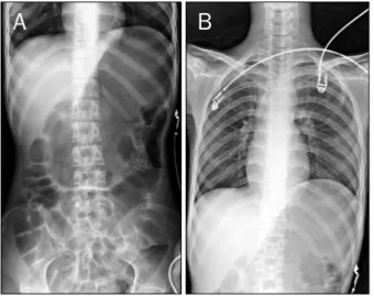
Fig. 1. (A) Severe gastric gaseous distention was noted on plain abdominal radiograph. (B) Consequently, abdominal left hemidi- aphragmatic elevation was visualized on plain chest radiograph.
Fig. 2. Contrast media enhanced abdominal computed tomography. (A) Multiple air (white arrows) densities were seen in the distended gastric wall. (B) Coronal image showed multi- ple air (white arrow) densities in the gastri wall. The second portion of the duodenum was dilated (white blank arrow). (C) Severe narrowing the space (white arrow head) between the aorta and superior mesenteric artery and markedly dilated second portion (white blank arrow) were also seen.
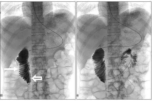
Nasogastric tube was inserted to decompress the distended stomach.
증 례
17세 여자가 속쓰림과 심한 구토 없이 구역질이 지속되어 외부 병원을 방문하였다. 의도적으로 식사 후 구토를 유발하 지는 않았으며 음식물을 삼키는 데도 문제는 없었다고 했다.
외부 병원에서 시행한 복부 단순촬영에서 심한 위 팽창이 보 였다. 복부 전산화단층촬영에서 심한 위 팽창에 의해 발생한 위기종으로 진단받고 그 원인으로 신경성 식욕부진증 및 상장 간막동맥 증후군을 의심하였다. 응급 수술을 권유받았으나 타 병원 자문을 원해 본원으로 전원되었다. 출생 및 발달 과정에
하였다고 한다. 과거력에서 결핵, 감염, 고혈압, 당뇨의 과거 력은 없었고 약물 복용이나 흡연, 음주는 하지 않았으며 가족 력에서도 특이사항 없었다. 하지만 환자는 내원 약 2년 전부 터 평소 체중 39 kg에서, 6개월 전 35 kg까지 감소하다가 최근 들어 32 kg까지 감소하였다. 내원 당시 키 157.8 cm, 몸무게 29.8 kg, 체질량지수(body mass index) 11.9 kg/m2 로 심한 저체중소견을 보였다. 진찰소견에서 혈압은 101/63 mmHg, 심박수 94회/분, 호흡수 16회/분, 체온 36.8oC였고, 의식은 명료하였으며 만성 병색을 보였다. 흉부 청진에서 호 흡음과 심음은 정상이었다. 복부 신체검사에서 복부는 팽만되 어 있었고 타진에서 공명음이 들렸으나 압통이나 반발통은 없 었다.
말초혈액검사에서 혈색소 12.6 g/dL, 백혈구 수 4,300/mm3, 혈소판 319,000/mm3, 생화학검사에서 혈청 총 단백질과 알부 민은 각각 6.1 g/dL, 3.6 g/dL, AST 31 IU/L, ALT 31/53 IU/L, ALP 214 U/L, 총 빌리루빈 0.3 mg/dL, 혈액요소질소 7.2 mg/dL, 크레아티닌 0.5 mg/dL, Na 140 mmol/L, K 4.1 mmol/L, Cl 105 mmol/L, CRP 0.4 mg/dL, 총 콜레스테롤 134 mg/dL, 고밀도 콜레스테롤 75 mg/dL, 저밀도 콜레스테 롤 49 mg/dL, 혈당은 98 mg/dL이었다. 혈액응고검사는 정 상이었고 소변검사에서도 이상 소견은 없었다. 복부 단순촬영 에서도 위가 요추 4번까지 심하게 팽창된 소견을 보였다(Fig.
1A). 흉부 단순촬영에서 폐 실질에는 이상이 없었으나 위가 심하게 팽창되어 좌측 횡경막이 우측 횡경막보다 상승된 소견 을 보였다(Fig. 1B). 원인 규명을 위해 복부 전산화단층촬영을 시행한 결과 공기와 액체로 위가 심하게 팽만되고 위 체부
Fig. 4. Upper gastrointestinal series on the hospital 27 days after nasogastric tube removal showed no delayed passage of barium on the third portion of the duodenum.
Fig. 3. Upper gastrointestinal series.
The contrast material-filled duodenal lumen (white arrow) showed abrupt vertical cut-off (white blank arrow) in front of the lumbar vertebrae due to superior mesenteric artery syndrome.
후벽의 공기음영이 전정부까지 위벽을 따라 산재된 소견이 보 였고(Fig. 2A, B) 십이지장의 제3부위가 상장간막동맥과 대동 맥 사이에서 눌려 있어 상장간막동맥 증후군을 의심하였다 (Fig. 2C). 위벽 내 공기 음영은 발열이나 감염을 시사하는 혈액검사 소견이 없어 위 기종으로 진단하고, 비위관으로 감 압 후 복부 단순촬영으로 감압 여부를 확인하였다(Fig. 3). 이 후 입원 2일째 성공적인 감압을 통해 위 팽창이 호전된 상태 를 복부 단순촬영을 통하여 확인하였고 체중 증가를 통한 상 간장막동맥 증후군의 호전을 위해 비경구적 영양법(parenter- al nutrition)을 병행하면서 비공장관을 통하여 400 kcal을 하
루 3번에 나눠 치료적 경관식이를 진행하였다. 입원 당시 감 염 증거가 적어 항생제를 투여하지는 않았다. 입원 5일째까지 비경구영양법을 병행하면서 서서히 하루 1,200 kcal목표로 증가하였으며 설사 등의 증상은 없었다. 입원 6일째 체중은 30.6 kg, 체질량지수12.4 kg/m2으로 상승되었으며 정상 대변 을 보며 걸어 다닐 정도로 전신상태가 호전되었다. 입원 21일 째 체중은 33.0 kg, 체질량지수 13.4 kg/m2로 지속적으로 증 가하였으며 상부위장관 조영술에서 상장간막동맥에 의해 눌 렸던 십이지장 제3부위의 십이지장 폐쇄부의 지연성 통과가 호전되었다(Fig. 4). 이후 입원 27일째 체중은 38.6 kg, 체질 량지수 15.7 kg/m2으로 상승되었으며 전신상태 호전 지속되 었다. 정상 대변양상을 확인 후 비공장관을 제거하였고, 구강 식이가 진행 가능하고 다른 소화기계 증상을 호소하지 않아 신경성 식욕부진증을 치료하기 위해 신경정신과로 전과되었 다. 이후 환자는 체중은 43.9kg, 체질량지수 17.6 kg/m2으로 지속적으로 체중증가 소견을 보이며 더 이상 구역, 구토 등 소화기계 증상 호소 없이 신경성 식욕부진증 치료를 위해 현 재 신경정신과 외래 추적 관찰 중이다.
고 찰
위벽 내에 공기가 존재하는 질환은 매우 드문 질환으로 원 인은 크게 비 감염성 원인인 위기종과 세균감염성 원인인 기 종성 위염으로 나눌 수 있다.1,2 비 감염성 원인인 위 기종은 위벽에 공기가 들어간 것인데, 주로 압력손상, 폐색 등의 원인 에 의해 생기며, 대체로 증상이 없는 경우가 많아 특별한 치료 없이 회복되고 예후도 좋다. 감염 원인인 기종성 위염 또한
로 미생물 감염에 의해 발생되며, 위벽 내에 가스형성 세균 (Escheria coli, Enterobacter species, Clostridium welchii, Staphylococcus aureus, C. perfringens, Pseudomonas ae- ruginosa) 등이 공기를 만들어내어 위벽에 공기가 존재하게 된다.6,7 위벽은 내강 쪽이 강한 산성이며, 풍부한 혈액 공급과 위 내부의 정맥물질의 방어효과가 견고하기 때문에 감염이 매 우 드문 장기 중에 하나이다.6,8,9 기종성 위염을 일으키는 원 인 중 가장 많은 것은 산이나 알칼리 등의 부식성 물질 섭취, 알코올 남용이었으나, 1990년 이후에는 당뇨, 투석환자, 면역 억제제를 사용한 환자에서 많이 발병한다고 보고되고 있다.6 기종성 위염은 1889년 처음으로 임상적인 기술을 하였고 1946년 최초로 방사선적 진단을 한 이후 수십 예가 보고되었 다.10 최근까지도 사망률이 60%까지 높은 질환이며 회복된 예 의 절반 이상에서 수술이 필요할 정도로 급성악화를 보이고 25%에서 회복 후 위 위축 등의 합병증이 동반되는 것으로 알려져 있다.4 따라서 진단과 함께 적극적인 항생제 투여와 전신 감염 상태를 안정화시키기 위한 다량의 수액요법, 그리 고 쇽 상태의 회복을 위한 치료가 필요하다.11
이에 비하여 위 기종은 위 폐쇄나 십이지장 폐쇄에 의해 위가 팽창되고 위 내압이 높아져 공기가 약해진 일부 위벽을 통해 위벽으로 침투하는 경우, 위벽에 갑작스러운 충격 또는 위장조루술(gastrostomy) 후 위 점막 결손부로 공기가 침투 하는 경우가 있다.1,3 이번 증례는 약 2년간의 지속적인 체중 감소를 보인 젊은 여자가 최근 들어 구역질을 호소하여 내원 후 신경성 식욕부진증으로 진단받은 경우이다. 신경성 식욕부 진증은 정신과 질환인 동시에 여러 장기에 영향을 미칠 수 있는 전신적인 질환이다. 신경성 식욕부진증 환자의 약 60%
에서 갑작스런 위 팽창같은 위 운동장애(gastric dysmotility) 를 보이는데 위 운동(gastric motility)과 위 배출(gastric emptyting)의 감소 및 위 용량(gastric capacity) 증가 등의 원인으로 급성 위 팽창이 발생한다.12,13 그리고 신경성 식욕부 진증에 의해 체질량지수가 지속적으로 감소할 경우 상장간막 동맥 증후군이 발생할 수 있다.14 상장간막동맥 증후군이 발생 할 경우 위 배출이 잘 되지 않아 섭취한 음식이 위 내에 저류 될 가능성이 높아지고, 이것이 위 팽만으로 이어지면 위 기종 이 발생하기 쉬운 조건이 된다.15 국내에서는 현재까지 보고된 예가 없으며 외국에서는 신경성 식욕부진증 환자에서 발생한 위기종이 1예 보고되었다.16
이번 증례에서는 내원 당시 감염을 시사하는 발열이나 백 혈구, C-반응성단백 상승 등의 소견이 없었으므로 기종성 위 염일 가능성이 적어, 위벽 내 공기 음영에 대해서 위 기종으로 진단하였다. 우선 보존적 치료로 상장간막동맥 증후군에 의한 지속적인 위 팽만으로 압력이 증가되어 발생할 수 있는 위벽
서는 비위관(nasogastric tube)을 삽입하여 위 감압을 시행하 였고 이후 비위관을 십이지장 제4부위에 위치하여 경관식이 를 하면서 상장간막동맥 증후군의 호전을 확인하였다. 상장간 막동맥 증후군에서 경관식이를 통한 치료 효과에 대해서는 많 은 논문에서 보고된 바 있다.17
이번 증례는 신경성 식욕부진 환자에서 위 기종이 발생한 국내 첫 증례로, 심한 급성 위 팽창을 보이는 환자에서 위 팽 창의 주된 유발 요인을 신경성 식욕부진증으로 인한 위운동장 애와 체중 감소에 의해 발생한 상장간막동맥 증후군으로 진단 하였으며, 비위관 감압으로 위 기종의 호전을 확인하고 비위 관을 십이지장 제4부위에 위치시켜 경관식이를 포함한 보존 적 치료로 호전된 증례를 경험하였기에 문헌고찰과 함께 보고 하는 바이다.
REFERENCES
1. Rosen A, Ariely D, Sorin E, Czerniak A. Emphysema of the stom- ach: a roentgenologic alarm. Report of a case and review of the literature. J Clin Gastroenterol 1996;23:211-213.
2. Reimunde E, Gutiérrez M, Balboa O, Espinel J, Rodríguez C.
Gastric pneumatosis. Gastroenterol Hepatol 2002;25:458- 461.
3. Low VH, Thompson RI. Gastric emphysema due to necrosis from massive gastric distention. Clin Imaging 1995;19:34-36.
4. Loi TH, See JY, Diddapur RK, Issac JR. Emphysematous gas- tritis: a case report and a review of literature. Ann Acad Med Singapore 2007;36:72-73.
5. Kussin SZ, Henry C, Navarro C, Stenson W, Clain DJ. Gas within the wall of the stomach report of a case and review of the literature. Dig Dis Sci 1982;27:949-954.
6. Huang CT, Liao WY. Emphysematous gastritis: a deadly in- fectious disease. Scand J Infect Dis 2009;41:317-319.
7. Ocepek A, Skok P, Virag M, Kamenik B, Horvat M. Emphysem- atous gastritis -- case report and review of the literature. Z Gastroenterol 2004;42:735-738.
8. Yalamanchili M, Cady W. Emphysematous gastritis in a hemo- dialysis patient. South Med J 2003;96:84-88.
9. Allan K, Barriga J, Afshani M, Davila R, Tombazzi C. Emphysem- atous gastritis. Am J Med Sci 2005;329:205-207.
10. Al-Jundi W, Shebl A. Emphysematous gastritis: case report and literature review. Int J Surg 2008;6:e63-e66.
11. Shipman PJ, Drury P. Emphysematous gastritis: case report and literature review. Australas Radiol 2001;45:64-66.
12. Hadley SJ, Walsh BT. Gastrointestinal disturbances in anorexia nervosa and bulimia nervosa. Curr Drug Targets CNS Neurol Disord 2003;2:1-9.
13. Benini L, Todesco T, Dalle Grave R, Deiorio F, Salandini L, Vantini I. Gastric emptying in patients with restricting and binge/purging subtypes of anorexia nervosa. Am J Gastroen- terol 2004;99:1448-1454.
14. Pentlow BD, Dent RG. Acute vascular compression of the duo- denum in anorexia nervosa. Br J Surg 1981;68:665-666.
15. Franken EA Jr, Fox M, Smith JA, Smith WL. Acute gastric dilata- tion in neglected children. AJR Am J Roentgenol 1978;130:
297-299.
16. Yokoi Y, Hirayama K. Gastric emphysema, a critical condition
accompanied by eating disorders: a case report. Nihon Shokakibyo Gakkai Zasshi 2010;107:1635-1640.
17. Lee YH, Park CH, Park CJ, Pai ST. A case of the superior mesen- teric artery syndrome. Korean J Gastroenterol 1992;24:
171-176.