INTRODUCTION
Advanced cancer of the abdomen and pelvic organs often causes urinary obstruction by direct extension or compression.
Urologists commonly consider an indwelling ureteral stent or percutaneous nephrostomy to treat obstructive uropathy in advanced cancer.
Primary ureter tumors can be epithelial or nonepithelial in origin. Fibroepithelial polyps (FEPs) are the most com- mon nonepithelial tumor and originate from mesodermal elements (1, 2). They have a fibrous core covered by normal urothelium and are smooth-margined, and cylindrical, ses- sile, or even frond-like (3). Ureteral FEPs are usually located in the proximal third of the ureter, and are predominately on the left side, where about 70% of such lesions occur (3, 4).
They are usually solitary, and reports of multiple, bilateral polyps are extremely rare (2). We report the seventh case of bilateral FEPs, which was diagnosed by ureteroscopy and proven by biopsy. All the polyps could not be removed since there were more than 20 small separate lesions in each ureter.
The patient showed a clinical picture of complete uretral ob- struction and acute renal failure that was not due to malig- nant ureteral obstruction, but to FEPs in both ureters.
CASE REPORT
A 67-yr-old man presented to the hospital, complaining of dark-colored urine intermittently for approximately 4 yr.
He had also experienced intermittent bilateral flank pain over the previous 2 months. His medical history included a total gastrectomy for advanced gastric cancer 2 yr earlier and che- motherapy with paclitaxel and cisplatin. There was no evi- dence of a radiologic abnormality in the urinary tract at that time or on the most recently obtained follow-up abdominal computed tomography (CT) image. The physical examina- tion of the patient was unremarkable. On laboratory exami- nation, there was microscopic hematuria. The blood chem- istry included serum creatinine of 10.7 mg/dL and serum potassium of 5.1 mM/L. Renal ultrasound (US) showed mod- erate hydronephrosis in both kidneys and dilatation of both upper ureters. It was thought that the bilateral obstructive uropathy was due to direct extension or external compression of both ureters by advanced gastric cancer metastases. There- fore, percutaneous nephrostomy catheters were placed bilat- erally on an emergency basis and antegrade pyleograms were obtained. The third day after the urinary diversion, the serum creatinine had decreased to 1.3 mg/dL. The antegrade pyleo- grams revealed moderate dilatation in both renal pelvises and upper ureters, and the mid to lower ureter could not be visu-
644
Sun-Ouck Kim, Chul Woong Youn, Taek Won Kang, Chang Min Im, Sung Il Jung, Kyung Jin Oh, Dongdeuk Kwon, Kwangsung Park, and Soo Bang Ryu
Department of Urology, Chonnam National University Medical School, Gwangju, Korea
Address for Correspondence Taek Won Kang, M.D.
Department of Urology, Chonnam National University Medical School, 26 Seonamdong-gil, Dong-gu, Gwangju 501-757, Korea
Tel : +82.62-220-6703, Fax : +82.62-227-1643 E-mail : [email protected]
J Korean Med Sci 2010; 25: 644-6 ISSN 1011-8934
DOI: 10.3346/jkms.2010.25.4.644
Multiple, Bilateral Fibroepithelial Polyps Causing Acute Renal Failure in a Gastric Cancer Patient
We report a case of primary fibroepithelial polyps (FEPs) in the middle of both ureters in a patient with advanced gastric cancer and acute renal failure. Ureteral FEPs are rare benign lesions, and multiple, bilateral lesions are extremely rare. To our knowl- edge, this report is the seventh case of bilateral FEPs in the literature. Our case has clinical implications because FEPs should be considered as a cause of ureter- al obstruction inducing acute renal failure in advanced gastric cancer.
Key Words : Neoplasms, Fibroepithelial; Polyp; Ureter
Received : 14 August 2008 Accepted : 18 December 2008
ⓒ 2010 The Korean Academy of Medical Sciences.
This is an Open Access article distributed under the terms of the Creative Commons Attribution Non-Commercial License (http://creativecommons.org/licenses/by-nc/3.0) which permits unrestricted non-commercial use, distribution, and reproduction in any medium, provided the original work is properly cited.
Bilateral Fibroepithelial Polyps in Ureter 645
alized, even after 1 hr, implying complete ureter obstruction (Fig. 1A). Bilateral retrograde ureterograms (RU) were att- empted after a normal cystoscopic examination, which reveal- ed a narrowed ureter caliber bilaterally, suggesting long seg- mental strictures from the upper to middle ureter bilateral- ly (Fig. 1B). Since the percutaneous nephrostomy tubes would reduce the patient’s quality of life and cause a number of prob- lems, such as discomfort and tube displacement, we inserted double-J ureteral stents in both ureters and then we clamped both nephrostomy catheters to ensure proper urine drainage
through the ureteral stent. Unfortunately, the patient devel- oped anuria immediately after removing both percutaneous nephrostomy tubes, and this progressed to hydronephrosis and flank pain. Urine cytology from the nephrostomy cathe- ters and both ureteral catheters was negative for malignancy.
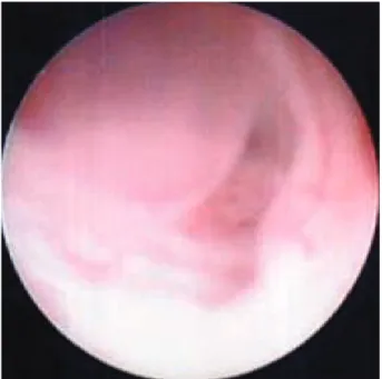
We could not find any ureteral or peri-ureteral lesion on either side, except for moderate hydronephrosis and dilatation of the ureter on abdominal CT. Surprisingly, ureteroscopic eval- uation showed over 20 small, separate FEPs from the mid- dle to upper ureters bilaterally (Fig. 2). Furthermore, on cys- toscopy, the lumens of the double-J stents were filled with small polyps without urine passage. We could not remove all of the lesions because the polyps were located diffusely over the entire ureter wall and we could not find any normal ureter mucosa. Furthermore, the ureter lumens were so nar- row that we could not resect the polypoid lesions while avoid- ing ureter injury. We were concerned about whether the rema- ining ureters would function normally without a long seg- mental ureteral stricture after the polyps were removed, even if it proved possible to remove all of the lesions. That is why we decided to maintain the percutaneous nephrostomy ca- theters. Histological examination of the resected specimen showed FEPs consisting of a core of fibrovascular tissue covered by a normal layer of transitional epithelium. Mild chronic inflammatory cell infiltration was seen in the stroma (Fig. 3).
DISCUSSION
Fibroepithelial polyps are mesodermal tumors that consist
Fig. 1. Radiologic images. (A) Antegrade pyelography through the nephrostomy catheter. The antegrade pyleograms revealed mod- erate dilatation in both renal pelvises and upper ureters, and the mid to lower ureter could not be visualized, even after 1 hr, imply- ing complete ureter obstruction. (B) Retrograde ureterogram show- ing a long segmental filling defect in the upper to middle ureter corresponding to the fibroepithelial polyps.
A B
Fig. 2. In situ visualization of small, multiple fibroepithelial polyps in the ureter. The ureteroscopic evaluation showed over 20 small, separate FEPs from the middle to upper ureters bilaterally.
Fig. 3. Histologic preparation of a fibroepithelial polyp. The stroma of fibroepithelial polyp is covered by a layer of normal transitional epithelial cells (H&E stain, ×200).
646 S.-O. Kim, C.W. Youn, T.W. Kang, et al.
of a fibrous core covered by normal urothelium (3). They often have smooth surfaces and are cylindrical, sessile, or even frond- like. FEPs can occur in newborns and adults older than 70 yr, but occur most commonly between the second and fourth decades (5). The male to female ratio is 3 to 2 and the left ureter is involved more commonly than the right, with about 70% of lesions occurring in the left ureter (3, 4). In adults, approximately 62% of these tumors occur at the ureteropelvic junction or in the upper ureter (3, 4). They can be smaller than 50 mm, ranging in size from 1 to 50 mm, with the largest reported polyp measuring 14 cm (6).
Macroscopically, FEPs can be divided into two categories:
long slender cylindrical masses that have a thin stalk connect- ed to the ureteral wall, or shorter, wider masses with multi- ple finger-like projections attached to a single base mass (7).
The etiology of these tumors is not clear. Chronic irritation, infection, hormonal imbalance, allergic factors, and develop- mental defects have all been suggested (8).
Gross or microscopic hematuria is the most common pre- senting clinical finding in adults (1, 9). The patients may be either completely asymptomatic or have unrelated symptoms.
Consequently, many patients are diagnosed late or inciden- tally for their persisting symptoms despite seeking medical care earlier in the course of the disease (1, 10).
It is not always easy to document radiological findings that would lead to a diagnosis of FEP on either intravenous urog- raphy (IVU) or RU, as in our case. On IVU, hydronephrosis is detected in 59% of cases. Ureteral filling defects are detect- ed in 26% and 48% of cases using IVU and RU, respectively (1). Our patient had an intermittent 4-yr history of dark-col- ored urine, which was probably hematuria. The renal US and abdominal CT findings from 4 yr ago to the most recent follow-up imaging study, obtained 6 months earlier, showed no specific abnormalities in the kidneys or ureters. These find- ings suggest that the slow-growing FEPs, which were prob- ably congenital in origin, could not be detected with ordi- nary radiological studies. This also warns us to evaluate pati- ents with hematuria thoroughly, including endoscopy in select- ed patients.
Multiple, bilateral FEPs are extremely rare. No previous studies have specifically reported bilateral, multiple FEPs located in mid-ureter. Our case has many other unusual fea- tures: the location in mid-ureter bilaterally; the clinical ap-
pearance with normal radiologic findings 6 months before the diagnosis was made and the delayed diagnosis on urete- roscopy; and the inability to remove the masses completely due to their small size and number. This report calls atten- tion to a rare manifestation of ureteral FEPs, which should be included in the differential diagnosis of long-lasting, undi- agnosed microscopic or gross hematuria with normal initial radiologic findings. In this situation, ureteroscopy could also be attempted to obtain a diagnosis and devise the proper man- agement. Moreover, bilateral FEPs should be included in the differential diagnosis of obstructive uropathy in advanced abdominal cancer patients.
REFERENCES
1. Franco I, Choudhury M, Eshghi M, Bhalodi A, Addonizio JC. Fib- roepithelial polyp associated with congenital ureteral diverticulum:
report of 2 cases. J Urol 1988; 140: 598-600.
2. Kiel H, Ullrich T, Roessler W, Wieland WF, Knuechel-Clarke R.
Benign ureteral tumors. Four case reports and a review of the liter- ature. Urol Int 1999; 63: 201-5.
3. Mariscal A, Mate JL, Guasch I, Casas D. Cystic transformation of a fibroepithelial polyp of the renal pelvis: radiologic and pathologic findings. AJR Am J Roentgenol 1995; 164: 1445-6.
4. Liddell RM, Weinberger E, Schofield DE, Pelman RS. Fibroepithe- lial polyp of the ureter in a child. AJR Am J Roentgenol 1991; 157:
1273-4.
5. Van Poppel H, Nuttin B, Oyen R, Stessens R, Van Damme B, Ver- duyn H. Fibroepithelial polyps of the ureter. Eur Urol 1986; 12:
174-9.
6. Lavelle JP, Knisely AS, Bellinger MF. Benign fibroepithelial polyps causing symptomatic bilateral intermittent hydroureteronephrosis. J Urol 1997; 158: 569.
7. Psihramis KE, Hartwick W. Ureteral fibroepithelial polyp with pos- itive urinary cytology. Urology 1993; 41: 387-91.
8. Cassar Delia E, Joseph VT, Sherwood W. Fibroepithelial polyps causing ureteropelvic junction obstruction in children-a case report and review article. Eur J Pediatr Surg 2007; 17: 142-6.
9. Bartone FF, Johansson SL, Markin RJ, Imray TJ. Bilateral fibroep- ithelial polyps of ureter in a child. Urology 1990; 35: 519-22.
10. Bolton D, Stoller ML, Irby P 3rd. Fibroepithelial ureteral polyps and urolithiasis. Urology 1994; 44: 582-7.